Comprehensive Overview of Spinal Cord Stimulation for Pain Management
1.
Spinal Cord Stimulation
Dr.Shirish Prasad Amatya, MD, FIPM, FIPP, CIPS
Fellowship in Musculoskeletal Ultrasound in Pain Medicine
Fellowship in Spine Endoscopy
Daradia: The Pain Clinic
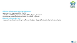
Alteration of Localneurochemistry (Modulation):
Suppresses the hyperexcitability of WDC
Activation of inhibitory neurotransmitter: GABA, Glycine, Serotonin
Inhibition of Excitatory neurotransmitter: Glutamate, Aspartate
Alteration of Sympathetic tone :
Increased vasodilatation and improve flow of Blood and Oxygen ( for Vascular Dz, Refractory Angina)
6.
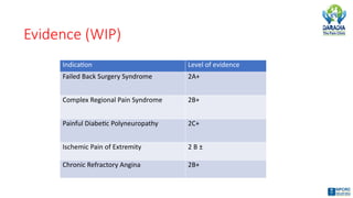
Evidence (WIP)
Indication Levelof evidence
Failed Back Surgery Syndrome 2A+
Complex Regional Pain Syndrome 2B+
Painful Diabetic Polyneuropathy 2C+
Ischemic Pain of Extremity 2 B ±
Chronic Refractory Angina 2B+
7.
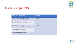
Evidence (ASIPP)
Spinal cordStimulation Evidence
Failed Back Surgery Syndrome Level II
Complex Regional Pain Syndrome
Diabetic Neuropathy Level IV
Abdominal/ Pelvic Pain
Ischemic Pain Syndrome
8.
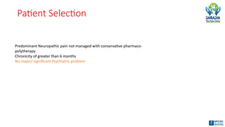
Patient Selection
Predominant Neuropathicpain not managed with conservative pharmaco-
polytherapy
Chronicity of greater than 6 months
No major/ significant Psychiatric problem
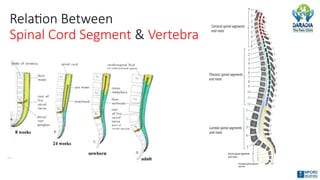
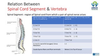
Relation Between
Spinal CordSegment & Vertebra
Spinal Segment : region of Spinal cord from which a pair of spinal nerve arises
Spinal cord segment ( 31 segments: C 8,T
12, L 5, S 5 and Co 1)
Vertebra level ( 33: C 7, T 12, L 5, S 5
and Co 4)
C 1 to C 4 C 1 to C 4 ( - 0 )
C 5 to T 4 C 4 to T 3 ( - 1)
T 5 to T 8 T 3 to T 6 ( - 2)
T 9 to T 12 T 6 to T 9 ( - 3 )
L 1 to L 5 T 9 to T 12
S 1 to C1 ( sacral & Coccygeal, Conus
Medullaris
L 1
Cauda Equina fibers and filum terminalis Below L1 to Tip of Coccyx
11.
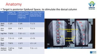
Anatomy
• Target isposterior Epidural Space, to stimulate the dorsal column
Pain Target Tip
of Catheter
Epidural Entry
Level ( 2 Level
below the
target tip )
Skin Entry level ( 2
level below the
Epidural entry)
Neck
Pain
C 2/3 C 5/6 T 1 / 2
Arm
Pain
C3/4 C 6/ 7 T 2 / 3
Leg Pain T 9/10 T 12 – L 1 L 2 /3
Foot
Pain
T 11- L1 L 3/4 L 5- S1
Back
Pain
T 8/11 L 1/ 2 L 3 – L 4
Abdomi
nal Pain
T 4/5 to
T5/6
T 8/9 T 11 – L 1
12.
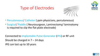
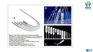
Type of Electrodes
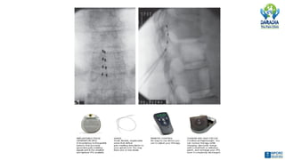
•Percutaneous/ Catheter ( pain physicians, percutaneous)
• Surgical/ Paddle ( Neurosurgeon, Laminectomy/ laminotomy
is required to slip the flat plate electrode)
Connected to Implantable Pulse Generator (IPG) or RF unit
Should be charged in 7 – 14 days
IPG can last up to 10 years
14.
Technical consideration
• Technicallychallenging interventional pain management technique.
• Involves placement of electrode leads in Posterior or Postero- lateral
epidural space under fluoroscopic guidance.
• Anchoring the lead, tunneling and connection of the connecting wires
• Trial period
• Positioning and implantation of Implantable Pulse Generator or
Radiofrequency receiver ( lower abdominal or posterior superior gluteal
area, should be located where patient can access with his/ her dominant
hand for adjustment of setting with remote control).
15.
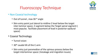
Fluoroscopy Technique
• NonCoaxial technology
• Out of tunnel , max 30 * angle
• Skin entry point just lateral to midline 2 level below the target
inter-laminar space; 2 segment below the Target spinal segment (
most popular, facilitate placement of lead in posterior epidural
space)
• Coaxial Technology
• Tunnel vision
• 30* caudal tilt of the C-arm
• Skin entry just paramedian of the spinous process below the
target interlaminar space ( breakage and migration issues)
16.
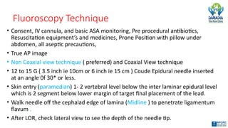
Fluoroscopy Technique
• Consent,IV cannula, and basic ASA monitoring, Pre procedural antibiotics,
Resuscitation equipment’s and medicines, Prone Position with pillow under
abdomen, all aseptic precautions,
• True AP image
• Non Coaxial view technique ( preferred) and Coaxial View technique
• 12 to 15 G ( 3.5 inch ie 10cm or 6 inch ie 15 cm ) Coude Epidural needle inserted
at an angle 0f 30* or less.
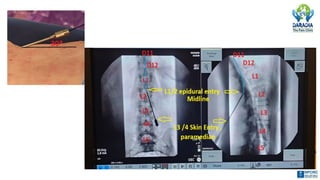
• Skin entry (paramedian) 1- 2 vertebral level below the inter laminar epidural level
which is 2 segment below lower margin of target final placement of the lead.
• Walk needle off the cephalad edge of lamina (Midline ) to penetrate ligamentum
flavum .
• After LOR, check lateral view to see the depth of the needle tip.
18.
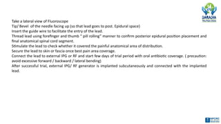
Take a lateralview of Fluoroscope
Tip/ Bevel of the needle facing up (so that lead goes to post. Epidural space)
Insert the guide wire to facilitate the entry of the lead.
Thread lead using forefinger and thumb “ pill rolling” manner to confirm posterior epidural position placement and
final anatomical spinal cord segment.
Stimulate the lead to check whether it covered the painful anatomical area of distribution.
Secure the lead to skin or fascia once best pain area coverage.
Connect the lead to external IPG or RF and start few days of trial period with oral antibiotic coverage. ( precaution:
avoid excessive forward / backward / lateral bending)
After successful trial, external IPG/ RF generator is implanted subcutaneously and connected with the implanted
lead.
20.
Trial Period
• Essentialto avoid the failed implant
• Trial period is about 3-14 days;
if < misleading success ,
if > risk of infection ( oral antibiotics during trial
stimulation)
normally 5-7 of trail period is better
21.
Program
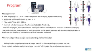
4 basic parameters
•Rate: frequency 20 – 120 Hz ( lower rate patient feel thumping, higher rate buzzing)
• Amplitude: intensity of current eg 0.1 – 10 V
• Pulse width/Time: 100 – 400 ms
• Electrode selection {electrons flow from cathode (-) to Anode (+) ,
selection is complex, electrode selection is changed until the patient obtains adequate anatomical coverage)
( electrode selection, rate and time of pulse is adjusted in trial period; patient is left with increase or decrease of
amplitude and duration of stimulation to achieve adequate analgesia)
All movement permitted: except: excessive bending and lateral rotation
Battery has to charged via external recharger every 7- 14 days depending upon mode and use.
Preset mode is available : patient can choose ; turn on/ off; increase the Amplitude or duration etc
22.
Goal / SuccessfulTrial
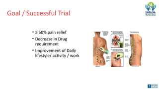
• ≥ 50% pain relief
• Decrease in Drug
requirement
• Improvement of Daily
lifestyle/ activity / work
23.
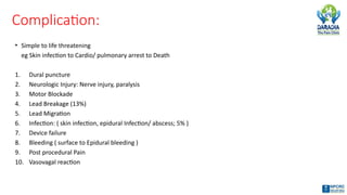
Complication:
• Simple tolife threatening
eg Skin infection to Cardio/ pulmonary arrest to Death
1. Dural puncture
2. Neurologic Injury: Nerve injury, paralysis
3. Motor Blockade
4. Lead Breakage (13%)
5. Lead Migration
6. Infection: ( skin infection, epidural Infection/ abscess; 5% )
7. Device failure
8. Bleeding ( surface to Epidural bleeding )
9. Post procedural Pain
10. Vasovagal reaction
#1 In SCS we use pulsed electrical energy near the spinal cord to control pain.
#2 Neurostimulaton;
1st placed in intrathecal space by Shealy; but now neurostimulation most commonly involves the implantation of leads in the epidural space to transmit pulsed energy across the spinal cord or near the desired nerve roots.
Same technology can be applied in deep brain stimulation, cortical brain stimulation and peripheral nerve stimulation.
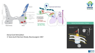
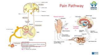
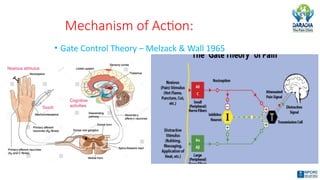
#4 Painful stimulation as well as sensation of touch and vibration from peripheral nerve has to pass through the same gate (substantia gelatinosa) of the dorsal horn of the spinal cord.
It was hypothesized that MANIPULATION OF INPUT; ie TOUCH AND VIBRATION CAN CLOSE THE GATE FOR TRANSMISSION OF PAIN.
Stimulation of Large Fibers (mylienated Aβfibers, carrying Touch/ Vibration), CLOSES THE GATE ( substantia gelatinosa located at dorsal Horn) by stimulation of Interneurons (gate keepers) to painful signals carried by Small Fibers ( unmyelinated C fibers and Thinly myelinated Aδfibers, carrying PAIN sensation )
#5 Recent studies has suggested changes in the local and supraspinal level and through dorsal horn interneuron and neurochemical mechanisms.
#8 50% of patient with neuropathic pain will have poor management with conservative therapy
Despite the recent increase in the number of agents available to treat neuropathic pain, a substantial number of patients still suffer from poorly controlled neuropathic pain.
Puig estimates as many as 50% of neuropathic pain patients have ineffective pain relief even with appropriate pharmacologic management.
Psychiatric screening to avoid failed implants
#11 Above levels reflect general concepts. The level of epidural entry and target level for the lead may vary based on patient size, anatomy, previous spinal surgery, pain location or other circumstances.
#12 Medtronic Inc
Advanced Bionic
American Neuromdulation System
#21 Stimulation should be applied with low intensity, just supra threshold for the activation of the low-threshold, large-diameter fibers, and should
be of non painful intensity. To be effective SCS must be applied continuously (or in cycles) for at least 20 minutes before the onset of analgesia.
This analgesia develops slowly and typically lasts several hours after cessation of the stimulation.
The patient is left with full control to (1) turn the stimulator on and off, (2) choose between numerous “programs” in their device (which have different
effects), and (3) adjust the intensity of stimulation up and down to comfort.
#23 Lead breakage/ migration : 13%; infection 5%
Staphyloccous infection common
 Most read