Comprehensive Overview of Membrane and Action Potentials in Neurons
Detailed exploration of membrane potentials, ion channels, action potential phases, and nerve impulse propagation including myelination and saltatory conduction.
Membrane potentials
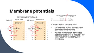
Caused byIon concentration
• Differences across a selectively
permeable membrane
• Normal mammalian nerve fiber,
potential difference is about 94 mv
with negativity inside the fiber
membrane
4.
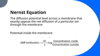
Nernst Equation
The diffusionpotential level across a membrane that
exactly oppose the net diffusion of a particular ion
through the membrane
Potential inside the membrane
5.
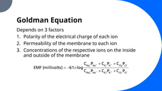
Goldman Equation
Depends on3 factors
1. Polarity of the electrical charge of each ion
2. Permeability of the membrane to each ion
3. Concentrations of the respective ions on the inside
and outside of the membrane
Origin of theNormal Resting
Membrane Potential
1. Contribution of the K+
Diffusion Potential
2. Contribution of Na+
Diffusion through the nerve
potential
3. Contribution of the Na+
-K+
Pump
8.
Origin of theNormal Resting
Membrane Potential
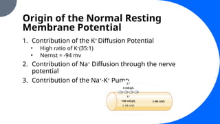
1. Contribution of the K+
Diffusion Potential
• High ratio of K+
(35:1)
• Nernst = -94 mv
2. Contribution of Na+
Diffusion through the nerve
potential
3. Contribution of the Na+
-K+
Pump
Origin of theNormal Resting
Membrane Potential
1. Contribution of the K+
Diffusion Potential
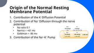
2. Contribution of Na+
Diffusion through the nerve
potential
• Na ratio 0.1
• Nernst = +61 mv
• Goldman = -86 mv
3. Contribution of the Na+
-K+
Pump
11.
Origin of theNormal Resting
Membrane Potential
1. Contribution of the K+
Diffusion Potential
2. Contribution of Na+
Diffusion through the nerve
potential
3. Contribution of the Na+
-K+
Pump
• Provides additional contribution to resting potential
• Causes continual loss of + charges from inside of the cell
• Additional -4 mv
12.
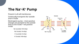
The Na+
-K+
Pump
Present inall cell membranes
Continually transports Na+
outside
and K+
inside
Electrogenic pump – more positive
charges are pumped to the outside
than to the inside = net positive
inside
Excitation
Process of elicitingthe action
potential
Any factor that causes Na+
to begin to
diffuse inward through the
membrane in sufficient numbers can
set off automatic regenerative
opening of the Na channels
May be mechanical, chemical, or
electrical
Mechanical – touch receptors,
mechanoreceptors
Chemical – neurotransmitters from
one neuron to the next in the brain
Electrical – electrical current to
transmit signals between successive
muscle cells in the heart and
intestine
16.
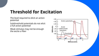
Threshold for Excitation
Thelevel required to elicit an action
potential
Subthreshold potentials do not elicit
a full action potential
Weak stimulus may not be enough
the excite a fiber
17.
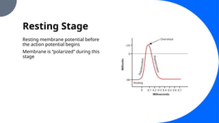
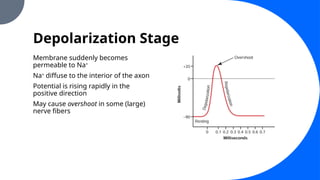
Depolarization Stage
Membrane suddenlybecomes
permeable to Na+
Na+
diffuse to the interior of the axon
Potential is rising rapidly in the
positive direction
May cause overshoot in some (large)
nerve fibers
18.
Refractory Period
• Whilethe membrane is still depolarized, a new action potential cannot occur
• Na and Ca channels are inactivated and no amount of excitatory signal
applied will open inactivation gates
• Absolute refractory period
• Period during which a second action potential cannot be elicited, even with
a strong stimulus
19.
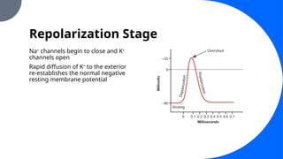
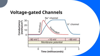
Repolarization Stage
Na+
channels beginto close and K+
channels open
Rapid diffusion of K+
to the exterior
re-establishes the normal negative
resting membrane potential
20.
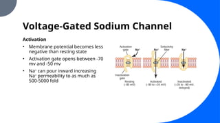
Voltage-Gated Sodium Channel
Activation
•Membrane potential becomes less
negative than resting state
• Activation gate opens between -70
mv and -50 mv
• Na+
can pour inward increasing
Na+
permeability to as much as
500-5000 fold
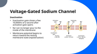
21.
Voltage-Gated Sodium Channel
Inactivation
•Inactivation gate closes a few
10,000ths of a second after
activation gate opens
• Na+
can no longer pour to the
inside of the membrane
• Membrane potential begins to
return toward the resting
membrane state (repolarization)
22.
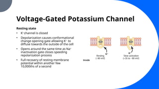
Voltage-Gated Potassium Channel
Restingstate
• K+
channel is closed
• Depolarization causes conformational
change opening gate allowing K+
to
diffuse towards the outside of the cell
• Opens around the same time as Na+
inactivation gate closes speeding
repolarization process
• Full recovery of resting membrane
potential within another few
10,000ths of a second
Voltage-gated Calcium Channel
•Serves along with (or instead of) Na+
in some cells to
cause most of the action potential
• Internal < external
• Depolarization causes influx of calcium into the cell
• Known as “slow channels”
• Slower than Na+
channels
• Provides a more sustained depolarization
25.
Voltage-gated Calcium Channel
•Deficit of calcium ions lead to hyperexcitability of Na+
channels
• Ca2+
binds to the exterior surfaces of the Na+
channel
protein molecule altering the electrical state of the
sodium channel, thereby lowering the voltage level
required to open the sodium gate
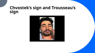
• Fall of 50% below normal may start causing
spontaneous discharges leading to muscle “tetany”
Initiation of theAction Potential
1. Positive-Feedback Cycle opens the Na+
channels
2. Threshold for initiation of the Action Potential
• Usually around -65 mv
“All or Nothing Principle”
- Once the threshold is reached, Action potential is
propagated regardless of the strength of the stimulus
28.
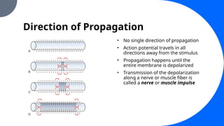
Direction of Propagation
•No single direction of propagation
• Action potential travels in all
directions away from the stimulus
• Propagation happens until the
entire membrane is depolarized
• Transmission of the depolarization
along a nerve or muscle fiber is
called a nerve or muscle impulse
29.
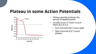
Plateau in someAction Potentials
• Plateau greatly prolongs the
period of depolarization
• Usually occurs in heart muscle
fibers (0.2-0.3 s)
• Fast channels (Na+
) cause spike
• Slow channels (Ca2+
) cause
plateau
30.
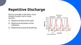
Repetitive Discharge
Occurs normallyin the heart, most
smooth muscle, and in many
neurons
1) Rhythmical beat of the heart
2) Rhythmical peristalsis of the
intestines
3) Rhythmical control of breathing
31.
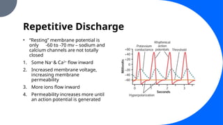
Repetitive Discharge
• “Resting”membrane potential is
only -60 to -70 mv – sodium and
calcium channels are not totally
closed
1. Some Na+
& Ca2+
flow inward
2. Increased membrane voltage,
increasing membrane
permeability
3. More ions flow inward
4. Permeability increases more until
an action potential is generated
32.
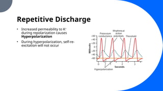
Repetitive Discharge
• Increasedpermeability to K+
during repolarization causes
Hyperpolarization
• During hyperpolarization, self-re-
excitation will not occur
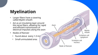
Myelination
• Larger fibershave a covering
called Myelin sheath
• Act as an insulating layer around
the nerve fibers, allowing for rapid
and efficient transmission of
electrical impulses along the axon
• Nodes of Ranvier
• found about every 1-3 mm
• Small uninsulated area
35.
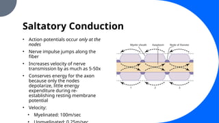
Saltatory Conduction
• Actionpotentials occur only at the
nodes
• Nerve impulse jumps along the
fiber
• Increases velocity of nerve
transmission by as much as 5-50x
• Conserves energy for the axon
because only the nodes
depolarize, little energy
expenditure during re-
establishing resting membrane
potential
• Velocity:
• Myelinated: 100m/sec
#3 K+ is great inside a nerve fiber but very low outside the membrane
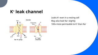
Strong tendency for extra numbers of K+ to diffuse outward through the membrane
Tendency to carry + charge outside of the cell thus creating electropositivity outside the membrane and electronegativity inside because of negative anions that remain behind
Diffusion potential the potential difference between the inside and outside becomes great enough to block further net K+ diffusion to the exterior despite high K+ concentration gradient
#4 Nernst potential is the potential inside the membrane
#5 Na, K, Cl – most important ions involved in nerve and muscle membrane potentials
#25 Calcium
Internal concentration 10,000 fold less than external concentration
Sustained depolarization in contrast to Na, initiation of AP
#26 Calcium
Internal concentration 10,000 fold less than external concentration
Sustained depolarization in contrast to Na, initiation of AP
#29 midportion suddenly develops increased permeability to sodium. The arrows show a “local circuit” of current flow from the depolarized areas of the membrane to the adjacent resting membrane areas. That is, positive electrical charges are carried by the inward-diffusing sodium ions through the depolarized membrane and then for several millimeters in both direc tions along the core of the axon. These positive charges increase the voltage for a distance of 1 to 3 millimeters inside the large myelinated fiber to above the threshold voltage value for initiating an action potential. Therefore, the sodium channels in these new areas immediately open, as shown in Figure 5-11C and D, and the explosive action potential spreads. These newly depolarized areas produce still more local circuits of current flow farther along the membrane, causing progressively more and more depolarization. Thus, the depolarization process travels along the entire length of the fiber. This transmis sion of the depolarization process along a nerve or muscle f iber is called a nerve or muscle impulse
#36 Electrical current flows through the surrounding extracellular fluid outside the myelin sheath as well as through the axoplasm inside the axon from node to node
Exciting successive nodes one after another