Comprehensive Overview of Culture-Dependent and Independent Microbial Analysis Methods
Explores key culture-dependent and culture-independent techniques for studying microorganisms, including molecular profiling, omics technologies, and microscopy, highlighting their advantages and limitations in microbial research.
Comprehensive Overview of Culture-Dependent and Independent Microbial Analysis Methods
2.
1. Culture-Dependent Methods
Thesemethods are foundational to
microbiology and rely on isolating and
growing microorganisms in pure or
mixed cultures. [1, 2]
Key Techniques
•Inoculation & Incubation: Introducing
specimens to nutrient media (agar,
broth) and maintaining optimal
temperature, pH, and oxygen levels for
growth. [1, 2]
•Isolation Techniques: Methods to
separate individual cells to form pure
colonies. Common examples include the
Streak Plate Method, Pour Plate
Method, and Spread Plate Method. [1,
2, 3, 4]
•Media Types: Using specialized media
to isolate specific types of microbes.
Common classifications include
Selective Media, Differential Media, and
Enrichment Media. [1, 2, 3]
3.
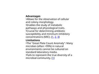
Advantages
•Allows for theobservation of cellular
and colony morphology.
•Enables the study of metabolic
pathways and physiological traits.
•Crucial for determining antibiotic
susceptibility and minimum inhibitory
concentrations (MIC). [1, 2, 3]
Limitations
•The "Great Plate Count Anomaly": Many
microbes (often >99%) in natural
environments cannot be cultured on
standard laboratory media.
•Fails to represent the true diversity of a
microbial community. [1]
4.
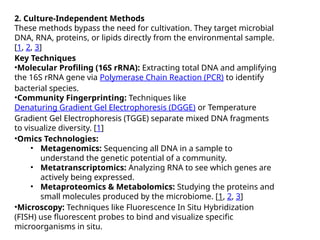
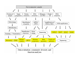
2. Culture-Independent Methods
Thesemethods bypass the need for cultivation. They target microbial
DNA, RNA, proteins, or lipids directly from the environmental sample.
[1, 2, 3]
Key Techniques
•Molecular Profiling (16S rRNA): Extracting total DNA and amplifying
the 16S rRNA gene via Polymerase Chain Reaction (PCR) to identify
bacterial species.
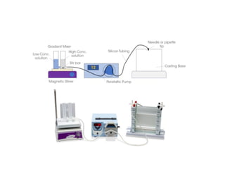
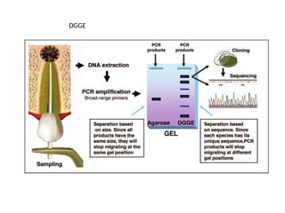
•Community Fingerprinting: Techniques like
Denaturing Gradient Gel Electrophoresis (DGGE) or Temperature
Gradient Gel Electrophoresis (TGGE) separate mixed DNA fragments
to visualize diversity. [1]
•Omics Technologies:
• Metagenomics: Sequencing all DNA in a sample to
understand the genetic potential of a community.
• Metatranscriptomics: Analyzing RNA to see which genes are
actively being expressed.
• Metaproteomics & Metabolomics: Studying the proteins and
small molecules produced by the microbiome. [1, 2, 3]
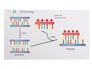
•Microscopy: Techniques like Fluorescence In Situ Hybridization
(FISH) use fluorescent probes to bind and visualize specific
microorganisms in situ.
5.
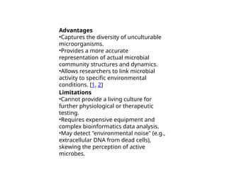
Advantages
•Captures the diversityof unculturable
microorganisms.
•Provides a more accurate
representation of actual microbial
community structures and dynamics.
•Allows researchers to link microbial
activity to specific environmental
conditions. [1, 2]
Limitations
•Cannot provide a living culture for
further physiological or therapeutic
testing.
•Requires expensive equipment and
complex bioinformatics data analysis.
•May detect "environmental noise" (e.g.,
extracellular DNA from dead cells),
skewing the perception of active
microbes.
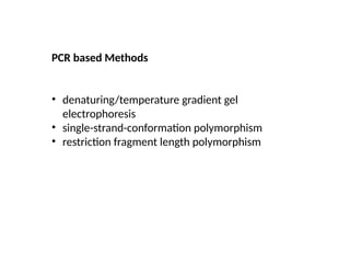
PCR based Methods
•denaturing/temperature gradient gel
electrophoresis
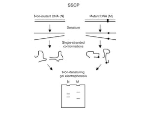
• single-strand-conformation polymorphism
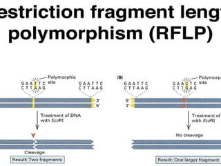
• restriction fragment length polymorphism
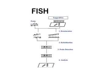
#10 Fluorescence in situ hybridization (abbreviated FISH) is a laboratory technique used to detect and locate a specific DNA sequence on a chromosome. In this technique, the full set of chromosomes from an individual is affixed to a glass slide and then exposed to a “probe”—a small piece of purified DNA tagged with a fluorescent dye. The fluorescently labeled probe finds and then binds to its matching sequence within the set of chromosomes. With the use of a special microscope, the chromosome and sub-chromosomal location where the fluorescent probe bound can be seen.

 Most read