OUR OBJECTIVE
Basictheory of circulatory.
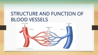
Structure and function of blood vessels
Blood Pressure intro and methods
Regulations of blood pressure.
Factor and physiology variation
Total peripheral resistance
Atrial Pressure
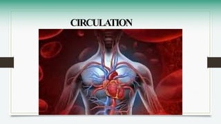
CIRCULATION
Circulation ishow the body moves blood, oxygen, and nutrients to every part of
the body.
• The heart works like a pump. It beats to push blood around the body.
• Blood travels through tubes called blood vessels — arteries, veins, and
capillaries.
• Arteries carry fresh, oxygen-rich blood away from the heart to the body.
• Veins carry blood back to the heart after the oxygen has been used.
• Capillaries are tiny vessels where oxygen and nutrients pass into body cells,
and waste (like carbon dioxide) is picked up.
BLOOD VESSELS
Bloodvessels are a closed network of tubes that transport blood throughout the
body, delivering oxygen and nutrients to tissues while removing metabolic waste.
General Structure of Blood Vessel Walls
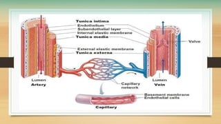
Most major blood vessels consist of three distinct tissue layers (tunics) surrounding a
central hollow space called the lumen:
• Tunica Intima (Inner Layer): Made of a smooth layer of endothelial cells that
minimize friction as blood flows through.
• Tunica Media (Middle Layer): Composed of smooth muscle fibers and elastic
tissue; controls vessel diameter through contraction (vasoconstriction) and
relaxation (vasodilation).
• Tunica Externa / Adventitia (Outer Layer): Made of strong collagen fibers that
protect and anchor the vessel to surrounding tissues.
12.
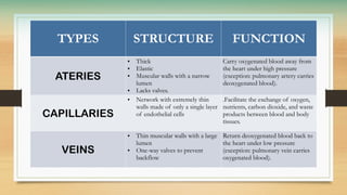
TYPES STRUCTURE FUNCTION
ATERIES
•Thick
• Elastic
• Muscular walls with a narrow
lumen
• Lacks valves.
Carry oxygenated blood away from
the heart under high pressure
(exception: pulmonary artery carries
deoxygenated blood).
CAPILLARIES
• Network with extremely thin
walls made of only a single layer
of endothelial cells
.Facilitate the exchange of oxygen,
nutrients, carbon dioxide, and waste
products between blood and body
tissues.
VEINS
• Thin muscular walls with a large
lumen
• One-way valves to prevent
backflow
Return deoxygenated blood back to
the heart under low pressure
(exception: pulmonary vein carries
oxygenated blood).
13.
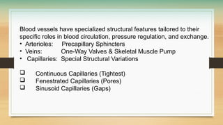
Blood vessels havespecialized structural features tailored to their
specific roles in blood circulation, pressure regulation, and exchange.
• Arterioles: Precapillary Sphincters
• Veins: One-Way Valves & Skeletal Muscle Pump
• Capillaries: Special Structural Variations
Continuous Capillaries (Tightest)
Fenestrated Capillaries (Pores)
Sinusoid Capillaries (Gaps)
14.
Another terminology
Vasa Vasorum(Vessels of the Vessels)A network of tiny blood
vessels located inside the outer layer (tunica externa) of very large
arteries and veins (like the Aorta and Vena Cava).
Function
• Large vessel walls are too thick for oxygen to diffuse all the way
through from the inside.
• The vasa vasorum acts as a personal blood supply to nourish the
outer layers of these massive vessels.
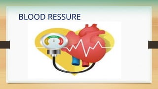
Blood pressure (BP)*is the pressure exerted by circulating blood against the
walls of blood vessels, especially arteries.
It is generated primarily by the pumping action of the left ventricle.
Blood pressure is essential for maintaining adequate blood flow to:
• Brain
• Heart
• Kidneys
• Muscles
• Other tissues
BP varies continuously throughout the cardiac cycle.
17.
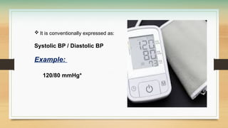
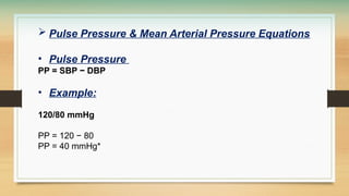
It isconventionally expressed as:
Systolic BP / Diastolic BP
Example:
120/80 mmHg*
18.
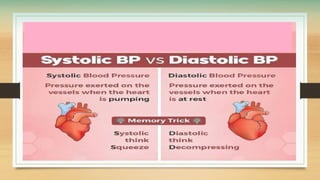
Systolic BP(SBP):
• Maximum arterial pressure during ventricular contraction.
• Represents the peak pressure generated by the heart.
Diastolic BP (DBP):
• Minimum arterial pressure during ventricular relaxation.
• Represents the pressure maintained in arteries between
heartbeats.
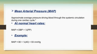
Mean ArterialPressure (MAP)
Approximate average pressure driving blood through the systemic circulation
during one cardiac cycle.*
• At normal heart rates:
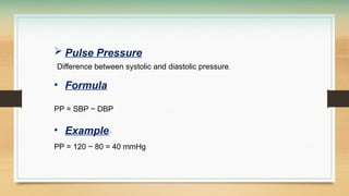
MAP ≈ DBP + ⅓(PP)
• Example:
MAP ≈ 80 + ⅓(40) = 93 mmHg
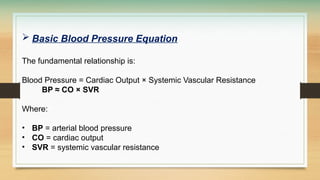
Basic BloodPressure Equation
The fundamental relationship is:
Blood Pressure = Cardiac Output × Systemic Vascular Resistance
BP ≈ CO × SVR
Where:
• BP = arterial blood pressure
• CO = cardiac output
• SVR = systemic vascular resistance
24.
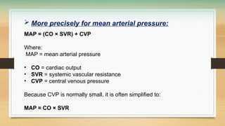
More preciselyfor mean arterial pressure:
MAP = (CO × SVR) + CVP
Where:
MAP = mean arterial pressure
• CO = cardiac output
• SVR = systemic vascular resistance
• CVP = central venous pressure
Because CVP is normally small, it is often simplified to:
MAP ≈ CO × SVR
25.
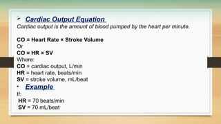
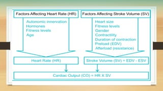
Cardiac OutputEquation
Cardiac output is the amount of blood pumped by the heart per minute.
CO = Heart Rate × Stroke Volume
Or
CO = HR × SV
Where:
CO = cardiac output, L/min
HR = heart rate, beats/min
SV = stroke volume, mL/beat
• Example
If:
HR = 70 beats/min
SV = 70 mL/beat
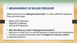
MEASUREMENT OFBLOOD PRESSURE
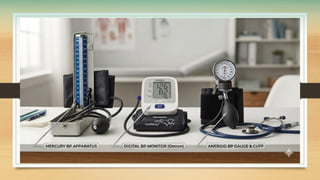
Blood pressure by the Sphygmomanometer. It is also called B.P apparatus.
There are three types:
• Mercury B.P apparatus
• Digital B.P apparatus
• Android B.P apparatus
Why Was Mercury Used in Sphygmomanometers?
• Mercury is a metal, but it is unusual because it is liquid at room temperature.
• A mercury sphygmomanometer uses the height of a mercury column
to represent pressure.
29.
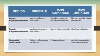
METHOD PRINCIPLE
MAIN
ADVANTAGE
MAIN
LIMITATION
Mercury
sphygmomanometer
Mercury column+
auscultation
Excellent reference
technique historically
Mercury toxicity; being
phased out
Aneroid
sphygmomanometer
Mechanical gauge +
auscultation
Mercury-free, portable Can lose calibration
Automated
oscillometric
Detects cuff pressure
oscillations
| Easy and rapid Algorithm-dependent;
validation important
CARDIAC OUTPUT
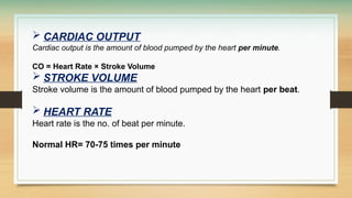
Cardiacoutput is the amount of blood pumped by the heart per minute.
CO = Heart Rate × Stroke Volume
STROKE VOLUME
Stroke volume is the amount of blood pumped by the heart per beat.
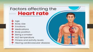
HEART RATE
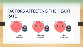
Heart rate is the no. of beat per minute.
Normal HR= 70-75 times per minute
34.
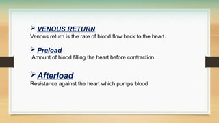
VENOUS RETURN
Venousreturn is the rate of blood flow back to the heart.
Preload
Amount of blood filling the heart before contraction
Afterload
Resistance against the heart which pumps blood
AUTONOMIC INNERVATION
Theautonomic nervous system regulates the heart rate, contraction
force, and electrical conduction through the Cardiac Plexus,
which contains both
• sympathetic
• parasympathetic fibers.
38.
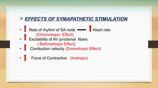
EFFECTS OFSYMAPATHETIC STIMULATION
• Rate of rhythm of SA node Heart rate
(Chronotropic Effect)
• Excitability of AV junctional fibers
( Bathmotropic Effect)
• Conduction velocity (Dromotropic Effect)
• Force of Contraction (Inotropic)
39.
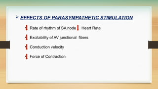
EFFECTS OFPARASYMPATHETIC STIMULATION
• Rate of rhythm of SA node Heart Rate
• Excitability of AV junctional fibers
• Conduction velocity
• Force of Contraction
42.
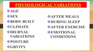
PHYSIOLOGICAL VARIATIONSOF BLOOD
PRESSURE
Physiological variation of blood pressure refers to the
normal, healthy ups and downs in blood pressure caused by
daily activities, age, and body functions. Blood pressure is
not fixed; it changes all the time to match what your body
needs.
44.
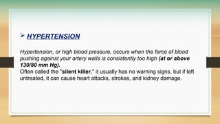
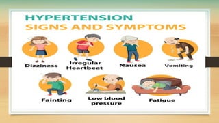
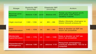
HYPERTENSION
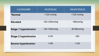
Hypertension, orhigh blood pressure, occurs when the force of blood
pushing against your artery walls is consistently too high (at or above
130/80 mm Hg).
Often called the "silent killer," it usually has no warning signs, but if left
untreated, it can cause heart attacks, strokes, and kidney damage.
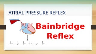
ATRIAL PRESSUREREFLEX
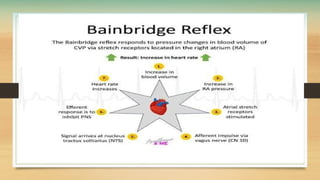
The atrial pressure reflex, commonly known as the Bainbridge Reflex, is a
physiological mechanism that increases heart rate in response to an elevated blood
volume and increased pressure within the right atrium of the heart.
How the Reflex Works
•Stimulus: High blood volume increases venous return, which stretches the walls
of the right atrium.
•Receptors: Low-pressure stretch mechanoreceptors in the atrial walls detect this
tension.
•Afferent Pathway: Signals travel via the vagus nerve to the brainstem (medulla
oblongata).
•Efferent Response: The brain decreases parasympathetic (vagal) tone and
increases sympathetic nervous system activity to the heart's sinoatrial (SA) node.
•Result: Heart rate speeds up (tachycardia) to pump the extra volume forward
TOTAL PERPHERALRESISTANCE
The resistance that blood must overcome to move through the systemic
blood vessels.
• It is a major factor in setting your blood pressure.
• Small muscular arteries called arterioles control most of this resistance by
changing their size.
Factors
•Vessel Radius: A smaller opening makes high resistance. A larger
opening makes low resistance.
•Blood Viscosity: Thicker blood increases resistance. Thinner blood
decreases it.
•Vessel Length: Longer blood vessels increase total resistance.
52.
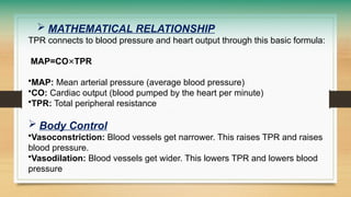
MATHEMATICAL RELATIONSHIP
TPRconnects to blood pressure and heart output through this basic formula:
MAP=CO×TPR
•MAP: Mean arterial pressure (average blood pressure)
•CO: Cardiac output (blood pumped by the heart per minute)
•TPR: Total peripheral resistance
Body Control
•Vasoconstriction: Blood vessels get narrower. This raises TPR and raises
blood pressure.
•Vasodilation: Blood vessels get wider. This lowers TPR and lowers blood
pressure




 Most read