Comprehensive Overview of Cellular Adaptations in Health and Disease
Detailed exploration of cellular adaptations including atrophy, hypertrophy, hyperplasia, metaplasia, and dysplasia with physiological and pathological examples.
Comprehensive Overview of Cellular Adaptations in Health and Disease
1.
CELLULAR ADAPTATIONS
PREPARED BY-MR. SUKHBIR SINGH TAMBER (ASST. PROFESSOR, PHARMACOLOGY)
UNIVERSITY SCHOOL OF PHARMACEUTICAL SCIENCES
2.
Definition
Adjustments madeby the cell when it encounters physiological or pathological stresses.
These adaptations are reversible in nature i.e. it gets reversed when stimulus is removed.
If stimulus persists for longer duration then cell may progress to die leads to cell death (apoptosis or necrosis).
Cellular adaptations can be physiological or pathological.
Physiological adaptations means response of cells to normal hormonal stimulations or endogenous chemicals e.g.-
enlargement of breast during puberty whereas Pathological adaptions are response to stress to modulate structure and
function of cells to escape injury.
3.
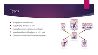
Types
Atrophy (Decreasein size)
Hypertrophy (Increase in size)
Hyperplasia (Increase in number of cells)
Metaplasia (Reversible change in cell type)
Dysplasia (disordered cellular development)
4.
ATROPHY
Term denotedfor decrease/ reduction cell size which leads to the shrinkage
of tissue or organ.
A) Physiologic atrophy -normal process of ageing in some tissues, For example:
i) Atrophy of lymphoid tissue with age.
ii) Atrophy of thymus in adult life.
iii) Atrophy of gonads after menopause.
iv) Atrophy of brain with ageing.
v) Osteoporosis with reduction in size of bony trabeculae due to ageing.
5.
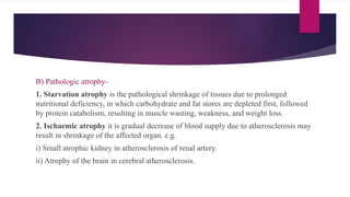
B) Pathologic atrophy-
1.Starvation atrophy is the pathological shrinkage of tissues due to prolonged
nutritional deficiency, in which carbohydrate and fat stores are depleted first, followed
by protein catabolism, resulting in muscle wasting, weakness, and weight loss.
2. Ischaemic atrophy it is gradual decrease of blood supply due to atherosclerosis may
result in shrinkage of the affected organ. e.g.
i) Small atrophic kidney in atherosclerosis of renal artery.
ii) Atrophy of the brain in cerebral atherosclerosis.
6.
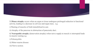
3. Disuse atrophyoccurs when an organ or tissue undergoes prolonged reduction in functional
activity, leading to a decrease in cell size and organ mass. e.g.
i) Wasting of muscles of limb immobilised in cast.
ii) Atrophy of the pancreas in obstruction of pancreatic duct.
4. Neuropathic atrophy (denervation atrophy) when nerve supply to muscle is interrupted leads
to muscle wasting e.g.
i) Poliomyelitis
ii) Motor neuron disease
iii) Nerve section.
7.
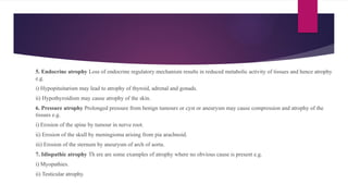
5. Endocrine atrophyLoss of endocrine regulatory mechanism results in reduced metabolic activity of tissues and hence atrophy
e.g.
i) Hypopituitarism may lead to atrophy of thyroid, adrenal and gonads.
ii) Hypothyroidism may cause atrophy of the skin.
6. Pressure atrophy Prolonged pressure from benign tumours or cyst or aneurysm may cause compression and atrophy of the
tissues e.g.
i) Erosion of the spine by tumour in nerve root.
ii) Erosion of the skull by meningioma arising from pia arachnoid.
iii) Erosion of the sternum by aneurysm of arch of aorta.
7. Idiopathic atrophy Th ere are some examples of atrophy where no obvious cause is present e.g.
i) Myopathies.
ii) Testicular atrophy.
8.
Hypertrophy
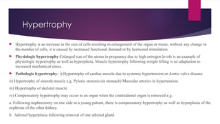
Hypertrophy isan increase in the size of cells resulting in enlargement of the organ or tissue, without any change in
the number of cells, it is caused by increased functional demand or by hormonal stimulation.
Physiologic hypertrophy-Enlarged size of the uterus in pregnancy due to high estrogen levels is an example of
physiologic hypertrophy as well as hyperplasia. Muscle hypertrophy following weight lifting is an adaptation to
increased mechanical stress.
Pathologic hypertrophy- i) Hypertrophy of cardiac muscle due to systemic hypertension or Aortic valve disease.
ii) Hypertrophy of smooth muscle e.g. Pyloric stenosis (in stomach) Muscular arteries in hypertension.
iii) Hypertrophy of skeletal muscle
iv) Compensatory hypertrophy may occur in an organ when the contralateral organ is removed e.g.
a. Following nephrectomy on one side in a young patient, there is compensatory hypertrophy as well as hyperplasia of the
nephrons of the other kidney.
b. Adrenal hyperplasia following removal of one adrenal gland.
9.
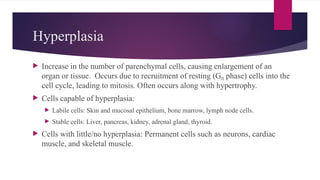
Hyperplasia
Increase inthe number of parenchymal cells, causing enlargement of an
organ or tissue. Occurs due to recruitment of resting (G phase) cells into the
₀
cell cycle, leading to mitosis. Often occurs along with hypertrophy.
Cells capable of hyperplasia:
Labile cells: Skin and mucosal epithelium, bone marrow, lymph node cells.
Stable cells: Liver, pancreas, kidney, adrenal gland, thyroid.
Cells with little/no hyperplasia: Permanent cells such as neurons, cardiac
muscle, and skeletal muscle.
10.
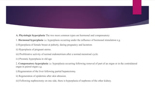
A. Physiologic hyperplasiaThe two most common types are hormonal and compensatory:
1. Hormonal hyperplasia i.e. hyperplasia occurring under the influence of hormonal stimulation e.g.
i) Hyperplasia of female breast at puberty, during pregnancy and lactation.
ii) Hyperplasia of pregnant uterus.
iii) Proliferative activity of normal endometrium after a normal menstrual cycle.
iv) Prostatic hyperplasia in old age.
2. Compensatory hyperplasia i.e. hyperplasia occurring following removal of part of an organ or in the contralateral
organ in paired organ e.g.
i) Regeneration of the liver following partial hepatectomy.
ii) Regeneration of epidermis after skin abrasion.
iii) Following nephrectomy on one side, there is hyperplasia of nephrons of the other kidney.
11.
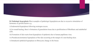
B. Pathologic hyperplasiaMost examples of pathologic hyperplasia are due to excessive stimulation of
hormones or growth factors e.g.
i) Endometrial hyperplasia following oestrogen excess.
ii) In wound healing, there is formation of granulation tissue due to proliferation of fibroblasts and endothelial
cells.
iii) Formation of skin warts from hyperplasia of epidermis due to human papilloma virus.
iv) Pseudocarcinomatous hyperplasia of the skin occurring at the margin of a non-healing ulcer.
v) Intraductal epithelial hyperplasia in fibrocystic change in the breast.
12.
Metaplasia
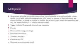
Metaplasia isdefined as a reversible change of one type of epithelial or mesenchymal adult cells to
another type of adult epithelial or mesenchymal cells, usually in response to abnormal stimuli, and
often reverts back to normal on removal of stimulus. The new cell type is usually less specialized and
may lose normal function (e.g., reduced mucus secretion).
Types- Epithelial Metaplasia & Mesenchymal Metaplasia
Causes:
a) Chronic irritation (e.g., smoking)
b) Persistent inflammation
c) Vitamin A deficiency
d) Chronic acid reflux
e) Hormonal or environmental stimuli
13.
Epithelial metaplasia
Epithelialmetaplasia is a reversible adaptive change in which one type of mature
epithelium is replaced by another mature epithelial type that is better able to withstand
chronic irritation or stress.
Types-
a) Squamous Metaplasia
b) Columnar Metaplasia
14.
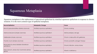
Squamous Metaplasia
Normal EpitheliumMetaplastic Change Cause
Bronchi (pseudostratified ciliated columnar) Stratified squamous epithelium Chronic smoking
Uterine endocervix (simple columnar) Stratified squamous epithelium Uterine prolapse, old age
Gallbladder (simple columnar) Stratified squamous epithelium Chronic cholecystitis with gallstones
Prostatic ducts (simple columnar) Stratified squamous epithelium Chronic prostatitis, estrogen therapy
Renal pelvis & urinary bladder (transitional
epithelium)
Stratified squamous epithelium Chronic infection, urinary stones
Nose, bronchi, urinary tract, lacrimal & salivary
glands
Stratified squamous epithelium Vitamin A deficiency
Squamous metaplasia is the replacement of specialized epithelium by stratified squamous epithelium in response to chronic
irritation. It is the most common type of epithelial metaplasia.
15.
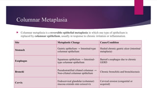
Columnar Metaplasia
Columnarmetaplasia is a reversible epithelial metaplasia in which one type of epithelium is
replaced by columnar epithelium, usually in response to chronic irritation or inflammation.
Site Metaplastic Change Cause/Condition
Stomach
Gastric epithelium → Intestinal-type
columnar epithelium
Healed chronic gastric ulcer (intestinal
metaplasia)
Esophagus
Squamous epithelium → Intestinal-
type columnar epithelium
Barrett's esophagus due to chronic
GERD
Bronchi
Pseudostratified ciliated columnar →
Non-ciliated columnar epithelium
Chronic bronchitis and bronchiectasis
Cervix
Endocervical glandular (columnar)
mucosa extends onto ectocervix
Cervical erosion (congenital or
acquired)
16.
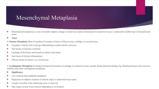
Mesenchymal Metaplasia
Mesenchymalmetaplasia is a rare, reversible adaptive change in which one mature mesenchymal (connective) tissue is replaced by another type of mesenchymal
tissue.
Types
1. Osseous Metaplasia (Bone Formation) Formation of bone in fibrous tissue, cartilage, or myxoid tissue.
Examples: Arterial wall in old age (Mönckeberg's medial calcific sclerosis)
Soft tissues in myositis ossificans
Cartilage of the larynx and bronchi in elderly individuals
Scar tissue of chronic inflammation
Fibrous stroma of tumors, e.g., leiomyoma
2. Cartilaginous Metaplasia (Cartilage Formation) Formation of cartilage in connective tissue, usually during abnormal healing. Eg.-Healing fractures with excessive
mobility may show cartilaginous metaplasia.
Significance
Less common than epithelial metaplasia.
Represents an adaptive response to chronic injury or abnormal tissue repair.
Usually reversible if the underlying cause is removed.
May impair normal tissue function depending on its location.
17.
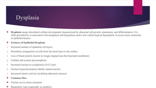
Dysplasia
Dysplasia meansdisordered cellular development characterized by abnormal cell growth, maturation, and differentiation. It is
often preceded by or associated with metaplasia and hyperplasia and is also called atypical hyperplasia. It occurs most commonly
in epithelial tissues.
Features of Epithelial Dysplasia
1. Increased number of epithelial cell layers
2. Disorderly arrangement of cells from the basal layer to the surface
3. Loss of basal polarity (nuclei no longer aligned near the basement membrane)
4. Cellular and nuclear pleomorphism
5. Increased nuclear-to-cytoplasmic (N:C) ratio
6. Nuclear hyperchromatism (darkly stained nuclei)
7. Increased mitotic activity (including abnormal mitoses)
Common Sites
Uterine cervix (most common)
Respiratory tract (especially in smokers)
18.
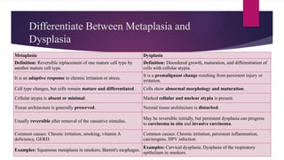
Differentiate Between Metaplasiaand
Dysplasia
Metaplasia Dysplasia
Definition: Reversible replacement of one mature cell type by
another mature cell type.
Definition: Disordered growth, maturation, and differentiation of
cells with cellular atypia.
It is an adaptive response to chronic irritation or stress.
It is a premalignant change resulting from persistent injury or
irritation.
Cell type changes, but cells remain mature and differentiated. Cells show abnormal morphology and maturation.
Cellular atypia is absent or minimal. Marked cellular and nuclear atypia is present.
Tissue architecture is generally preserved. Normal tissue architecture is disturbed.
Usually reversible after removal of the causative stimulus.
May be reversible initially, but persistent dysplasia can progress
to carcinoma in situ and invasive carcinoma.
Common causes: Chronic irritation, smoking, vitamin A
deficiency, GERD.
Common causes: Chronic irritation, persistent inflammation,
carcinogens, HPV infection.
Examples: Squamous metaplasia in smokers; Barrett's esophagus.
Examples: Cervical dysplasia; Dysplasia of the respiratory
epithelium in smokers.
19.
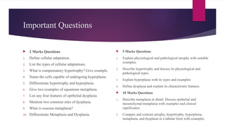
Important Questions
2Marks Questions
1. Define cellular adaptation.
2. List the types of cellular adaptations.
3. What is compensatory hypertrophy? Give example.
4. Name the cells capable of undergoing hyperplasia.
5. Differentiate hypertrophy and hyperplasia.
6. Give two examples of squamous metaplasia.
7. List any four features of epithelial dysplasia.
8. Mention two common sites of dysplasia.
9. What is osseous metaplasia?
10. Differentiate Metaplasia and Dysplasia.
5 Marks Questions
1. Explain physiological and pathological atrophy with suitable
examples.
2. Describe hypertrophy and discuss its physiological and
pathological types.
3. Explain hyperplasia with its types and examples.
4. Define dysplasia and explain its characteristic features.
10 Marks Questions
1. Describe metaplasia in detail. Discuss epithelial and
mesenchymal metaplasia with examples and clinical
significance.
2. Compare and contrast atrophy, hypertrophy, hyperplasia,
metaplasia, and dysplasia in a tabular form with examples.