Comprehensive Overview of Cell Injury: Mechanisms, Causes, and Cellular Responses
Detailed exploration of cell injury including causes like hypoxia and ischemia, reversible and irreversible injury mechanisms, ATP depletion, calcium influx, and cellular adaptations.
Comprehensive Overview of Cell Injury: Mechanisms, Causes, and Cellular Responses
1.
CELL INJURY
PREPARED BY-MR. SUKHBIR SINGH TAMBER (ASST. PROFESSOR, PHARMACOLOGY)
UNIVERSITY SCHOOL OF PHARMACEUTICAL SCIENCES
2.
Introduction
Cell injuryis defined as structural and functional damage that occurs when cells are exposed to harmful stimuli
(physical, chemical, infectious, or biological), resulting in changes in their internal and external environment.
Cells can tolerate stress up to a certain limit through adaptive mechanisms. When the stress exceeds this
capacity, cell injury occurs.
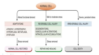
Cellular Responses to Cell Injury
1. Cellular Adaptation- Occurs when there is increased functional demand or mild stress. Cells undergo reversible
adaptive changes (e.g., atrophy, hypertrophy, hyperplasia, metaplasia). The cell returns to normal once the stress is
removed.
2. Reversible Cell Injury- Occurs due to mild to moderate injury. Structural and functional changes are temporary.
The cell recovers completely if the injurious stimulus is removed.
3. Irreversible Cell Injury- Occurs when the injury is severe or prolonged. The damage is permanent. Results
in cell death by necrosis or apoptosis.
4.
Etiology of CellInjury
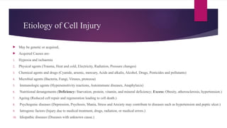
May be genetic or acquired,
Acquired Causes are-
1. Hypoxia and ischaemia
2. Physical agents (Trauma, Heat and cold, Electricity, Radiation, Pressure changes)
3. Chemical agents and drugs (Cyanide, arsenic, mercury, Acids and alkalis, Alcohol, Drugs, Pesticides and pollutants)
4. Microbial agents (Bacteria, Fungi, Viruses, protozoa)
5. Immunologic agents (Hypersensitivity reactions, Autoimmune diseases, Anaphylaxis)
6. Nutritional derangements (Deficiency: Starvation, protein, vitamin, and mineral deficiency; Excess: Obesity, atherosclerosis, hypertension.)
7. Ageing (Reduced cell repair and regeneration leading to cell death.)
8. Psychogenic diseases (Depression, Psychosis, Mania, Stress and Anxiety may contribute to diseases such as hypertension and peptic ulcer.)
9. Iatrogenic factors (Injury due to medical treatment, drugs, radiation, or medical errors.)
10. Idiopathic diseases (Diseases with unknown cause.)
5.
Hypoxia & Ischemia
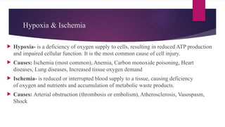
Hypoxia- is a deficiency of oxygen supply to cells, resulting in reduced ATP production
and impaired cellular function. It is the most common cause of cell injury.
Causes: Ischemia (most common), Anemia, Carbon monoxide poisoning, Heart
diseases, Lung diseases, Increased tissue oxygen demand
Ischemia- is reduced or interrupted blood supply to a tissue, causing deficiency
of oxygen and nutrients and accumulation of metabolic waste products.
Causes: Arterial obstruction (thrombosis or embolism), Atherosclerosis, Vasospasm,
Shock
6.
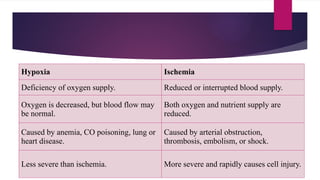
Hypoxia Ischemia
Deficiency ofoxygen supply. Reduced or interrupted blood supply.
Oxygen is decreased, but blood flow may
be normal.
Both oxygen and nutrient supply are
reduced.
Caused by anemia, CO poisoning, lung or
heart disease.
Caused by arterial obstruction,
thrombosis, embolism, or shock.
Less severe than ischemia. More severe and rapidly causes cell injury.
7.
Types of cellinjury
Reversible and Irreversible
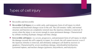
Reversible Cell Injury-it is a mild, early, and temporary form of cell injury in which
structural and functional changes occur in the cell. The cell remains viable, and normal
structure and function are completely restored once the injurious stimulus is removed. It
occurs when the injury is not severe enough to cause permanent damage. Characterized
by cellular swelling (hydropic change) and fatty change.
Irreversible cell injury- is a severe, progressive, and permanent form of cell injury in which
the damage exceeds the cell's capacity to repair. As a result, the cell cannot recover, even if
the injurious stimulus is removed, and ultimately undergoes cell death by necrosis or
apoptosis. Characterized by severe membrane damage, mitochondrial dysfunction,
lysosomal rupture, and nuclear changes (pyknosis, karyorrhexis, and karyolysis).
8.
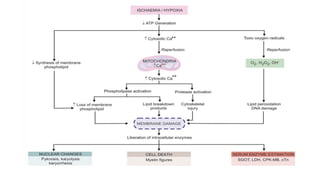
Pathogenesis of Ischaemicand
Hypoxic Injury
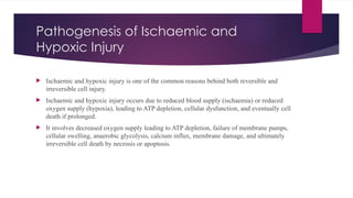
Ischaemic and hypoxic injury is one of the common reasons behind both reversible and
irreversible cell injury.
Ischaemic and hypoxic injury occurs due to reduced blood supply (ischaemia) or reduced
oxygen supply (hypoxia), leading to ATP depletion, cellular dysfunction, and eventually cell
death if prolonged.
It involves decreased oxygen supply leading to ATP depletion, failure of membrane pumps,
cellular swelling, anaerobic glycolysis, calcium influx, membrane damage, and ultimately
irreversible cell death by necrosis or apoptosis.
9.
Reversible Cell Injury
If ischemia (reduced blood supply) or hypoxia (reduced oxygen supply) lasts only for a short time,
the damage to the cell is reversible. When normal blood circulation and oxygen supply are restored
quickly, the cell regains its normal structure and function.
In Simple Words
• Short-duration ischemia/hypoxia → Temporary cell injury.
• Blood flow restored quickly → ATP production resumes.
• Cell recovers completely → No permanent damage.
11.
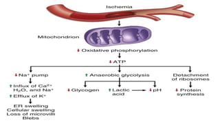
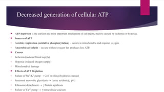
Decreased generation ofcellular ATP
ATP depletion is the earliest and most important mechanism of cell injury, mainly caused by ischemia or hypoxia.
Sources of ATP
o Aerobic respiration (oxidative phosphorylation) – occurs in mitochondria and requires oxygen.
o Anaerobic glycolysis – occurs without oxygen but produces less ATP.
Causes
o Ischemia (reduced blood supply)
o Hypoxia (reduced oxygen supply)
o Mitochondrial damage
Effects of ATP Depletion
o Failure of Na /K pump → Cell swelling (hydropic change)
⁺ ⁺
o Increased anaerobic glycolysis → Lactic acidosis (↓ pH)
o Ribosome detachment → ↓ Protein synthesis
o Failure of Ca² pump → ↑ Intracellular calcium
⁺
12.
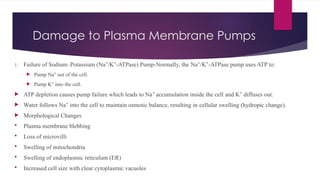
Damage to PlasmaMembrane Pumps
1. Failure of Sodium–Potassium (Na /K -ATPase) Pump-Normally, the Na /K -ATPase pump uses ATP to:
⁺ ⁺ ⁺ ⁺
Pump Na out of the cell.
⁺
Pump K into the cell.
⁺
ATP depletion causes pump failure which leads to Na accumulation inside the cell and K diffuses out.
⁺ ⁺
Water follows Na into the cell to maintain osmotic balance, resulting in cellular swelling (hydropic change).
⁺
Morphological Changes
Plasma membrane blebbing
Loss of microvilli
Swelling of mitochondria
Swelling of endoplasmic reticulum (ER)
Increased cell size with clear cytoplasmic vacuoles
13.
2. Failure ofCalcium (Ca² ) Pump-ATP depletion and membrane damage impair the Ca²
⁺ ⁺
pump.
Ca² enters the cell (calcium influx) and accumulates, especially in mitochondria.
⁺
Increased intracellular calcium:
Causes mitochondrial swelling.
Leads to deposition of phospholipid-rich amorphous densities.
14.
Intracellular lactic acidosis/Increased
anaerobic glycolysis
Intracellular lactic acidosis is a reversible change in cell injury caused by hypoxia or ischemia, in which cells
switch from aerobic respiration to anaerobic glycolysis, leading to lactic acid accumulation and a fall in
intracellular pH.
Pathogenesis
1. Hypoxia/Ischemia → ↓ Oxygen supply.
2. Aerobic respiration in mitochondria fails.
3. Cells switch to anaerobic glycolysis for ATP production.
4. Glycogen stores are depleted.
5. Lactic acid accumulates inside the cell.
6. Intracellular pH decreases (lactic acidosis).
7. Acidic pH causes clumping of nuclear chromatin (early reversible nuclear change).
15.
Reduced protein synthesis
/Ribosome detachment
Reduced protein synthesis is a reversible change in cell injury caused by prolonged hypoxia, resulting in damage to the rough endoplasmic reticulum
(RER) and Golgi apparatus, leading to impaired protein production.
Pathogenesis
1. Prolonged hypoxia → ↓ ATP production.
2. Swelling of rough endoplasmic reticulum (RER) and Golgi apparatus.
3. Ribosomes detach from the rough ER.
4. Polysomes break down into monosomes.
5. Ribosomes become dispersed in the cytoplasm.
6. Protein synthesis decreases.
7. Golgi apparatus also shows reduced protein synthesis.
Ultrastructural Changes
1. Loss of microvilli
2. Membrane blebbing
3. Loss of intramembranous particles
4. Formation of myelin figures (membranous whorls from damaged plasma and organelle membranes)
16.
IRREVERSIBLE CELL INJURY
Irreversible cell injury is a severe and permanent form of cell injury in which the cell cannot recover, even after restoration of blood supply or oxygen.
It ultimately results in cell death (necrosis or apoptosis).
Causes
1. Prolonged ischemia
2. Severe hypoxia
3. Persistent toxins or infections
Hallmarks (Features)
1. Irreversible mitochondrial dysfunction-Mitochondria cannot produce ATP even after reperfusion or reoxygenation.
2. Loss of plasma membrane integrity-Plasma membrane becomes permanently damaged & cellular contents leak out, leading to cell death.
Other Changes
1. Severe ATP depletion
2. Continued protein depletion
3. Reduced intracellular pH (acidosis)
4. Leakage of lysosomal enzymes into the cytoplasm, causing autodigestion of the cell
5. Extensive membrane and organelle damage
18.
Calcium influx
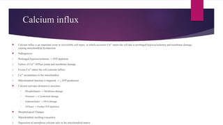
Calciuminflux is an important event in irreversible cell injury, in which excessive Ca² enters the cell due to prolonged hypoxia/ischemia and membrane damage,
⁺
causing mitochondrial dysfunction.
Pathogenesis
1. Prolonged hypoxia/ischemia → ATP depletion.
2. Failure of Ca² -ATPase pump and membrane damage.
⁺
3. Excess Ca² enters the cell (calcium influx).
⁺
4. Ca² accumulates in the mitochondria.
⁺
5. Mitochondrial function is impaired → ↓ ATP production.
Calcium activates destructive enzymes:
o Phospholipases → Membrane damage
o Proteases → Cytoskeletal damage
o Endonucleases → DNA damage
o ATPases → Further ATP depletion
Morphological Changes
1. Mitochondrial swelling (vacuoles)
2. Deposition of amorphous calcium salts in the mitochondrial matrix
19.
Role of destructiveenzymes
Phospholipases- Activated phospholipases are enzymes activated by increased intracellular
Ca² during irreversible cell injury. They degrade membrane phospholipids, leading to damage
⁺
of the plasma membrane, mitochondrial membrane, and lysosomal membrane. This
causes leakage of cellular contents and lysosomal enzymes, resulting in further membrane
destruction and progression to irreversible cell injury and cell death.
Intracellular proteases are enzymes activated by increased intracellular
Ca² during irreversible cell injury. They degrade cellular proteins, particularly cytoskeletal
⁺
proteins (Microfilaments (Actin filaments); Intermediate filaments; Microtubules),
causing loss of cell shape and structural support. This weakens the plasma membrane, leading
to increased membrane damage and progression to irreversible cell injury.
20.
Activated endonucleasesare enzymes activated by increased intracellular Ca² during irreversible cell injury.
⁺
They degrade nuclear DNA and nucleoproteins, causing DNA fragmentation and irreversible nuclear damage.
The damaged DNA activates pro-apoptotic proteins, triggering apoptosis and leading to irreversible cell injury
and cell death. Irreversible damage to the nucleus can be in three forms:
1. Pyknosis – Condensation and shrinkage of the nucleus, which becomes dark and basophilic (deep blue-purple).
2. Karyorrhexis – Fragmentation of the nucleus into small pieces dispersed in the cytoplasm.
3. Karyolysis – Dissolution (fading) of the nucleus due to DNA degradation.
21.
Lysosomal Hydrolytic Enzymes
Lysosomal hydrolytic enzymes are released due to lysosomal membrane damage during irreversible cell injury which leads to -
1. Release and activation of hydrolytic enzymes (proteases, lipases, DNases, RNases, phosphatases, etc.)
2. Enzymatic digestion (autolysis) of cellular components
3. Cell death (necrosis)
4. Formation of myelin figures
5. Phagocytosis by macrophages or calcium soap formation
Clinical Significance-Leakage of intracellular enzymes into the blood serves as markers of tissue injury:
1. AST (SGOT)
2. LDH
3. CK-MB
4. Cardiac troponins (cTn) – markers of myocardial infarction
23.
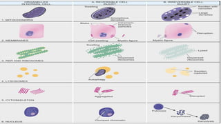
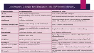
Ultrastructural Changes duringReversible and irreversible cell injury.
Organelle/Structure Reversible Cell Injury Irreversible Cell Injury
Cell size Cellular swelling (hydropic change) Severe swelling followed by cell rupture
Plasma membrane
Membrane blebbing; loss of microvilli; membrane remains
intact
Severe membrane disruption and rupture with leakage of cellular contents
Mitochondria Swelling of mitochondria
Marked mitochondrial swelling with large vacuoles and amorphous
calcium-rich densities; irreversible mitochondrial dysfunction
Endoplasmic reticulum (ER)
Dilatation/swelling of rough ER with detachment of
ribosomes
Severe ER damage with fragmentation
Ribosomes
Detachment from rough ER leading to reduced protein
synthesis
Complete loss of ribosomes due to extensive membrane damage
Golgi apparatus Swelling with decreased protein synthesis Severe disruption and degeneration
Lysosomes Membranes remain intact
Rupture of lysosomal membranes with release of hydrolytic enzymes
causing autodigestion
Cytoskeleton Mild disorganization; loss of microvilli
Extensive degradation by activated proteases causing collapse of cell
architecture
Myelin figures Small membranous whorls may appear Numerous large myelin figures due to extensive membrane breakdown
Intramembranous particles Decreased/loss of membrane particles Extensive membrane destruction
Calcium deposition Usually absent
Calcium accumulation within mitochondria and formation of amorphous
densities
Cell integrity Structural integrity maintained; changes are reversible
Loss of structural integrity; changes are irreversible leading to necrosis or
apoptosis
24.
Ischaemia-Reperfusion Injury
Ischaemia–reperfusioninjury is the additional cell and tissue damage that occurs when blood supply
is restored (reperfusion) to an ischemic tissue. Rather than restoring structure and function of the cell,
paradoxically deteriorates the already injured cell and leads it to cell death.
Causes (Mechanisms)
1. Generation of reactive oxygen species (ROS) causing oxidative damage.
2. Calcium influx into cells leading to mitochondrial dysfunction.
3. Inflammation due to activation of neutrophils and complement.
4. Mitochondrial damage resulting in decreased ATP production and apoptosis.
Examples
1. Myocardial infarction after reopening a blocked coronary artery
2. Ischemic stroke after restoration of cerebral blood flow
25.
Free Radical-Mediated CellInjury
Oxidative stress is a condition in which the production of reactive oxygen species (ROS)
exceeds the body's antioxidant defense mechanisms, resulting in damage to lipids,
proteins, and DNA, leading to cell injury and death.
Free radicals are highly reactive molecules with one unpaired electron. They steal
electrons from other molecules, causing oxidative damage.
Free radical-mediated cell injury is caused by reactive oxygen species (ROS), which are
highly reactive molecules containing an unpaired electron. They damage cell membranes,
proteins, DNA, and cytoskeleton, leading to cell injury and death.
26.
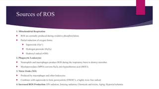
Sources of ROS
1.Mitochondrial Respiration
ROS are normally produced during oxidative phosphorylation.
Partial reduction of oxygen forms:
Superoxide (O • )
₂ ⁻
Hydrogen peroxide (H O )
₂ ₂
Hydroxyl radical (•OH)
2. Phagocytic Leukocytes
Neutrophils and macrophages produce ROS during the respiratory burst to destroy microbes.
Myeloperoxidase (MPO) converts H O into hypochlorous acid (HOCl).
₂ ₂
3. Nitric Oxide (NO)
Produced by macrophages and other leukocytes.
Combines with superoxide to form peroxynitrite (ONOO ), a highly toxic free radical.
⁻
4. Increased ROS Production- UV radiation, Ionizing radiation, Chemicals and toxins, Aging, Hypoxia/ischemia
27.
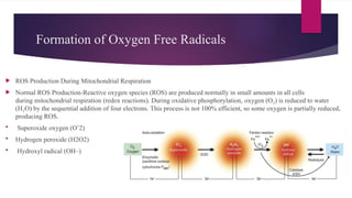
Formation of OxygenFree Radicals
ROS Production During Mitochondrial Respiration
Normal ROS Production-Reactive oxygen species (ROS) are produced normally in small amounts in all cells
during mitochondrial respiration (redox reactions). During oxidative phosphorylation, oxygen (O ) is reduced to water
₂
(H O) by the sequential addition of four electrons. This process is not 100% efficient, so some oxygen is partially reduced,
₂
producing ROS.
Superoxide oxygen (O’2)
Hydrogen peroxide (H2O2)
Hydroxyl radical (OH–)
28.
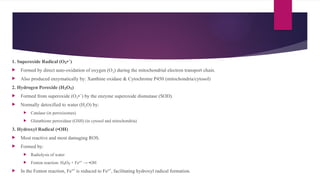
1. Superoxide Radical(O • )
₂ ⁻
Formed by direct auto-oxidation of oxygen (O ) during the mitochondrial electron transport chain.
₂
Also produced enzymatically by: Xanthine oxidase & Cytochrome P450 (mitochondria/cytosol)
2. Hydrogen Peroxide (H O )
₂ ₂
Formed from superoxide (O • ) by the enzyme superoxide dismutase (SOD).
₂ ⁻
Normally detoxified to water (H O) by:
₂
Catalase (in peroxisomes)
Glutathione peroxidase (GSH) (in cytosol and mitochondria)
3. Hydroxyl Radical (•OH)
Most reactive and most damaging ROS.
Formed by:
Radiolysis of water
Fenton reaction: H O + Fe² → •OH
₂ ₂ ⁺
In the Fenton reaction, Fe³ is reduced to Fe² , facilitating hydroxyl radical formation.
⁺ ⁺
29.
ROS Production inPhagocytic Leukocytes
Reactive oxygen species (ROS) are produced by phagocytic leukocytes, mainly neutrophils and macrophages,
during inflammation to destroy ingested microbes. This process is called the respiratory (oxidative) burst.
Mechanism
1. During phagocytosis, the enzyme NADPH oxidase on the phagosome membrane generates superoxide (O • ).
₂ ⁻
2. Superoxide (O • ) is converted into hydrogen peroxide (H O ).
₂ ⁻ ₂ ₂
3. Myeloperoxidase (MPO), present in leukocyte granules, converts H O into hypochlorous acid (HOCl).
₂ ₂
4. HOCl is a highly reactive antimicrobial agent that kills bacteria and other pathogens.
Effects
1. Destroys ingested microorganisms
2. Helps in host defense during inflammation
3. Excess ROS and HOCl may also damage surrounding normal tissues
30.
Effects of FreeRadicals
1. Lipid Peroxidation- Free radicals attack polyunsaturated fatty acids (PUFAs) in cell membranes. Forms lipid
peroxides and lipid hydroperoxy radicals these causes loss of membrane integrity, increased permeability,
and destruction of organelles.
2. Protein Oxidation
Free radicals oxidize cellular proteins, causing:
Cross-linking of amino acids
Fragmentation of polypeptides
Results in enzyme inactivation, protein degradation, and cell destruction.
3. DNA Damage-Free radicals cause single-strand breaks in nuclear and mitochondrial DNA. This leads to cell
injury, mutations, apoptosis, and may cause malignant transformation.
4. Cytoskeletal Damage-ROS damage cytoskeletal proteins. Interfere with mitochondrial oxidative phosphorylation,
causing ATP depletion. Leads to loss of cell shape, membrane instability, and cell injury.
31.
Antioxidants
Antioxidants aresubstances that neutralize or remove free radicals (ROS), thereby protecting cells from oxidative stress and free radical–mediated cell injury.
Types of Antioxidants
1. Enzymatic Antioxidants
Superoxide dismutase (SOD): Converts superoxide (O • ) into hydrogen peroxide (H O ).
₂ ⁻ ₂ ₂
Catalase: Converts H O into water (H O) and oxygen (O ).
₂ ₂ ₂ ₂
Glutathione peroxidase (GSH): Converts H O and lipid peroxides into harmless products.
₂ ₂
2. Non-Enzymatic Antioxidants
• Vitamin E (α-tocopherol): Prevents lipid peroxidation of cell membranes.
• Vitamin C (Ascorbic acid): Scavenges free radicals and regenerates vitamin E.
• Vitamin A and β-carotene: Quench free radicals and protect cell membranes.
• Glutathione (GSH): Major intracellular antioxidant.
Functions
Neutralize ROS and free radicals
Prevent lipid peroxidation
Protect proteins, lipids, and DNA
Reduce oxidative stress
Prevent cell injury and death
32.
Morphological Changes inCell injury
Reversible Cell Injury
1. Hydropic change
2. Hyaline change
3. Mucoid change
4. Fatty change
Irreversible Cell Injury
1. local or focal change (i.e. autolysis, necrosis and apoptosis)
2. and the changes that follow it (i.e. gangrene and pathologic calcification)
33.
Hydropic Change
Hydropicchange is the accumulation of water within the cytoplasm of the cell due to cell
injury. It is the earliest and most common form of reversible cell injury. Also called cloudy
swelling due to gross appearance of the affected organ or vacuolar degeneration due to
formation of cytoplasmic vacuoles.
Etiology (Causes)- Hypoxia/ischemia, Bacterial toxins, Chemicals and poisons, Burns,
High fever, Intravenous hypertonic glucose or saline, Other toxic or metabolic injuries
Pathogenesis- Cell injury causes a decrease in ATP production, leading to failure of
the Na /K -ATPase pump. As a result, sodium (Na ) accumulates inside the cell,
⁺ ⁺ ⁺
while potassium (K ) diffuses out. The increased intracellular sodium draws water into the
⁺
cell by osmosis to maintain iso-osmotic balance, resulting in cellular swelling (hydropic
change). In addition, calcium (Ca² ) enters the cell, which further contributes to cellular
⁺
injury. Hydropic swelling is reversible if the injurious stimulus is removed early.
34.
Hyaline change
Hyalinechange is the accumulation of glassy, homogeneous, eosinophilic (pink-staining) proteinaceous material within or outside cells. It is a descriptive
histological term and does not refer to a specific substance.
Types of Hyaline Change
1. Intracellular Hyaline- Occurs mainly in epithelial cells. Examples:
a. Hyaline droplets in proximal renal tubular cells due to proteinuria.
b. Zenker's degeneration – Hyaline degeneration of rectus abdominis muscle in typhoid fever.
c. Mallory hyaline (Mallory bodies) – Aggregates of intermediate filaments in hepatocytes in alcoholic liver disease.
d. Viral hyaline inclusions – Nuclear or cytoplasmic inclusions in viral infections.
e. Russell bodies – Accumulation of immunoglobulins in the rough ER of plasma cells.
2. Extracellular Hyaline (Hyalinization)- Occurs mainly in connective tissues. Examples:
f. Hyaline degeneration in uterine leiomyoma.
g. Hyalinized old scars (fibrocollagenous tissue).
h. Hyaline arteriolosclerosis in hypertension and diabetes mellitus.
i. Hyalinized glomeruli in chronic glomerulonephritis.
j. Corpora amylacea in the prostate, brain, spinal cord, and old lung infarcts.
35.
Mucoid change
Mucoidchange is the accumulation of mucus-like (mucinous) material in epithelial cells or connective tissue. Mucus = Secretory product of mucous glands
containing proteins and mucopolysaccharides.; Mucin = Major glycoprotein component of mucus.; Connective tissue mucin is called myxoid.
Staining- Epithelial mucin: PAS positive and Alcian blue positive & Connective tissue (myxoid) mucin: PAS negative, Alcian blue positive, and Colloidal iron
positive.
Types of Mucoid Change
1. Epithelial Mucin- Occurs due to excess mucus secretion by epithelial cells.Examples:
a. Catarrhal inflammation (respiratory tract, alimentary tract, uterus)
b. Mucocele due to duct obstruction (oral cavity, gallbladder)
c. Cystic fibrosis of the pancreas
d. Mucin-secreting tumors (ovary, stomach, large intestine)
2. Connective Tissue Mucin (Myxoid Change)- Occurs due to accumulation of mucin in connective tissue. Examples:
e. Myxoid tumors: Myxoma, neurofibroma, fibroadenoma, soft tissue sarcomas
f. Dissecting aortic aneurysm (Erdheim's medial degeneration, Marfan syndrome)
g. Myxedema (myxomatous change in dermis)
h. Ganglion cyst of the wrist (myxoid change in synovium)
36.
Intracellular Accumulations
Intracellularaccumulation is the abnormal accumulation of substances within the cytoplasm (especially
lysosomes) or nucleus of cells due to disturbances in cellular metabolism.
Mild accumulation → Causes reversible cell injury.
Severe or persistent accumulation → Leads to irreversible cell injury and cell death.
Abnormal intracellular accumulations can be divided into 3 groups:
1. Accumulation of constituents of normal cell metabolism produced in excess e.g. accumulations of lipids (fatty
change, cholesterol deposits), proteins and carbohydrates.
2. Accumulation of abnormal substances produced as a result of abnormal metabolism due to lack of some enzymes
e.g. storage diseases or inborn errors of metabolism.
3. Accumulation of pigments e.g. endogenous pigments under special circumstances, and exogenous pigments due to
lack of enzymatic mechanisms to degrade the substances or transport them to other sites.
37.
Fatty Change (Steatosis/FattyMetamorphosis)
Fatty change (steatosis or fatty metamorphosis) is the intracellular accumulation of neutral fat (triglycerides) within
the cytoplasm of parenchymal cells. It represents an absolute increase in intracellular lipids. Fatty change is most
common in the liver, but may also occur in the heart, skeletal muscle, kidneys, and other organs.
Fatty Liver (Hepatic Steatosis)- The liver is the commonest site of fatty change because it plays a central role in fat
metabolism.
i. Mild fatty change → Reversible
ii. Severe fatty change → May lead to irreversible cell injury and cell death
Etiology (Causes)-Fatty liver develops due to two major groups of causes:
1. Conditions with Excess Fat-These conditions increase the amount of fat reaching the liver beyond its metabolic
capacity like Obesity, Diabetes mellitus, Congenital hyperlipidaemia
2. Liver Cell Damage- These conditions impair the liver's ability to metabolize and export fat. Like Alcoholic liver
disease, Protein-calorie malnutrition, Chronic illnesses (e.g., tuberculosis), Acute fatty liver of pregnancy, Hypoxia (e.g.,
anaemia, cardiac failure), Hepatotoxins (Carbon tetrachloride (CCl ), Chloroform, Ether, Aflatoxins), Drug-induced
₄
liver injury (Methotrexate, Steroids, Halothane, Tetracycline)
38.
Pathogenesis of Fattyliver
Fatty liver occurs due to defects in normal fat metabolism leading to intracellular triglyceride
accumulation.
Six mechanisms are responsible:
1. Increased free fatty acid entry
2. Increased fatty acid synthesis
3. Decreased conversion of fatty acids into ketone bodies resulting in increased esterification of
fatty acids to triglycerides.
4. Increased α-glycerophosphate causing increased esterification of fatty acids to triglycerides.
5. Decreased lipid acceptor protein (apoprotein) synthesis
6. Block in lipoprotein secretion
39.
Cholesterol depositsare the intracellular accumulation of cholesterol and cholesterol esters within
macrophages, usually due to hypercholesterolaemia. These lipid-laden macrophages are
called foam cells. Examples- Fibrofatty plaques of atherosclerosis, Xanthomas (foam cells
form tumour-like masses ) and Xanthelasma (yellow cholesterol-rich plaques, commonly seen
around the eyelids.)
Stromal fatty infiltration is the deposition of mature adipose (fat) cells in the stromal connective
tissue of an organ. Unlike fatty change (steatosis), fat is not accumulated inside parenchymal cells.
Most commonly occurs in obesity. Commonly Affected Organs are Heart (epicardial fat
infiltration) & Pancreas.
40.
INTRACELLULAR ACCUMULATION OF
PROTEINS
Definition:
Abnormal accumulation of proteins within the cytoplasm of cells due to excessive synthesis, reabsorption, or
defective metabolism.
Examples
Proteinuria – Excess plasma proteins are reabsorbed by proximal renal tubular cells, forming pink hyaline
droplets (reversible).
Russell's bodies – Accumulation of immunoglobulins in the cytoplasm of plasma cells.
α -Antitrypsin deficiency
₁ – Mutant α -antitrypsin protein accumulates as eosinophilic globules in
₁
hepatocytes.
Mallory bodies (Alcoholic hyaline) – Accumulation of cytokeratin intermediate filaments in hepatocytes,
seen in alcoholic liver disease.
41.
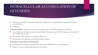
INTRACELLULAR ACCUMULATION OF
GLYCOGEN
Abnormal accumulation of glycogen within the cytoplasm due to impaired glucose metabolism or genetic
enzyme defects.
Conditions
1. Diabetes Mellitus- Impaired cellular uptake of glucose causes intracellular glycogen accumulation.
Sites: Epithelium of distal proximal convoluted tubule, Descending loop of Henle, Hepatocytes, Pancreatic β-
cells, Cardiac muscle cells.
Microscopy: Clear cytoplasmic vacuoles on H&E stain.
Special stains: PAS (Periodic acid–Schiff) and Best's carmine.
2. Glycogen Storage Diseases (Glycogenosis)
Inherited genetic disorders causing defective glycogen metabolism due to enzyme deficiency.
Results in excessive intracellular glycogen accumulation.
42.
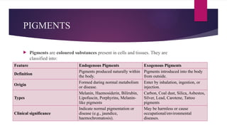
PIGMENTS
Pigments arecoloured substances present in cells and tissues. They are
classified into:
Feature Endogenous Pigments Exogenous Pigments
Definition
Pigments produced naturally within
the body.
Pigments introduced into the body
from outside.
Origin
Formed during normal metabolism
or disease.
Enter by inhalation, ingestion, or
injection.
Types
Melanin, Haemosiderin, Bilirubin,
Lipofuscin, Porphyrins, Melanin-
like pigments
Carbon, Coal dust, Silica, Asbestos,
Silver, Lead, Carotene, Tattoo
pigments
Clinical significance
Indicate normal pigmentation or
disease (e.g., jaundice,
haemochromatosis).
May be harmless or cause
occupational/environmental
diseases.
43.
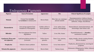
Endogenous Pigments
Pigment Source/FormationAppearance Site Clinical Significance/Examples
Melanin
Formed from tyrosine
by tyrosinase in melanocytes
Brown-black
Skin, hair, eye, meninges,
adrenal medulla
Hyperpigmentation (Addison disease,
chloasma, melanoma); Hypopigmentation
(albinism, vitiligo, leucoderma)
Haemosiderin
Iron-storage pigment from
breakdown of hemoglobin
Golden yellow to
brown granules
Liver, spleen, bone
marrow, macrophages
Haemosiderosis, haemochromatosis, old
hemorrhage, heart failure cells
Bilirubin
Non-iron pigment from heme
degradation
Yellow Liver, bile, tissues
Hyperbilirubinemia → Jaundice,
kernicterus in infants
Lipofuscin (Wear-and-
tear pigment)
Lipid peroxidation products in
lysosomes
Yellow-brown
granules
Heart, liver, neurons,
testes
Aging, atrophy, chronic wasting diseases
(brown atrophy)
Porphyrins Defective heme synthesis Red-brown Liver, bone marrow
Porphyrias, photosensitivity, neurological
symptoms
Melanin-like pigments Abnormal metabolism Brown-black Various tissues
Ochronosis (Alkaptonuria), Dubin–Johnson
syndrome
44.
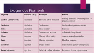
Exogenous Pigments
Pigment Routeof Entry Examples Effects
Carbon (Anthracosis) Inhalation Smokers, urban pollution
Usually harmless; severe exposure →
pneumoconiosis
Coal dust Inhalation Coal miners Coal workers' pneumoconiosis
Silica Inhalation Stone workers Silicosis
Asbestos Inhalation Construction workers Asbestosis, lung fibrosis
Silver Ingestion Chronic silver intake Argyria (gray pigmentation)
Lead Ingestion Chronic poisoning Blue line on gums
Carotene Ingestion Excess carrots Carotenemia (yellow-orange skin)
Tattoo pigments Injection India ink, carbon, cinnabar Permanent dermal pigmentation
45.
Important 2 MarksQuestion
1. Mention two hallmarks of irreversible cell injury.
2. Name the enzymes activated by intracellular calcium during irreversible
cell injury.
3. What is oxidative stress?
4. What are reactive oxygen species (ROS)? Give examples.
5. What is hydropic change? Mention its underlying mechanism.
6. How does failure of the Na /K -ATPase pump lead to cellular swelling?
⁺ ⁺
7. Give the role of anaerobic glycolysis in hypoxic cell injury.
8. What are the three nuclear changes seen in irreversible cell injury?
9. What is ischemia–reperfusion injury? Mention its major mechanisms.
10. Mention any four harmful effects of free radicals on cells.
11. What are the consequences of intracellular lactic acidosis?
12. Why does protein synthesis decrease during hypoxic cell injury?
13. Enlist the etiology of cell injury.
14. Name the calcium-activated enzymes involved in irreversible cell injury
and state one function of each.
15. What are foam cells? Mention two conditions in which they are found.
16. Define hyaline change. Give two examples.
17. What is mucoid change? Differentiate epithelial mucin from connective
tissue mucin.
18. Differentiate between hypoxia and ischemia.
19. What is fatty change (steatosis)? Mention two common causes.
20. List the mechanisms responsible for fatty liver.
21. Enlist 4 endogenous/ exogenous pigments.
22. What are Russell bodies? Mention their pathological significance.
23. Explain the significance of Mallory bodies.
24. What is glycogen accumulation? Mention two conditions in which it
occurs.
25. Define Antioxidants with example.
46.
5 Marksquestion
1. Describe the sequence of events occurring during reversible cell
injury.
2. Explain free radical-mediated cell injury.
3. Define oxidative stress. Explain the generation of reactive
oxygen species (ROS) and their role in cellular injury.
4. Describe the mechanisms responsible for irreversible cell injury.
5. Describe the morphological changes observed in reversible cell
injury with suitable examples.
6. Describe fatty change (steatosis) and enlist its causes. Highlight
the pathogenesis of fatty liver.
7. Discuss ATP depletion as the central mechanism of cell injury.
10 Marks question
1. Describe the pathogenesis of ischemic and hypoxic
cell injury with a suitable flowcharts.
2. Discuss intracellular accumulations. Classify them
and explain the mechanisms of lipid, protein,
glycogen, and pigment accumulation.
3. Differentiate reversible cell injury & irreversible cell
injury on the basis of sequence of biochemical,
ultrastructural, and nuclear changes occurring during
.
 Most read
 Most read