Dr. NIKHILKUMARS S SAKLE
Assistant Professor
Department of Pharmacology
Y. B. Chavan College of Pharmacy, Auranagabad
Basic Principles of Cell Injury and
Adaptation
Unit I
Introduction, definitions, Homeostasis, Components
and Types of Feedback systems, Causes of cellular
injury, Pathogenesis (Cell membrane damage,
Mitochondrial damage, Ribosome damage, Nuclear
damage), Morphology of cell injury – Adaptive
changes (Atrophy, Hypertrophy, hyperplasia,
Metaplasia, Dysplasia), Cell swelling, Intra cellular
accumulation, Calcification, Enzyme leakage and Cell
Death Acidosis & Alkalosis, Electrolyte imbalance
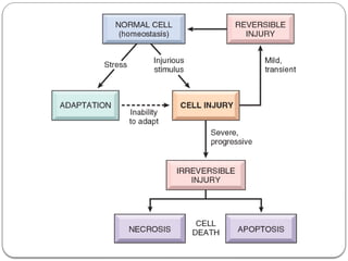
Cellular injury
 Cell injury is defined as the effect of a variety of
stresses due to etiologic agents a cell encounters
resulting in changes in its internal and external
environment.
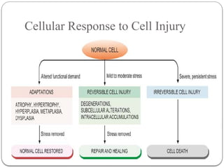
 The cellular response to stress may vary and depends
upon following two variables:
 i) Host factors i.e. the type of cell and tissue involved.
 ii) Factors pertaining to injurious agent i.e. extent and
type of cell injury.
Cellular Responses to cell injury
1. When there is increased functional demand, the cell
may adapt to the changes which are expressed
morphologically, which then revert back to normal
after the stress is removed (Cellular adaptation)
2. When the stress is mild to moderate, the injured cell
may recover (reversible cell injury), while persistent
and severe form of cell injury may cause cell death
(irreversible cell injury).
Etiology (Causes) of cellular injury
 The cells may be broadly injured by two major ways:
 A. Genetic causes:
 B. Acquired causes:
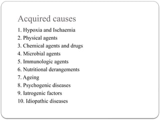
Acquired causes
1. Hypoxia and Ischaemia
2. Physical agents
3. Chemical agents and drugs
4. Microbial agents
5. Immunologic agents
6. Nutritional derangements
7. Ageing
8. Psychogenic diseases
9. Iatrogenic factors
10. Idiopathic diseases
Hypoxia and Ischaemia
 Hypoxia is the most common cause of cell injury.
 Hypoxia may result from the following 2 ways:
 The most common mechanism of hypoxic cell injury is
by reduced supply of blood to cells due to interruption
i.e. ischaemia.
 Hypoxia may also result from impaired blood supply
from causes other than interruption e.g. disorders of
oxygen carrying RBCs (e.g. anaemia, carbon
monoxide poisoning), heart diseases, lung diseases
and increased demand of tissues.
Physical agents
 Physical agents in causation of disease are as under:
i) mechanical trauma (e.g. road accidents);
ii) thermal trauma (e.g. by heat and cold);
iii) electricity;
iv) radiation (e.g. ultraviolet and ionising); and
v) rapid changes in atmospheric pressure.
Chemical agents and drugs
 An ever-increasing list of chemical agents and drugs may
cause cell injury. Important examples include the following:
i) chemical poisons such as cyanide, arsenic, mercury;
ii) strong acids and alkalis;
iii) environmental pollutants;
iv) insecticides and pesticides;
v) oxygen at high concentrations;
vi) hypertonic glucose and salt;
vii) social agents such as alcohol and narcotic drugs; and
viii) therapeutic administration of drugs.
Microbial agents
 Injuries by microbes include infections caused by
bacteria, rickettsiae, viruses, fungi, protozoa, metazoa,
and other parasites.
Immunologic agents
 Immunity is a ‘double-edged sword’—it protects the
host against various injurious agents but it may also
turn lethal and cause cell injury
 e.g.
i) hypersensitivity reactions;
ii) anaphylactic reactions; and
iii) autoimmune diseases.
Nutritional derangements
 A deficiency or an excess of nutrients may result in
nutritional imbalances.
  Nutritional deficiency diseases may be due to
overall deficiency of nutrients (e.g. starvation), of
protein calorie (e.g. marasmus, kwashiorkor), of
minerals (e.g. anaemia), or of trace elements.
  Nutritional excess is a problem of affluent societies
resulting in obesity, atherosclerosis, heart disease and
hypertension.
Ageing
 Cellular ageing or senescence leads to impaired ability
of the cells to undergo replication and repair, and
ultimately lead to cell death culminating in death of the
individual.
Psychogenic diseases
 There are no specific biochemical or morphologic
changes in common acquired mental diseases due to
mental stress, strain, anxiety, overwork and frustration
e.g. depression, schizophrenia. However, problems of
drug addiction, alcoholism, and smoking result in
various organic diseases such as liver damage, chronic
bronchitis, lung cancer, peptic ulcer, hypertension,
ischaemic heart disease etc.
Iatrogenic factors
 Although as per Hippocratic oath, every physician is
bound not to do or administer anything that causes
harm to the patient, there are some diseases as well as
deaths attributed to iatrogenic causes (owing to
physician).
 Examples include occurrence of disease or death due
to error in judgement by the physician and untoward
effects of administered therapy (drugs, radiation).
Idiopathic diseases
 Idiopathic means “of unknown cause”. Finally,
although so much is known about the etiology of
diseases, there still remain many diseases for which
exact cause is undetermined. For example, most
common form of hypertension (90%) is idiopathic (or
essential) hypertension.
 Similarly, exact etiology of many cancers is still
incompletely known.
Cellular Response to Cell Injury
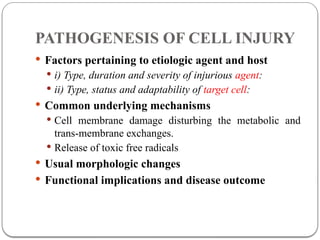
PATHOGENESIS OF CELL INJURY
 Factors pertaining to etiologic agent and host
 i) Type, duration and severity of injurious agent:
 ii) Type, status and adaptability of target cell:
 Common underlying mechanisms
 Cell membrane damage disturbing the metabolic and
trans-membrane exchanges.
 Release of toxic free radicals
 Usual morphologic changes
 Functional implications and disease outcome
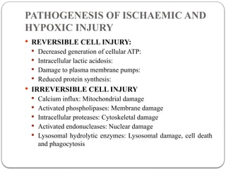
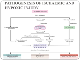
PATHOGENESIS OF ISCHAEMIC AND
HYPOXIC INJURY
 REVERSIBLE CELL INJURY:
 Decreased generation of cellular ATP:
 Intracellular lactic acidosis:
 Damage to plasma membrane pumps:
 Reduced protein synthesis:
 IRREVERSIBLE CELL INJURY
 Calcium influx: Mitochondrial damage
 Activated phospholipases: Membrane damage
 Intracellular proteases: Cytoskeletal damage
 Activated endonucleases: Nuclear damage
 Lysosomal hydrolytic enzymes: Lysosomal damage, cell death
and phagocytosis
PATHOGENESIS OF ISCHAEMIC AND
HYPOXIC INJURY
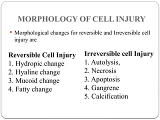
MORPHOLOGY OF CELL INJURY
 Morphological changes for reversible and Irreversible cell
injury are
Reversible Cell Injury
1. Hydropic change
2. Hyaline change
3. Mucoid change
4. Fatty change
Irreversible cell Injury
1. Autolysis,
2. Necrosis
3. Apoptosis
4. Gangrene
5. Calcification
MORPHOLOGY OF REVERSIBLE
CELL INJURY
 Common examples of morphologic forms of reversible
cell injury are as under:
1. Hydropic change
2. Hyaline change
3. Mucoid change
4. Fatty change
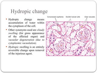
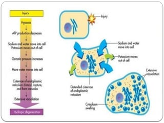
Hydropic change
 Hydropic change means
accumulation of water within
the cytoplasm of the cell.
 Other synonyms used are cloudy
swelling (for gross appearance
of the affected organ) and
vacuolar degeneration (due to
cytoplasmic vacuolation).
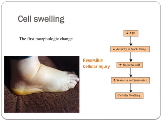
 Hydropic swelling is an entirely
reversible change upon removal
of the injurious agent.
Cell swelling
The first morphologic change
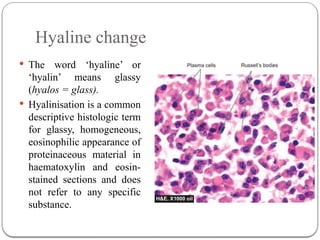
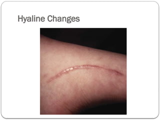
Hyaline change
 The word ‘hyaline’ or
‘hyalin’ means glassy
(hyalos = glass).
 Hyalinisation is a common
descriptive histologic term
for glassy, homogeneous,
eosinophilic appearance of
proteinaceous material in
haematoxylin and eosin-
stained sections and does
not refer to any specific
substance.
Hyaline Changes
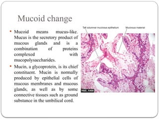
Mucoid change
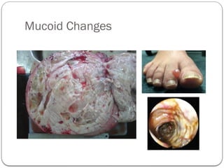
 Mucoid means mucus-like.
Mucus is the secretory product of
mucous glands and is a
combination of proteins
complexed with
mucopolysaccharides.
 Mucin, a glycoprotein, is its chief
constituent. Mucin is normally
produced by epithelial cells of
mucous membranes and mucous
glands, as well as by some
connective tissues such as ground
substance in the umbilical cord.
Mucoid Changes
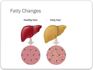
Fatty change
 Steatosis or fatty metamorphosis
 Intracellular accumulation of neutral fat within
parenchymal cells.
 It includes the older, now abandoned, terms of fatty
degeneration and fatty infiltration
 Fatty change is particularly common in the liver but
may occur in other non-fatty tissues as well
 e.g. in the heart, skeletal muscle, kidneys and other
organs
Fatty Liver
 ETIOLOGY
 Fatty change in the liver may result from one of the two types of causes:
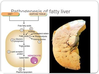
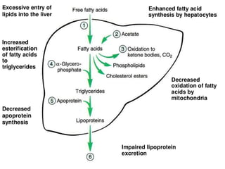
1. Conditions with excess fat Th ese are conditions in which the capacity of the liver to metabolise
fat is exceeded e.g.
 i) Obesity
 ii) Diabetes mellitus
 iii) Congenital hyperlipidaemia
2. Liver cell damage Th ese are conditions in which fat cannot be metabolised due to liver cell
injury e.g.
 i) Alcoholic liver disease (most common)
 ii) Starvation
 iii) Protein calorie malnutrition
 iv) Chronic illnesses (e.g. tuberculosis)
 v) Acute fatty liver in late pregnancy
 vi) Hypoxia (e.g. anaemia, cardiac failure)
 vii) Hepatotoxins (e.g. carbon tetrachloride, chloroform,
 ether, afl atoxins and other poisons)
 viii) Drug-induced liver cell injury (e.g. administration of methotrexate, steroids, CCl4, halothane
anaesthetic, tetracycline etc)
 ix) Reye’s syndrome
Fatty Changes
Pathogenesis of fatty liver
MORPHOLOGY OF IRREVERSIBLE
CELL INJURY (CELL DEATH)
 Autolysis
 Necrosis
 Apoptosis
 Gangrene
 Calcification
Autolysis
 Autolysis (i.e. self-digestion) is disintegration of the cell
by its own hydrolytic enzymes liberated from lysosomes.
 Autolysis can occur in the living body when it is
surrounded by inflammatory reaction (vital reaction),
but the term is generally used for postmortem change in
which there is complete absence of surrounding
inflammatory response.
 Autolysis is rapid in some tissues rich in hydrolytic
enzymes such as in the pancreas, and gastric mucosa;
intermediate in tissues like the heart, liver and kidney;
and slow in fibrous tissue.
Necrosis
 Necrosis is defined as a localised area of death of tissue followed
later by degradation of tissue by hydrolytic enzymes liberated
from dead cells; it is invariably accompanied by inflammatory
reaction.
 Necrosis can be caused by various agents such as hypoxia,
chemical and physical agents, microbial agents, immunological
injury, etc.
 Based on etiology and morphologic appearance, there are 5 types
of necrosis:
 Coagulative Necrosis
 Liquefaction (colliquative) Necrosis
 Caseous Necrosis
 Fat Necrosis
 Fibrinoid Necrosis.
Apoptosis
 Apoptosis is a form of ‘coordinated and internally
programmed cell death’ having significance in a variety of
physiologic and pathologic conditions (apoptosis=falling off
or dropping off , as that of leaves or petals).
 The term was first introduced in 1972 distinct from necrosis
by being controlled and regulated cell death, and opposed to
that of mitosis by having regulated size of the cell turn over.
 When the cell is not needed, pathway of cell death is
activated (‘cell suicide’).
 Unlike necrosis, apoptosis is not accompanied by any
inflammation and collateral tissue damage.
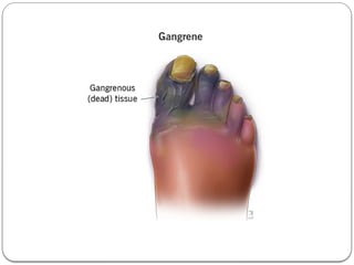
Gangrene
 Gangrene is necrosis of tissue associated with superadded
putrefaction, most often following coagulative necrosis due to
ischaemia (e.g. in gangrene of the bowel, gangrene of limb).
 On the other hand, gangrenous or necrotising inflammation is
characterised primarily by inflammation provoked by virulent
bacteria resulting in massive tissue necrosis. Thus, the end
result of necrotising inflammation and gangrene is the same
but the way the two are produced, is different. The examples
of necrotising inflammation are: gangrenous appendicitis,
gangrenous stomatitis (noma, cancrum oris).
 There are 2 main types of gangrene—Dry and Wet, and a
variant of wet gangrene called gas gangrene.
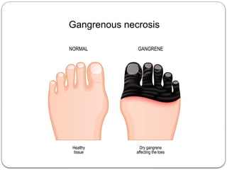
Dry Gangrene
 This form of gangrene begins in the distal part of a limb due to
ischaemia. The typical example is the dry gangrene in the toes and
feet of an old patient due to severe atherossclerosis.
 Other causes of dry gangrene foot include thromboangiitis
obliterans (Buerger’s disease), Raynaud’s disease, trauma, ergot
poisoning.
 It is usually initiated in one of the toes which is farthest from the
blood supply, containing so little blood that even the invading
bacteria find it hard to grow in the necrosed tissue.
 The gangrene spreads slowly upwards until it reaches a point
where the blood supply is adequate to keep the tissue viable.
 A line of separation is formed at this point between the gangrenous
part and the viable part
Wet Gangrene
 Wet gangrene occurs in naturally moist tissues and organs such as the
bowel, lung, mouth, cervix, vulva etc. To other examples of wet
gangrene having clinical significance are as follows:
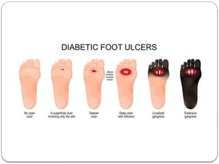
  Diabetic foot which is due to high glucose content in the necrosed
tissue which favours growth of bacteria.
  Bed sores occurring in a bed-ridden patient due to pressure on sites
like the sacrum, buttocks and heel.
 Wet gangrene usually develops due to blockage of both. The affected
part is stuffed with blood which favours the rapid growth of putrefactive
bacteria.
 The toxic products formed by bacteria are absorbed causing profound
systemic manifestations of septicaemia, and finally death. The spreading
wet gangrene generally lacks clear-cut line of demarcation and may
spread to peritoneal cavity causing peritonitis.
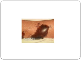
Gas Gangrene
 It is a special form of wet gangrene caused by gas-
forming clostridia (gram-positive anaerobic bacteria-
Clostridium perfringens) which gain entry into the tissues
through open contaminated wounds, especially in the
muscles, or as a complication of operation on colon
which normally contains clostridia.
 Clostridia produce various toxins which produce
necrosis and oedema locally and are also absorbed
producing profound systemic manifestations.
Calcification
 Deposition of calcium salts in tissues other than osteoid
or enamel is called pathologic or heterotopic calcification.
 Two distinct types of pathologic calcification are
recognised:
  Dystrophic calcification is characterised by deposition
of calcium salts in dead or degenerated tissues with
normal calcium metabolism and normal serum calcium
level.
  Metastatic calcification, on the other hand, occurs in
apparently normal tissues and is associated with deranged
calcium metabolism and hypercalcaemia.
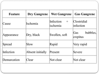
Feature Dry Gangrene Wet Gangrene Gas Gangrene
Cause Ischemia
Infection +
ischemia
Clostridial
infection
Appearance Dry, black Swollen, soft
Gas bubbles,
crepitus
Spread Slow Rapid Very rapid
Infection Absent initially Present Severe
Demarcation Clear Not clear Not clear
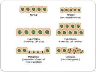
ADAPTIVE DISORDERS
 Adjustments which the cells make in response to stresses
 Physiologic adaptation
 Pathologic adaptation
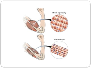
  Decreasing or increasing their size i.e. atrophy and
hypertrophy respectively, or by increasing their number
i.e. hyperplasia (postfix word -trophy means nourishment;
-plasia means growth of new cells).
  Changing the pathway of phenotypic differentiation of
cells i.e. metaplasia and dysplasia (prefix word meta-
means transformation; dys- means bad development).
 Adaptive responses are reversible on withdrawal of
stimulus. However, if the irritant stimulus persists for
long time, the cell may not be able to survive and may
either die or progress further e.g. cell death may occur
in sustained atrophy; dysplasia may progress into
carcinoma in situ.
ATROPHY
 Reduction of the number and size of parenchymal cells
of an organ or its parts
 The organ is small, often shrunken
 Cell become smaller in size but not dead cells
 Shrinkage in cell size is due to reduction in cell
organelles.
 CAUSES
 A. Physiologic atrophy e.g. Atrophy of brain with
ageing
 B. Pathologic atrophy e.g. Ischaemic atrophy
HYPERTROPHY
 Hypertrophy is an increase in the size of parenchymal
cells resulting in enlargement of the organ or tissue,
without any change in the number of cells.
 CAUSES
 Physiologic hypertrophy e.g. Enlarged size of the
uterus in pregnancy
 Pathologic hypertrophy e.g. Hypertrophy of skeletal
muscle in athletes and manual labourers
HYPERPLASIA
 Hyperplasia is an increase in the number of
parenchymal cells resulting in enlargement of the
organ or tissue.
 Hyperplasia persists so long as stimulus is present.
 CAUSE
 A. Physiologic hyperplasia e.g. Hyperplasia of
pregnant uterus
 B. Pathologic hyperplasia e.g. In wound healing, there
is formation of granulation tissue
METAPLASIA
 Metaplasia is defined as a reversible change of one
type of epithelial or mesenchymal adult cells to
another type of adult epithelial or mesenchymal cells,
usually in response to abnormal stimuli, and often
reverts back to normal on removal of stimulus.
 Metaplasia is broadly divided into 2 types: epithelial
and mesenchymal.
DYSPLASIA
 Dysplasia means ‘disordered cellular development’,
often preceded or accompanied with metaplasia and
hyperplasia; it is therefore also referred to as atypical
hyperplasia. Dysplasia occurs most often in epithelial
cells.
 Dysplastic changes often occur due to chronic
irritation or prolonged inflammation.
Comprehensive Overview of Cell Injury and Adaptation Mechanisms in Pathophysiology