Comprehensive Guide to Patient Feeding and Therapeutic Diets in Nursing Care
Detailed overview of patient feeding responsibilities, types of diets including normal and modified therapeutic diets, feeding methods, and nursing procedures to ensure proper nutrition and care.
It is theresponsibility of a nurse to ensure nutritional needs of all
patients are met
This may involve preparation, serving and feeding depending on
the hospital policy
Nutrition is a vital component in management of many conditions
Diet is categorized into two categories:
Normal diet
Modified diet
3
MR CHEGE .B.M
4.
1. Normal Diet
Itis normally taken with no modification. However it should be
balanced
It is the most frequently ordered diet
The patient should be able to choose food of their choice
2. Modified Diet
Food can be altered to meet various goals e.g. suit activity, due to
culture and religion and to meet therapeutic needs
When food is altered to meet health needs, it is referred to as
therapeutic diet
4
MR CHEGE .B.M
5.
Reasons for GivingTherapeutic Diet
Rising or reducing body weight
Regulate amount of certain nutrients in some disease
Aiding in digestion, malabsorption and maldigestion
Eliminating food that the body can not tolerate e.g. allergic
conditions
Therapeutic diet can be modified in the following ways:
According to Texture: Liquid, Soft
According to Constituent of Nutrients: High protein; Low
Carbohydrates; Low Calcium
According to Energy Content: Low calories; High calories
5
MR CHEGE .B.M
6.
1. MODIFICATION ACCORDINGTO TEXTURE
a) Liquid Diet
These are foods that are liquid at room temperature.
Mostly used in post – operative patients
Also indicated in patients with difficulty in chewing
Easily absorbed
Do not stimulate peristalsis, hence can be used in patients with
diarrhea
6
MR CHEGE .B.M
7.
They include:
i) ClearLiquid
Shouldn’t be given for more than three days
A clear liquid diet consists of clear liquids, such as water, broth and
plain gelatin, that are easily digested and leave no undigested
residue in the intestinal tract.
It may be prescribed before surgery or if you have certain
digestive problems.
A clear liquid diet can't provide adequate calories and nutrients
7
MR CHEGE .B.M
8.
Purpose
Used before tests,procedures or surgeries that require no food in the stomach or
intestines, such as before colonoscopy.
Recommended as a short-term diet if you have certain digestive problems, such
as nausea, vomiting or diarrhea, or after certain types of surgery.
8
MR CHEGE .B.M
9.
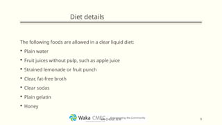
Diet details
The followingfoods are allowed in a clear liquid diet:
Plain water
Fruit juices without pulp, such as apple juice
Strained lemonade or fruit punch
Clear, fat-free broth
Clear sodas
Plain gelatin
Honey
9
MR CHEGE .B.M
10.
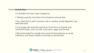
b) Soft Diet
Isdivided into two major categories
Mostly used for transition from liquid to normal diet
Its a diet that is soft in texture, low in residue, easily digested, and
well tolerated.
It provides the essential nutrients in the form of liquids and
semisolid foods, such as milk, fruit juices, eggs and cheese.
Recommended for people who have GI disturbances or acute
infections and those unable to tolerate a normal diet.
10
MR CHEGE .B.M
11.
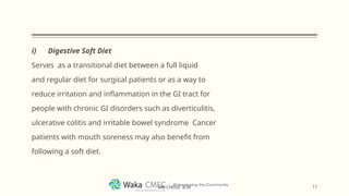
i) Digestive SoftDiet
Serves as a transitional diet between a full liquid
and regular diet for surgical patients or as a way to
reduce irritation and inflammation in the GI tract for
people with chronic GI disorders such as diverticulitis,
ulcerative colitis and irritable bowel syndrome Cancer
patients with mouth soreness may also benefit from
following a soft diet.
11
MR CHEGE .B.M
12.
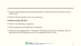
Food is easilydigested and easily absorbed. Contains less fat, less fibre and no
seasoning.
Should contain protein but in less amounts
ii) Mechanically Soft Diet
Little or no chewing is required.
Used in patients with difficulty in chewing
Foods can be adapted for a "full liquid" diet by processing in a blender with an
appropriate thinning liquid such as a vegetable, fruit or milk.
12
MR CHEGE .B.M
13.
2.
MODIFICATION ACCORDING TOCONSTITUENT OF NUTRIENTS
a) Carbohydrate Controlled Diet
The amount of carbohydrates is reduced e.g. diabetes mellitus
A specific amount is recommended in such patients depending on
blood glucose
Some carbohydrates may be restricted e.g. In lactose intolerance
13
MR CHEGE .B.M
14.
b) Fat ControlledDiet
A low fat diet contains very little or no fats
Carbohydrates are given in such cases to provide energy
A diet can also be modified in types of foods and patients are
discouraged from taking saturated fats, which are common in
animal fats as they increase risk of cardiovascular disease
A high fat diet (ketogenic diet) is indicated for patients with
seizures. It is unfit for patients with malabsorption and pancreatic
disease. In such cases, a low fat diet is used
14
MR CHEGE .B.M
15.
c) Protein ModifiedDiet
High protein diet used in patients who require tissue repair or
those in active growth (children, neonates, adolescence), patients
with burns and those who have undergone major surgery
Low protein diet (restricted protein diet) is mostly prescribed in
patients with liver and some kidney disorders
A specific type of protein may also be restricted in some conditions
e.g. gluten in cases of celiac disease
15
MR CHEGE .B.M
16.
d) Electrolyte ModifiedDiet
i) Sodium Restricted Diet
Indicated in patients with cardiovascular disease and renal disease
Mild Restrictions
Patient should take 3000 – 4000 mls/day
Salt can be added when cooking in little amount. No salt addition
on the table
Moderate Restrictions
Patient should take 1000 – 2000 mls/day
No salt should be added while cooking nor on the table
16
MR CHEGE .B.M
17.
Strict Restrictions
Patient shouldn’ttake more than 50 mls. No salt addition to food
and any salty foods are eliminated from the diet
Used in severe hypertension
ii) Calcium Modified Diet
It is increased in osteoporosis and reduced in urinary tract stones
e.g. renal stones
iii) Phosphorus Modified Diet
It is reduced in patients with renal failure
iv) Potassium Modified Diet
Increased in patients taking diuretics and reduced in patients with
kidney failure
17
MR CHEGE .B.M
18.
e) Fibre ModifiedDiet
Can be high fibre diet (high fibre content) or low fibre diet (low
fibre content).
Soluble fibre decreases amounts of cholesterol in blood and
reduce transit time while insoluble fibre increases bulk in stool
while increasing peristalsis
Low fibre diet is less stimulating to the GIT. Indicated in severe
diarrhea and post – operatively
f) Bland Diet
Indicated in patients with peptic ulcer disease (PUD) and gastritis.
It is high in calories but limits foods that stimulate gastro –
intestinal tract to produce more acid
18
MR CHEGE .B.M
19.
3. MODIFICATION ACCORDINGTO ENERGY CONTENT
a) High Calorie Diet
Used for under nourished patients and those with increased
thyroid function
Indicated in patients with burns and those undergoing major
surgery since they need extra sources of energy
Has high contents of all nutrients
b) Low Calorie Diet
Used to reduce weight especially in obese patients
Has low contents of all nutrients
19
MR CHEGE .B.M
20.
METHODS OF FEEDINGPATIENTS
i) Self Feeding
Patient is served but feeds self
ii) Oral Feeding
Patient unable to feed self but can swallow. They are therefore
assisted during feeds
20
MR CHEGE .B.M
21.
iii) Tube Feeding
Theadministration of nutritionally balanced liquefied foods or
nutrients through a tube inserted into the esophagus, stomach,
duodenum, or jejunum.
Indicated in patients unable to swallow for various reasons
iv)Parenteral Feeding
Its feeding via intravenous route
21
MR CHEGE .B.M
22.
Points to Considerbefore Feeding Patients
Make patients comfortable
Give a bedpan or urinal before meals to prevent interruption
Cover any wounds, blood or urine bags
Remove all unnecessary items
Avoid unpleasant drugs or painful injections during meals
Analgesics should be given 30 minutes before meals to ensure
patient is pain free
22
MR CHEGE .B.M
23.
Position patients appropriatelyin Fowler’s position unless the
condition doesn’t allow
Serve food in small frequent feeds
Fill glasses and cups ¾ full to avoid spilling the content
Be pleasant while serving food
Take time to chat with the patient
23
MR CHEGE .B.M
24.
Procedure of ServingPatients who can Feed themselves
Requirements
Know the number of patients and those on special diet
Arrange food with covers on a trolley
Take a bed table or a locker bedside
Steps
Wash hands
Start from one corner of the ward and be alternating the starting
side
Inform patients that food is available and allow them to make
choices
24
MR CHEGE .B.M
25.
Explain to thepatient the reason they are on a given diet
Serve food on plates according to the patient’s request
Place it on the bed table/locker where the patient can reach with
ease
Apply serving guidelines
When done, go round checking on patients who have not fed and
enquire why
When patients are done with eating, clear equipment and ensure
they are washed
25
MR CHEGE .B.M
26.
Oral Feeding
Requirements
Food ona tray
Utensils
Mouth wash
Feeding towel
Cup of warm water
Preparation
Wash hands and dry them
Position the patient
26
MR CHEGE .B.M
27.
Steps
Place the feedingtowel on patient’s chest
Using a spoon, give patient food, giving him/her time to chew and
swallow
Give him sips of water when he needs it
At the end of the meal, help patient wash the mouth
Leave patient comfortable
Clear the equipment
27
MR CHEGE .B.M
28.
Parenteral Feeding
Parenteral nutrition(PN) is feeding a person intravenously,
bypassing the usual process of eating and digestion.
Food is given directly through veins.
Is provided when the gastrointestinal tract is nonfunctional
because of an interruption in its continuity (it is blocked, or has a
leak - a fistula) or because its absorptive capacity is impaired.
28
MR CHEGE .B.M
29.
TUBE FEEDING/ENTERAL NUTRITION
Indications
Unconsciouspatients
Oral trauma or surgery
Types of Tubes
Nasal – Gastric Tube: Inserted through the nose to the stomach
Nasal – Jejunal Tube: Inserted through the nose to the jejunum
Gastric tube/Gastrostomy: Inserted through the abdominal wall
to the stomach
Jejunal Tube: Inserted through the abdominal wall to the jejunum
29
MR CHEGE .B.M
30.
Nasal – GastricTube
Indications
Feeding
Drug administration
Gastric lavage/washout
Management of gastric retention
Diagnostic purposes
Pre – operative and post - operative management of gastric
operations
30
MR CHEGE .B.M
Procedure of NGTube Insertion and Feeding
Requirements
A tray containing the following:
Gloves
Towel – wet (face towel)
Bowl or kidney dish with nasal – gastric tube
Lubricant in a gallipot
20ml and 50ml syringes
Spigot
Receiver with blue litmus paper
Strapping and scissors
30 ml clean water 32
MR CHEGE .B.M
33.
Additional for feeding
Requiredfeed in a jar in a bowl of warm water
Towel
Spoon
Steps
Greet the patient and explain the procedure
Screen the bed
Bring the set tray at bedside and place on the locker
In conscious put patient in upright position. If unconscious place
patient in lateral position.
Wipe the face with wet towel (flannel) and clean the nostrils if
necessary
33
MR CHEGE .B.M
34.
Drape the patient’sneck and chest with a towel
Wash and dry hands and put on gloves
Remove the tube from its sterile package and place it on sterile
kidney dish
Measure the length of the tube to be inserted from tip of nose to
earlobe and down to xiphoid process (tip of sternum) identify the
mark
Lubricate the end of the tube
Explain about insertion and request patient assist to hyperextend
the head to allow the nurse visualize nasal passage for easier
insertion
34
MR CHEGE .B.M
35.
Pass the tubethrough the nostril and down the nasopharynx.
When it touches the oral pharynx ask the patient to swallow it or
give sips of water to help him swallow and prevent vomiting
Look inside the patient’s mouth to see if the tube is passing down
Pass the length as indicated by previously identified mark on the
tube
Aspirate some stomach content, this confirms tube in stomach. If
you are not sure test for acidity on blue litmus paper, which should
turn red
35
MR CHEGE .B.M
36.
Other ways ofconfirming this is by:
Using a syringe push 5ml of air once and listen over epigastric
area with a stethoscope, a whooshing sound indicates air in the
stomach
Immersing the free end tip of the tube in sterile water in a
medicine measure, if air bubbles are observed, it indicates the
tube is in the lungs
Attach the spigot to the tube and secure with a strapping
36
MR CHEGE .B.M
37.
Feeding
If the tubeis for feeding test the temperature of the feed and
measure the amount to be used
Pinch the tube and remove the spigot
Fix the syringe and pour 15 ml warm water into the barrel and
allow it to run into the tube slowly
Pinch the tube when the water reaches the bottom of the syringe
but it is still in the syringe
Pour the feed and allow to run slowly by gravity until the total feed
has been given
37
MR CHEGE .B.M
38.
Cleanse the tubewith the remaining water and pinch it before the
water finishes to prevent air entry
Spigot the tube and leave it well secured
Remove towel from the neck and chest
Thank the patient for co – operation and make him/her
comfortable
Enter the amount of feed and time of feeding in feeding/fluid chart
Note: A funnel may be used for feeding instead of a syringe
38
MR CHEGE .B.M
39.
For subsequent feedsaspirate stomach contents to determine the
amount of feed retained
If more than 15 ml is aspirated from an adult or 10 from an infant
check with the in charge before proceeding with feeds
Clear, decontaminate and clean the equipment and sore them
appropriately for the next feed
Wash and dry hands
Report as necessary
39
MR CHEGE .B.M
40.
Procedure of Aspiration
Thisis the process of removing gastric content by suction and use
of a syringe
Purposes
Relieve abdominal discomfort
Maintain patency of the tube
Maintain gastric decompression after surgery
Measure and monitor gastric bleeding and feeds
Indications
Poisoning and Bleeding ulcers
Intestinal obstruction
Before every subsequent feeds
40
MR CHEGE .B.M
41.
Steps
Pinch the tube,remove spigot and fix the syringe with a plunger
and release the pinch
Gently withdraw plunger of the syringe, this will cause content to
come out
When syringe is almost full, pinch tube again, remove syringe,
replace spigot then release the pinch and empty syringe in a
container
Repeat this process till no aspirate is drawn
Leave patient comfortable and ensure spigot is in place
Measure amount of aspirate and record in fluid chart
Examine aspirate for content, color and smell
Record and report
41
MR CHEGE .B.M
42.
Removal of NGTube
Requirements
Mouthwash
Receiver
Gauze swabs
Steps
Explain procedure to the patient
Flush the tube with 10 – 30 mls of water to remove any debris and
release the tube from stomach lining
Remove strapping
Remove 15 – 20 cm of the tube slowly. This ensures that the tube
has reached esophagus
42
MR CHEGE .B.M
43.
Remove the remainingpart as you guard it with a gauze
As you remove, instruct the patient to hold the breath, this helps in
closing the epiglottis
Discard the tube and give the patient mouthwash
Leave patient comfortable
43
MR CHEGE .B.M
44.
Complications of TubeFeeding
1. Mechanical Complications
These are problems associated with the tube. They include:
i) Tube Clogging
This is blockage of tube by feeds or medication given
Commonly occurs in smaller tubes and can be prevented by
flushing the tube with warm water after every feed
If it occurs the tube should be removed and another one inserted
44
MR CHEGE .B.M
45.
ii) Tube Dislocation
Thisis when tube changes location. It may move to the intestines or
come out through the esophagus
This can be prevented by proper anchoring of the tube
If it occurs, the tube should be repositioned properly
iii) Nasal Irritation
This is trauma caused by the tube.
It can be prevented by:
Use of a lubricant during insertion
Use of a small tube
Properly securing it to prevent movement
Oral and nasal care
45
MR CHEGE .B.M
46.
iv) Aspiration Pneumonia
Thisis when gastric contents enters into the respiration system
during feeding
It may result from improper positioning of the patient; due to lack
of cough reflex or due to incompetence of esophageal sphincters
It can be prevented by proper positioning of the patient; tube and
elevating patient for at least 1 hour after feeds
If unconscious, place them in left lateral position
46
MR CHEGE .B.M
47.
2. Metabolic Complications
Resultdue to change of diet. They include:
Hypoglycemia
Fluid electrolyte imbalance – aspiration and discarding the
aspirates causes increased loss of electrolytes especially hydrogen
Renal dysfunction – results from electrolyte imbalance and fluid
deficit
47
MR CHEGE .B.M
48.
3. GIT Complications
Theyinclude: Nausea; Vomiting; Infection; Diarrhea
They result from: Lack of fibre in diet; Poor fluid intake; Fast
administration of feeds
4. Psychological Complications
The presence of tube especially in conscious patients causes
psychological trauma and a feeling of worthlessness as it affects
their body image
48
MR CHEGE .B.M
49.
Procedure of GastricLavage
It involves the removal of gastric contents through an NG tube or
oral gastric tube
Indications
Poisoning with non corrosive substances
Cleaning prior to surgery
Obtaining a specimen for laboratory analysis
Contraindications
Patients with a compromised, unprotected airway
Patients at risk of gastrointestinal hemorrhage or perforation
49
MR CHEGE .B.M
50.
Requirements
A Clean Trolleyarranged as follows
Top Shelf
Bottom Shelf
Funnel with rubber tubing
Bowl, containing disposable
gastric tubes
Lotion thermometer
3 litres irrigating fluid at
body temperature
Cotton wool swabs in a
galipot
Clean gloves
Gallipot with lubricant
Jar for taking sample of
stomach content
Small tray with spatula (for
unconscious patients)
Receiver for dirty swabs
Vomiting bowl and tissue
paper
Bucket
Blue litmus
Mouthwash in a cup
Specimen container
50
MR CHEGE .B.M
51.
Steps
Explain the procedureto the patient to ensure co – operation
Screen the bed and close the windows nearby
Ensure adequate working space
Clean, rinse and dry trolley
Ensure fluid is at the right temperature
Arrange equipment neatly
Wheel the trolley to the bedside of the patient
If conscious sit the patient upright
If unconscious put the patient in lateral position
51
MR CHEGE .B.M
52.
Place protective mackintoshand towel appropriately
Ensure the patient does not have any dentures
Wash hands and dry them
Don gloves
Fill the jug with a solution at 37 degrees Celsius
Connect tubing to funnel and connect glass tubing and clip
Lubricate the end of the stomach tube adequately
Keep vomit bowl at hand, place bucket on the floor
Expel air from the tube
Estimate size of the tube
52
MR CHEGE .B.M
53.
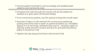
Instruct patient tobreath in and out deeply and swallow when
asked to and not bite the tube
Introduce the tube through the mouth and ask the patient to
swallow as it goes down and breath deeply
In an unconscious patient, use the spatula to keep the mouth open
Ascertain if tube is in the stomach for unconscious patient by
putting end of the tube in water as patient breathes out. If bubbles
appear, it is in the lungs, withdraw tube and re – introduce again or
aspirate a little stomach content and test for acid with blue litmus
paper. It should turn red
Tighten the clip and put the fluid in the funnel ¾ full
53
MR CHEGE .B.M
54.
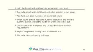
Hold the funnelwith left hand above patient’s head level
Open clip slowly with right hand and allow solution to run slowly
Add fluid as it goes in, do not let funnel get empty
When 300ml of fluid has gone in, lower the funnel and invert it
over the bucket and let the fluid flow until none comes out
Obtain specimen if required and take to the laboratory with
request form
Repeat the process till only clear fluid comes out
Pinch the tube and gently pull it out
54
MR CHEGE .B.M
55.
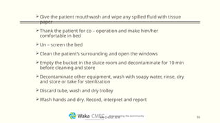
Give the patientmouthwash and wipe any spilled fluid with tissue
paper
Thank the patient for co – operation and make him/her
comfortable in bed
Un – screen the bed
Clean the patient’s surrounding and open the windows
Empty the bucket in the sluice room and decontaminate for 10 min
before cleaning and store
Decontaminate other equipment, wash with soapy water, rinse, dry
and store or take for sterilization
Discard tube, wash and dry trolley
Wash hands and dry. Record, interpret and report
55
MR CHEGE .B.M
56.
Title and ContentLayout with List
Add your first bullet point here
Add your second bullet point here
Add your third bullet point here
Editor's Notes
#1 NOTE:
To change the image on this slide, select the picture and delete it. Then click the Pictures icon in the placeholder to insert your own image.