Comprehensive Guide to Amputation: Surgical Management and Rehabilitation
Detailed overview of amputation epidemiology, surgical principles, nerve and muscle management, post-op care, complications, prosthetics, and psychological aspects for medical professionals.
Epidemiology & Etiology
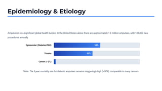
Amputationis a significant global health burden. In the United States alone, there are approximately 1.6 million amputees, with 185,000 new
procedures annually.
*Note: The 3-year mortality rate for diabetic amputees remains staggeringly high (~50%), comparable to many cancers.
3.
Pre-operative Assessment
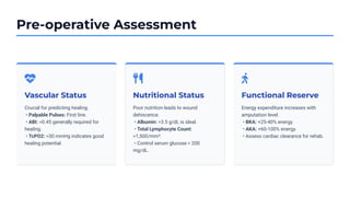
Vascular Status
Crucialfor predicting healing.
• Palpable Pulses: First line.
• ABI: >0.45 generally required for
healing.
• TcPO2: >30 mmHg indicates good
healing potential.
Nutritional Status
Poor nutrition leads to wound
dehiscence.
• Albumin: >3.5 g/dL is ideal.
• Total Lymphocyte Count:
>1,500/mm³.
• Control serum glucose < 200
mg/dL.
Functional Reserve
Energy expenditure increases with
amputation level.
• BKA: +25-40% energy.
• AKA: +60-100% energy.
• Assess cardiac clearance for rehab.
4.
Anatomic Levels: LowerLimb
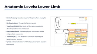
• Hemipelvectomy: Resection of part of the pelvis. Rare, usually for
tumors.
• Hip Disarticulation: Through the hip joint capsule.
• Transfemoral (AKA): Ideal length is 12cm above knee joint to
allow for prosthetic knee mechanism.
• Knee Disarticulation: End-bearing stump, but cosmetic issues
with prosthetic knee center.
• Transtibial (BKA): "The Workhorse". Preserves the knee joint,
vastly improving function.
• Syme’s: Ankle disarticulation with heel pad preservation. Excellent
end-bearing.
5.
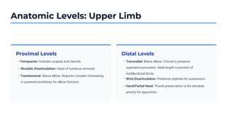
Anatomic Levels: UpperLimb
Proximal Levels Distal Levels
• Forequarter: Includes scapula and clavicle.
• Shoulder Disarticulation: Head of humerus removed.
• Transhumeral: Above elbow. Requires complex harnessing
or powered prosthesis for elbow function.
• Transradial: Below elbow. Critical to preserve
supination/pronation. Ideal length is junction of
middle/distal thirds.
• Wrist Disarticulation: Preserves styloids for suspension.
• Hand/Partial Hand: Thumb preservation is the absolute
priority for opposition.
6.
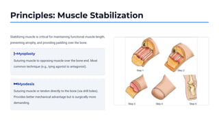
Principles: Muscle Stabilization
Stabilizingmuscle is critical for maintaining functional muscle length,
preventing atrophy, and providing padding over the bone.
Myoplasty
Suturing muscle to opposing muscle over the bone end. Most
common technique (e.g., tying agonist to antagonist).
Myodesis
Suturing muscle or tendon directly to the bone (via drill holes).
Provides better mechanical advantage but is surgically more
demanding.
7.
Principles: Nerves andBone
Nerve Management
Goal: Prevent painful neuromas.
Standard: Traction Neurectomy (pull distal, cut sharp, allow to
retract into soft tissue bed).
Modern (TMR): Targeted Muscle Reinnervation. Transferring
cut nerve endings to motor points of nearby muscles to reduce
phantom pain and enable bionic control.
Bone Management
Beveling: Sharp bone edges must be smoothed (rasped) to
prevent skin breakdown from the inside out.
Ertl Procedure: A bone bridge is created between tibia and
fibula in BKA to increase end-bearing capability (specialized).
8.
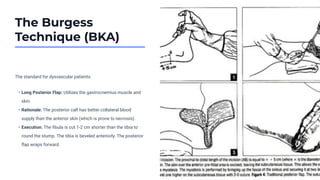
The Burgess
Technique (BKA)
Thestandard for dysvascular patients.
• Long Posterior Flap: Utilizes the gastrocnemius muscle and
skin.
• Rationale: The posterior calf has better collateral blood
supply than the anterior skin (which is prone to necrosis).
• Execution: The fibula is cut 1-2 cm shorter than the tibia to
round the stump. The tibia is beveled anteriorly. The posterior
flap wraps forward.
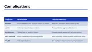
Complications
Complication Pathophysiology Prevention/Management
HematomaAccumulated blood acts as culture medium for bacteria. Meticulous hemostasis, drains (remove @ 48h).
Infection Higher risk in diabetic/trauma patients. Periop antibiotics, aggressive debridement.
Wound Necrosis Poor perfusion or tension on closure. Adequate vascular assessment, no tension sutures.
Joint Contracture Muscle imbalance/poor positioning (Flexion). Prone positioning 20 mins/day. Avoid pillows under stump.
DVT / PE Virchow's triad (stasis). DVT prophylaxis (Heparin/Lovenox), early mobilization.
11.
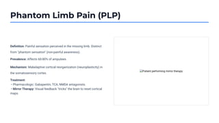
Phantom Limb Pain(PLP)
Definition: Painful sensation perceived in the missing limb. Distinct
from "phantom sensation" (non-painful awareness).
Prevalence: Affects 60-80% of amputees.
Mechanism: Maladaptive cortical reorganization (neuroplasticity) in
the somatosensory cortex.
Treatment:
• Pharmacologic: Gabapentin, TCA, NMDA antagonists.
• Mirror Therapy: Visual feedback "tricks" the brain to reset cortical
maps.
12.
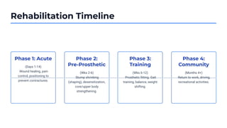
Rehabilitation Timeline
Phase 1:Acute
(Days 1-14)
Wound healing, pain
control, positioning to
prevent contractures.
Phase 2:
Pre-Prosthetic
(Wks 2-6)
Stump shrinking
(shaping), desensitization,
core/upper body
strengthening.
Phase 3:
Training
(Wks 6-12)
Prosthetic fitting. Gait
training, balance, weight
shifting.
Phase 4:
Community
(Months 4+)
Return to work, driving,
recreational activities.
13.
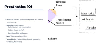
Prosthetics 101
• Socket:The interface. Must distribute pressure (e.g., Patellar
Tendon Bearing).
• Suspension: How it stays on.
• Pylon: The structural frame (shin).
• Terminal Device: The Foot (SACH, Dynamic Response) or
Hand (Hook, Myoelectric).
○ Suction/Vacuum: Best proprioception.
○ Pin Lock: Easy to don/doff.
○ Belts/Straps: Older, auxiliary use.
14.
Psychological Considerations
Body Image
Amputationis a permanent alteration of self. Patients often
experience a "disintegration" of their body schema. Early
viewing and touching of the residual limb helps reintegration.
Grief & Loss
Patients go through stages similar to grieving a death
(Kubler-Ross): Denial, Anger, Bargaining, Depression,
Acceptance. Clinical depression affects ~30% of amputees.