Biochemistry
Unit – 1
Carbohydrates
:- Mr. Priyanshu Anand
%%%%%%%%%%%%%%%%%%%%%%%%%%%%%%%%%%%%%%%%%%%%%%%%%%%%%%%%%%
Contents :-
 Introduction, Digestion, absorption and metabolism of carbohydrates and related disorders
 Regulation of blood glucose
 Diabetes Mellitus – type 1 and type 2, symptoms, complications & management in brief
 Investigations of Diabetes Mellitus
o OGTT – Indications, Procedure, Interpretation and types of GTT curve
o Mini GTT, extended GTT, GCT, IV GTT
o HbA1c (Only definition)
 Hypoglycemia – Definition & causes
%%%%%%%%%%%%%%%%%%%%%%%%%%%%%%%%%%%%%%%%%%%%%%%%%%%%%%%%%%
Introduction
The term carbohydrate was originally used to describe compounds that were literally
"hydrates of carbon" because they had the empirical formula CH2O. In recent years,
carbohydrates have been classified on the basis of their structures, not their formulas. They
are now defined as polyhydroxy aldehydes and ketones.
TYPES OF CARBOHYDRATES
1. Monosaccharide (Simple Carbohydrate)
These are used only for energy in living organisms. Simple carbohydrates are also known
as "Monosaccharides".
The chemical formula for all the monosaccharides is CnH2nOn.
e.g. - , glucose, fructose, galactose.
2. Disaccharides (Compound Carbohydrate)
These are used by living organisms for energy. They are composed of two
monosaccharides joined together by the process known as dehydration synthesis. In this
process, one molecule loses one hydrogen atom, while the other loses one hydrogen
atom and one oxygen atom.
Disaccharides have the chemical equation C12H22O11.
Ex. Maltose, lactose and Sucrose.
3. Polysaccharide (Complex Carbohydrate)
Polysaccharides, also called as glycanes, are high molecular weight which on hydrolysis
yield monosaccharides. Complex carbohydrates contain hundreds of sugar units. While
monosaccharides and disaccharides can be used for energy immediately,
polysaccharides release their energy slowly.
Chemically, the polysaccharide may be distinguished into homopolysachharide &
heteropolysachharide.
Homopolysachharides which on hydrolysis yield only single monosachharide. Eg. starch,
cellulose, etc.
Hetropolysachharide which on hydrolysis yield two or more monosaccharide. Eg.
heparin, chondroitin, etc.
DIGESTION, ABSORPTION & METABOLISM OF CARBOHYDRATES
During digestion, carbohydrates are broken down into simple, soluble sugars that can be
transported across the intestinal wall into the circulatory system to be transported throughout
the body.
Carbohydrate digestion begins in the mouth with the action of salivary amylase on starches
and ends with monosaccharides being absorbed across the epithelium of the small intestine.
Once the absorbed monosaccharides are transported to the tissues, the process of cellular
respiration begins.
There are several pathways known for the metabolism of carbohydrates. These pathways are
as follows :-
 Glycolysis
 Kreb’s Cycle / Citric acid cycle / Tricarboxylic acid cycle
 Gluconeogenesis
 Cori Cycle
 Glycogenolysis
 Pentose phosphate pathway
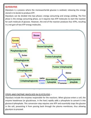
GLYCOLYSIS
Glycolysis is a process where the monosaccharide glucose is oxidized, releasing the energy
stored in its bonds to produce ATP.
Glycolysis can be divided into two phases: energy consuming and energy yielding. The first
phase is the energy consuming phase, so it requires two ATP molecules to start the reaction
for each molecule of glucose. However, the end of the reaction produces four ATPs, resulting
in a net gain of two ATP energy molecules.
STEPS AND ENZYME INVOLVED IN GLYCOLYSIS :-
Glycolysis include the enzymes responsible for the reactions. When glucose enters a cell, the
enzyme hexokinase (or glucokinase, in the liver) rapidly adds a phosphate to convert it into
glucose-6-phosphate. This conversion step requires one ATP and essentially traps the glucose
in the cell, preventing it from passing back through the plasma membrane, thus allowing
glycolysis to proceed.
In the next step of the first phase of glycolysis, the enzyme glucose-6 phosphate isomerase
converts glucose-6-phosphate into fructose-6 phosphate. Like glucose, fructose is also a six
carbon-containing sugar. The enzyme phosphofructokinase-1 then adds one more phosphate
to convert fructose-6-phosphate into fructose-1-6-bisphosphate, another six-carbon sugar,
using another ATP molecule. Aldolase then breaks down this fructose 1- 6-bisphosphate into
two three-carbon molecules, glyceraldehyde- 3 phosphate and dihydroxyacetone phosphate.
The triosephosphate isomerase enzyme then converts dihydroxyacetone phosphate into a
second glyceraldehyde-3- phosphate molecule. Therefore, by the end of this chemical- priming
or energy-consuming phase, one glucose molecule is broken down into two glyceraldehyde-3-
phosphate molecules.
The second phase of glycolysis, the energy-yielding phase, creates the energy that is the
product of glycolysis.
Glyceraldehyde-3-phosphate dehydrogenase converts each three-carbon glyceraldehyde-3-
phosphate produced during the energy-consuming phase into 1,3-bisphosphoglycerate. This
reaction releases an electron that is then picked up by NAD+ to create an NADH molecule.
NADH is a high-energy molecule, like ATP, but unlike ATP, it is not used as energy currency by
the cell. Because there are two glyceraldehyde-3-phosphate molecules, two NADH molecules
are synthesized during this step. Each 1,3 bisphosphoglycerate is subsequently
dephosphorylated (i.e., a phosphate is removed) by phosphoglycerate kinase into 3-
phosphoglycerate. Each phosphate released in this reaction can convert one molecule of ADP
into one high- energy ATP molecule, resulting in a gain of two ATP molecules.
The enzyme phosphoglycerate mutase then converts the 3 phosphoglycerate molecules into 2-
phosphoglycerate. The enolase enzyme then acts upon the 2-phosphoglycerate molecules to
convert them into phosphoenolpyruvate molecules. The last step of glycolysis involves the
dephosphorylation of the two phosphoenolpyruvate molecules by pyruvate kinase to create
two pyruvate molecules and two ATP molecules.
In summary, one glucose molecule breaks down into two pyruvate molecules, and creates two
net ATP molecules and two NADH molecules by glycolysis. Therefore, glycolysis generates
energy for the cell and creates pyruvate molecules that can be processed further through the
aerobic Krebs cycle (also called the citric acid cycle or tricarboxylic acid cycle); converted into
lactic acid or alcohol (in yeast) by fermentation; or used later for the synthesis of glucose
through gluconeogenesis.
ANAEROBIC RESPIRATION
When oxygen is limited or absent, pyruvate enters an anaerobic pathway. In these reactions,
pyruvate can be converted into lactic acid. In addition to generating an additional ATP, this
pathway serves to keep the pyruvate concentration low so glycolysis continues, and it oxidizes
NADH into the NAD+ needed by glycolysis. In this reaction, lactic acid replaces oxygen as the
final electron acceptor. Anaerobic respiration occurs in most cells of the body when oxygen is
limited or mitochondria are absent or nonfunctional. For example, because erythrocytes (red
blood cells) lack mitochondria, they must produce their ATP from anaerobic respiration. This is
an effective pathway of ATP production for short periods of time, ranging from seconds to a
few minutes. The lactic acid produced diffuses into the plasma and is carried to the liver,
where it is converted back into pyruvate or glucose via the Cori cycle. Similarly, when a person
exercises, muscles use ATP faster than oxygen can be delivered to them.
They depend on glycolysis and lactic acid production for rapid ATP production.
AEROBIC RESPIRATION
In the presence of oxygen, pyruvate can enter the Krebs cycle where additional energy is
extracted as electrons are transferred from the pyruvate to the receptors NAD+, GDP, and
FAD, with carbon dioxide being a “waste product”. The NADH and FADH2 pass electrons on to
the electron transport chain, which uses the transferred energy to produce ATP. As the
terminal step in the electron transport chain, oxygen is the terminal electron acceptor and
creates water inside the mitochondria.
KREB’S CYCLE / CITRIC ACID CYCLE / TRICARBOXYLIC ACID CYCLE
The pyruvate molecules generated during glycolysis are transported across the mitochondrial
membrane into the inner mitochondrial matrix, where they are metabolized by enzymes in a
pathway called the Krebs cycle. The Krebs cycle is also commonly called the citric acid cycle or
the tricarboxylic acid (TCA) cycle. During the Krebs cycle, high-energy molecules, including ATP,
NADH, and FADH2, are created. NADH and FADH2 then pass electrons through the electron
transport chain in the mitochondria to generate more ATP molecules.
 TCA is the most important metabolic pathway for the energy supply to the body. About
65–70% of the ATP is synthesized by Krebs cycle.
 Citric acid cycle essentially involves the oxidative decarboxylation of Acetyl Co-enzyme A
to CO₂ and H₂O. This cycle utilizes about 2/3 of total oxygen consumed by the body.
Location of TCA cycle: The enzymes of TCA cycle are located in the mitochondrial matrix in
close proximity to the electron transport chain.
Reactions of TCA cycle
Oxidative decarboxylation of Pyruvate to Acetyl Co-A by pyruvate dehydrogenase complex.
This step is a connecting link between glycolysis and TCA cycle.
1. Formation of Citrate – Krebs cycle proper starts with the condensation of Acetyl Co-A
and oxaloacetate catalyzed by enzyme citrate synthase.
2. Citrate is isomerized to Isocitrate by the enzyme aconitase. This is achieved in a
two-stage reaction by dehydration and rehydration.
3. Formation of Oxalosuccinate – Isocitrate is converted into oxalosuccinate by isocitrate
dehydrogenase, producing NADH₂.
4. Formation of α-Ketoglutarate – Oxalosuccinate is further converted into
α-ketoglutarate.
5. Conversion of α-Ketoglutarate to Succinyl Co-A – via oxidative decarboxylation catalyzed
by α-ketoglutarate dehydrogenase complex. This enzyme requires multiple cofactors.
6. Formation of Succinate – Succinyl Co-A is converted to succinate by succinyl-CoA
thiokinase. This reaction is coupled with phosphorylation of GDP → GTP.
7. Conversion of Succinate to Fumarate – Succinate is oxidized by succinate
dehydrogenase to fumarate, producing FADH₂.
8. Formation of Malate – Fumarate is hydrated to malate by fumarase.
9. Conversion of Malate to Oxaloacetate – Malate is oxidized by malate dehydrogenase,
producing NADH. This regenerates oxaloacetate, completing the cycle.
GLUCONEOGENESIS
Gluconeogenesis is the synthesis of new glucose molecules from pyruvate, lactate, glycerol, or
the amino acids alanine or glutamine. This process takes place primarily in the liver during
periods of low glucose, that is, under conditions of fasting, starvation, and low carbohydrate
diets.
First, the pyruvate is converted into oxaloacetate by the help of enzyme pyruvate carboxylase
present in mitochondria. Oxaloacetate then serves as a substrate for the enzyme
phosphoenolpyruvate carboxykinase (PEPCK), which transforms oxaloacetate into
phosphoenolpyruvate (PEP). From this step, gluconeogenesis is nearly the reverse of
glycolysis. PEP is converted back into 2- phosphoglycerate, which is converted into 3-
phosphoglycerate. Then, 3-phosphoglycerate is converted into 1,3 bisphosphoglycerate and
then into glyceraldehyde-3-phosphate. Two molecules of glyceraldehyde-3 phosphate then
combine to form fructose-1-6-bisphosphate, which is converted into fructose 6-phosphate and
then into glucose-6-phosphate. Finally, a series of reactions generates glucose itself.
In gluconeogenesis (as compared to glycolysis), the enzyme hexokinase is replaced by glucose-
6 phosphatase, and the enzyme phosphofructokinase-1 is replaced by fructose 1,6-
bisphosphatase. This helps the cell to regulate glycolysis and gluconeogenesis independently
of each other.
As part of lipolysis, fats can be broken down into glycerol, which can be phosphorylated to
form dihydroxyacetone phosphate or DHAP. DHAP can either enter the glycolytic pathway or
be used by the liver as a substrate for gluconeogenesis.
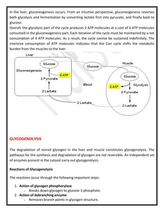
CORI CYCLE
The Cori cycle (also known as the Lactic acid cycle), named after its discoverers, Carl Ferdinand
Cori and Gerty Cori, refers to the metabolic pathway in which lactate produced by anaerobic
glycolysis in the muscles moves to the liver and is converted to glucose, which then returns to
the muscles and is metabolized back to lactate.
Muscular activity requires ATP, which is provided by the breakdown of glycogen in the skeletal
muscles.
The breakdown of glycogen, a process known as glycogenolysis, releases glucose in the form of
glucose-1 phosphate (G-1-P).
The G-1-P is converted to G-6-P by the enzyme phosphoglucomutase.
G-6-P is readily fed into glycolysis, (or can go into the pentose phosphate pathway if G-6-P
concentration is high) a process that provides ATP to the muscle cells as an energy source.
During muscular activity, the store of ATP needs to be constantly replenished. When the
supply of oxygen is sufficient, this energy comes from feeding pyruvate into the Krebs cycle.
When oxygen supply is insufficient, typically during intense muscular activity, energy must be
released through anaerobic metabolism.
Lactic acid fermentation converts pyruvate to lactate by lactate dehydrogenase. Most
importantly, fermentation regenerates NAD+, maintaining the NAD+ concentration so that
additional glycolysis reactions can occur. The fermentation step oxidizes the NADH produced
by glycolysis back to NAD+, transferring two electrons from NADH to reduce pyruvate into
lactate. Instead of accumulating inside the muscle cells, lactate produced by anaerobic
fermentation is taken up by the liver. This initiates the other half of the Cori cycle.
In the liver, gluconeogenesis occurs. From an intuitive perspective, gluconeogenesis reverses
both glycolysis and fermentation by converting lactate first into pyruvate, and finally back to
glucose.
Overall, the glycolysis part of the cycle produces 2 ATP molecules at a cost of 6 ATP molecules
consumed in the gluconeogenesis part. Each iteration of the cycle must be maintained by a net
consumption of 4 ATP molecules. As a result, the cycle cannot be sustained indefinitely. The
intensive consumption of ATP molecules indicates that the Cori cycle shifts the metabolic
burden from the muscles to the liver.
GLYCOGENOLYSIS
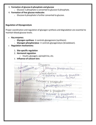
The degradation of stored glycogen in the liver and muscle constitutes glycogenolysis. The
pathways for the synthesis and degradation of glycogen are not reversible. An independent set
of enzymes present in the cytosol carry out glycogenolysis.
Reactions of Glycogenolysis
The reactions occur through the following important steps:
1. Action of glycogen phosphorylase
o Breaks down glycogen to glucose-1-phosphate.
2. Action of debranching enzyme
o Removes branch points in glycogen structure.
3. Formation of glucose-6-phosphate and glucose
o Glucose-1-phosphate is converted to glucose-6-phosphate.
4. Formation of free glucose molecules
o Glucose-6-phosphate is further converted to glucose.
Regulation of Glycogenolysis
Proper coordination and regulation of glycogen synthesis and degradation are essential to
maintain blood glucose levels.
 Key enzymes:
o Glycogen synthase → controls glycogenesis (synthesis).
o Glycogen phosphorylase → controls glycogenolysis (breakdown).
 Regulation mechanisms:
1. Site-specific regulation
2. Hormonal regulation
 Insulin, glucagon, epinephrine, etc.
3. Influence of calcium ions
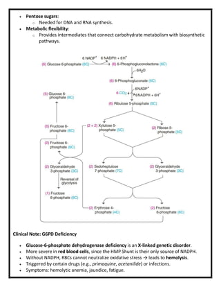
Hexose Monophosphate (HMP) Shunt / Pentose
Phosphate Pathway
 The HMP Shunt (also called the pentose phosphate pathway or phosphogluconate
pathway) is an alternative pathway to glycolysis and the TCA cycle for the oxidation of
glucose.
 Unlike glycolysis, it is anabolic in nature, since it produces NADPH and pentose sugars
(like ribose-5-phosphate).
Location
 Enzymes of the HMP Shunt are located in the cytosol.
 Active in tissues such as:
o Liver
o Adipose tissue
o Adrenal glands
o Erythrocytes
o Testes
o Lactating mammary glands
Phases
1. Oxidative Phase
o Generates NADPH and ribulose-5-phosphate.
o Key for biosynthesis and antioxidant defense.
2. Non-Oxidative Phase
o Interconverts sugars (3, 4, 5, 7 carbons).
o Produces ribose-5-phosphate for nucleotides.
o Links back to glycolysis via fructose-6-phosphate and glyceraldehyde-3-
phosphate.
Importance
 NADPH production:
o Essential for fatty acid synthesis, cholesterol synthesis, and maintaining reduced
glutathione in RBCs.
 Pentose sugars:
o Needed for DNA and RNA synthesis.
 Metabolic flexibility:
o Provides intermediates that connect carbohydrate metabolism with biosynthetic
pathways.
Clinical Note: G6PD Deficiency
 Glucose-6-phosphate dehydrogenase deficiency is an X-linked genetic disorder.
 More severe in red blood cells, since the HMP Shunt is their only source of NADPH.
 Without NADPH, RBCs cannot neutralize oxidative stress → leads to hemolysis.
 Triggered by certain drugs (e.g., primaquine, acetanilide) or infections.
 Symptoms: hemolytic anemia, jaundice, fatigue.
REGULATION OF BLOOD GLUCOSE LEVEL
Blood glucose regulation involves maintaining blood glucose levels at constant levels in the
face of dynamic glucose intake and energy use by the body.
On average this target range is 60-100 mg/dL for an adult although people can be
asymptomatic at much more varied levels. In order to maintain this range there are two main
hormones that control blood glucose levels: insulin and glucagon.
Insulin is released when there are high amounts of glucose in the blood stream. Glucagon is
released when there are low levels of glucose in the blood stream. There are other hormones
that effect glucose regulation and are mainly controlled by the sympathetic nervous system.
The brain doesn’t have any energy storage of its own and as a result needs a constant flow of
glucose, using about 120 grams of glucose daily or about 60% of total glucose used by the
body at resting state. Without proper blood glucose regulation the brain and other organs
could starve leading to death.
INSULIN
A key regulatory pathway to control blood glucose levels is the hormone insulin. Insulin is
released from the beta cells in the islets of Langerhans found in the pancreas. Insulin is
released when there is a high concentration of glucose in the blood stream.
GLUCAGON
When blood glucose levels are low, glucagon is released which inhibits the break down of
glucose (glycolysis) and activates the formation of glucose (gluconeogenesis). Gluconeogenesis
creates glucose from such molecules as pyruvate, lactate, glycerol, and glucogenic amino
acids. Glucagon is released from the alpha cells of the islets of Langerhans in the pancreas.
EPINEPHRINE
During extreme stimuli the sympathetic nervous system kicks into action. The brain sends
signals throughout the body to increase heart rate, cause bronchial dilation and hepatic
glucose release. The brain also sends signals to the adrenal glands. Epinephrine is released
from the adrenal glands. This hormone is important to the prolonged sympathetic response.
Epinephrine travels through the blood stream and causes the liver to release glucose thus
increasing blood glucose levels in order to be ready for the threat.
DIABETES MELLITUS
When there is a issue with the ability of the human body to use glucose due to a problem with
the insulin pathway diabetes mellitus may be at fault.
Diabetes Mellitus is a metabolic disorder characterized by chronic hyperglycemia and
excessive urine excretion.
TYPES OF DIABETES MELLITUS
Type 1 Type 1diabetes mellitus is often discovered early in a persons’ life and has also been
called juvenile diabetes mellitus. Type 1 diabetes mellitus is where no insulin is made. This is
due to damage to the pancreatic beta cells, often due to an autoimmune disorder. Without
insulin the glucose cannot go into the cells and will not make energy. As a result treatment for
type 1 diabetes mellitus is regular injections of insulin.
Type 2 Type diabetes mellitus is a condition where the body becomes resistant to insulin or
not enough insulin is made. People who are obese have a high risk for contracting type 2
Diabetes mellitus. Although there is no cure for type 2 careful monitoring of exercise and
nutrition can help. Other effects of the body are in increased rate of stroke and heart issues.
Also damage to small blood vessels can occur. This happens particularly in the eyes (diabetic
retinopathy) and kidneys (renalopathy). Also due to increased amounts of sugar in the blood
stream a diabetic is at a higher risk of infection and wounds will take longer to heal.
Other forms of diabetes include diabetes insipidus, brittle diabetes, and bronze diabetes.
Diabetes insipidus is characterized by the excess output (3 20 liters per day; normal is less than
3 liters per day) of dilute urine. There are four major classifications of diabetes insipidus, each
of which is related to the primary site/cause of the excess urine output. These forms are
referred to as central, nephrogenic, dipsogenic, and gestational.
Central diabetes insipidus relates to pathologies that affect the hypothalamus or the pituitary
resulting in defects in vasopressin (antidiuretic hormone, ADH) synthesis or release.
Nephrogenic diabetes insipidus results from the loss of, or abnormal, responses of the kidneys
to the actions of vasopressin.
Dipsogenic diabetes insipidus relates to disturbances in the hypothalamic control of thirst.
Gestational diabetes insipidus results from excess maternal production of prostaglandins or it
is due to placental synthesis of an enzyme called vasopressinase (leucyl cystinyl
aminopeptidase) that results in degradation of vasopressin.
SYMPTOMS OF DIABETES MELLITUS
 Frequent urination
 Excessive thirst
 Unexplained weight loss
 Extreme hunger
 Sudden vision changes
 Tingling or numbness in the hands or feet
 Feeling very tired much of the time
 Very dry skin
 Sores that is slow to heal
 More infections than usual
COMPLICATIONS OF DIABETES MELLITUS
Long-term complications of diabetes develop gradually. The longer you have diabetes — and
the less controlled your blood sugar — the higher the risk of complications.
 Cardiovascular disease :- Diabetes dramatically increases the risk of various
cardiovascular problems, including coronary artery disease with chest pain (angina),
heart attack, stroke and narrowing of arteries (atherosclerosis). If you have diabetes,
you're more likely to have heart disease or stroke.
 Nerve damage (neuropathy) :- Excess sugar can injure the walls of the tiny blood vessels
(capillaries) that nourish your nerves, especially in your legs. This can cause tingling,
numbness, burning or pain that usually begins at the tips of the toes or fingers and
gradually spreads upward. If left untreated, you could lose all sense of feeling in the
affected limbs. Damage to the nerves related to digestion can cause problems with
nausea, vomiting, diarrhea or constipation. For men, it may lead to erectile dysfunction.
 Kidney damage (nephropathy) :- The kidneys contain millions of tiny blood vessel
clusters (glomeruli) that filter waste from your blood. Diabetes can damage this delicate
filtering system. Severe damage can lead to kidney failure or irreversible end-stage
kidney disease, which may require dialysis or a kidney transplant.
 Eye damage (retinopathy :- Diabetes can damage the blood vessels of the retina
(diabetic retinopathy), potentially leading to blindness. Diabetes also increases the risk
of other serious vision conditions, such as cataracts and glaucoma.
 Foot damage :- Nerve damage in the feet or poor blood flow to the feet increases the
risk of various foot complications. Left untreated, cuts and blisters can develop serious
infections, which often heal poorly. These infections may ultimately require toe, foot or
leg amputation.
 Skin condition :- Diabetes may leave you more susceptible to skin problems, including
bacterial and fungal infections.
 Hearing impairmen :- Hearing problems are more common in people with diabetes.
 Alzheimer's diseas :- Type 2 diabetes may increase the risk of dementia, such as
Alzheimer's disease. The poorer your blood sugar control, the greater the risk appears to
be. Although there are theories as to how these disorders might be connected, none has
yet been proved.
 Depression :- Depression symptoms are common in people with type 1 and type 2
diabetes. Depression can affect diabetes management.
 Complications of gestational diabetes :-
o Complications in your baby can occur as a result of gestational diabetes, including:
 Excess growth. Extra glucose can cross the placenta, which triggers your
baby's pancreas to make extra insulin. This can cause your baby to grow too
large (macrosomia). Very large babies are more likely to require a C-section
birth.
 Low blood sugar. Sometimes babies of mothers with gestational diabetes
develop low blood sugar (hypoglycemia) shortly after birth because their
own insulin production is high. Prompt feedings and sometimes an
intravenous glucose solution can return the baby's blood sugar level to
normal.
 Type 2 diabetes later in life. Babies of mothers who have gestational
diabetes have a higher risk of developing obesity and type 2 diabetes later
in life.
 Death. Untreated gestational diabetes can result in a baby's death either
before or shortly after birth.
o Complications in the mother also can occur as a result of gestational diabetes,
including:
 Preeclampsia. This condition is characterized by high blood pressure, excess
protein in the urine, and swelling in the legs and feet. Preeclampsia can
lead to serious or even life-threatening complications for both mother and
baby.
 Subsequent gestational diabetes. Once you've had gestational diabetes in
one pregnancy, you're more likely to have it again with the next pregnancy.
You're also more likely to develop diabetes — typically type 2 diabetes —
as you get older.
o Complications of prediabetes :- Prediabetes may develop into type 2 diabetes.
MANAGEMENT OF DIABETES MELLITUS
 Normalize insulin activity. This is the main goal of diabetes treatment — normalization
of blood glucose levels to reduce the development of vascular and neuropathic
complications.
 Intensive treatment. Intensive treatment is three to four insulin injections per day or
continuous subcutaneous insulin infusion, insulin pump therapy plus frequent blood
glucose monitoring and weekly contacts with diabetes educators.
 Nutrition, meal planning, and weight control are the foundations of diabetes
management.
 Weight loss. This is the key treatment for obese patients with type 2 diabetes.
 Diet education, behavioral therapy, group support, and ongoing nutritional counselling
should be encouraged.
INVESTIGATION OF BLOOD GLUCOSE LEVEL
Oral Glucose Tolerance Test (OGTT)
Indications
 Diagnosis of Diabetes Mellitus and Impaired Glucose Tolerance (IGT).
 Screening in pregnancy (Gestational Diabetes Mellitus).
 When fasting plasma glucose is borderline.
Procedure
 Patient fasts overnight (8–12 hrs).
 Baseline fasting blood glucose sample taken.
 Administer 75 g oral glucose (adults).
 Blood samples at 0, 30, 60, 90, and 120 minutes.
Interpretation
 Normal: Fasting < 100 mg/dL; 2-hr < 140 mg/dL.
 Impaired Glucose Tolerance (IGT): 2-hr 140–199 mg/dL.
 Diabetes Mellitus: Fasting ≥ 126 mg/dL OR 2-hr ≥ 200 mg/dL.
Types of GTT Curve
 Normal curve: Peak at 30–60 min, returns to baseline by 2 hrs.
 Diabetic curve: Higher peak, delayed return, remains elevated at 2 hrs.
 Lag curve: Delayed peak, returns to normal later (seen in mild cases).
 Flat curve: No significant rise (seen in malabsorption, hypothyroidism).
Variants of GTT
 Mini GTT: Shortened test, fewer samples (screening purpose).
 Extended GTT: Samples taken up to 3–5 hrs (used in research/rare cases).
 Glucose Challenge Test (GCT): Screening in pregnancy; 50 g glucose load, 1-hr plasma
glucose measured.
 IV GTT: Glucose given intravenously; used when patient cannot tolerate oral glucose
(rare, research settings).
HbA1c
 HbA1c = Glycated Hemoglobin, formed when glucose binds irreversibly to hemoglobin.
 Reflects average blood glucose over 2–3 months.
HYPOGLYCEMIA
INTRODUCTION
Hypoglycemia is a clinical condition characterized by abnormally low blood glucose levels.
Clinically significant when plasma glucose < 70 mg/dL (though symptoms often appear below
55 mg/dL).
Severe hypoglycemia: when neuroglycopenic symptoms occur (confusion, seizures, coma).
Causes of Hypoglycemia
1. Drug-related
 Insulin therapy (overdose, mismatch with meals).
 Oral hypoglycemic agents (sulfonylureas, meglitinides).
 Alcohol (inhibits gluconeogenesis).
2. Endocrine/Metabolic
 Adrenal insufficiency (↓ cortisol).
 Hypopituitarism (↓ ACTH, GH).
 Severe liver disease (↓ glycogen stores, impaired gluconeogenesis).
 Renal failure (↓ clearance of insulin).
3. Reactive/Post-prandial
 Occurs after meals due to exaggerated insulin response.
 Seen after gastric surgery (dumping syndrome).
4. Fasting Hypoglycemia
 Prolonged fasting or starvation.
 Inborn errors of metabolism (glycogen storage diseases, fatty acid oxidation defects).
5. Critical Illness
 Sepsis, severe infections.
 Malnutrition.