Biotechnology
unit-4
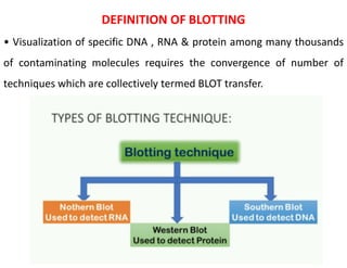
DEFINITION OF BLOTTING
• Visualization of specific DNA , RNA & protein among many thousands
of contaminating molecules requires the convergence of number of
techniques which are collectively termed BLOT transfer.
IMMUNOBLOTTING
• Viral antigens are detected with a polyclonal or an MAb onto
nitrocellulose paper.
• After incubation, the protein bands (immune complexes) are
visualized with peroxides-conjugated protein and a color reagent.
• A color develops in the bands where the antibody binds to the
antigen.
• Immunoblotting assay mixture of these two technique.
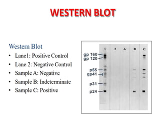
WESTERN BLOTTING
•Western blotting is based on the principles of immunochromatography,
where proteins are separated into polyacrylamide gel according to the
isoelectric point and molecular weight.
•A technique for detecting specific proteins separated by
electrophoresis by use of labeled antibodies.
•Immunoblotting is performed chiefly in diagnostic laboratories to
identify the desirable protein antigens in complex mixtures.
• An improved immunoblot method Zestern analysis, is able to address
this issue without the electrophoresis step, thus significantly improving
the efficiency of protein analysis.
• Other related techniques include dot blot analysis, zestern analysis,
immunohistochemistry where antibodies are used to detect proteins in
tissues and cells by immunostaining and enzyme-linked immunosorbent
assay (ELISA)
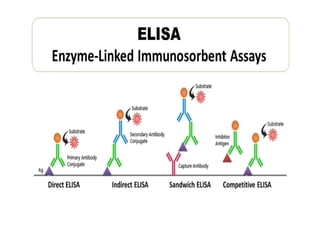
ELISA
Enzyme-linked immunosorbent assay (ELISA): ELISA stands for "enzyme-
linked immunosorbent assay." This is a rapid immunochemical test that
involves an enzyme (a protein that catalyzes a biochemical reaction). It
also involves an antibody or antigen (immunologic molecules). ELISA
tests are utilized to detect substances that have antigenic properties,
primarily proteins (as opposed to small molecules and ions such as
glucose and potassium). Some of these include hormones, bacterial
antigens and antibodies.
TYPES OF ELISA
1- Direct ELISA 2- Indirect ELISA
3- Sandwich ELISA 4- Competitive ELISA
Diseases That Can Be Diagnosed Using ELISA
ELISA can be used to detect some of these conditions:
•Ebola
•Pernicious anaemia
•AIDS
•Rotavirus
•Lyme disease
•Syphilis
•Toxoplasmosis
•Zika virus
•Carcinoma of the epithelial cells
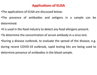
Applications of ELISA
•The applications of ELISA are discussed below:
•The presence of antibodies and antigens in a sample can be
determined.
•It is used in the food industry to detect any food allergens present.
•To determine the concentration of serum antibody in a virus test.
•During a disease outbreak, to evaluate the spread of the disease, e.g.
during recent COVID-19 outbreak, rapid testing kits are being used to
determine presence of antibodies in the blood sample.
The technique was first described by Harry Towbin in 1979 and named
by W. Neal Burnette in 1981. The procedure of western blotting can be
understood by the following points:
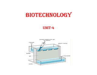
Gel Electrophoresis: The protein samples are separated using gel
electrophoresis. SDS-PAGE is commonly used for this step.
Transfer: The separated proteins are transferred on a solid support as
nitrocellulose or polyvinylidene difluoride membrane.
Total Protein Staining: The proteins transferred on the membrane are
visualised by staining the membrane using dyes such as Ponceau S,
Coomasie R-350 or amido black.
Blocking: This step is done to block the interaction between the
membrane and the antibody that will be applied in the next step. Since
the target molecule and antibodies are both proteins, there are chances
that the membrane might interact with the antibody.
Incubation: An antibody with a reporter enzyme is probed on the
membrane. A substrate that is specific to the enzyme is applied on the
surface. If binding happens, a colourimetric reaction is produced.
Detection and Visualization: The unbound probes are washed away and
the gel is visualised for the bound proteins.
Applications
•Detection of particular protein from a mixture of proteins.
•Size and amount estimate of proteins in the mixture.
•Verification following a high sensitivity ELISA test for diagnosis of Lyme,
HIV infection, BSE, HBV and so on.
•Detect condensed isoforms of proteins as well as tagged proteins.
Advantage
•Effective early diagnostic tool.
•Detect minimal immunogenic response form virus or bacteria.
•Requires fewer antibodies for testing.
•Detect specific protein from a large mixture of different proteins. (Even
more than 300,000).
Disadvantages
•Requires specific primary antibodies to perform test on desired protein
of interest.
•Challenging and hence requires well trained staffs.
•Poorer results as antibodies may revel off-target bindings.
•Detecting and imaging the results can be expensive as equipment cost
is high.
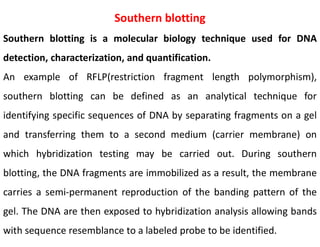
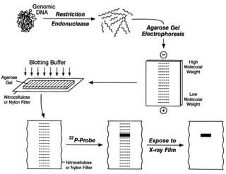
Southern blotting
Southern blotting is a molecular biology technique used for DNA
detection, characterization, and quantification.
An example of RFLP(restriction fragment length polymorphism),
southern blotting can be defined as an analytical technique for
identifying specific sequences of DNA by separating fragments on a gel
and transferring them to a second medium (carrier membrane) on
which hybridization testing may be carried out. During southern
blotting, the DNA fragments are immobilized as a result, the membrane
carries a semi-permanent reproduction of the banding pattern of the
gel. The DNA are then exposed to hybridization analysis allowing bands
with sequence resemblance to a labeled probe to be identified.
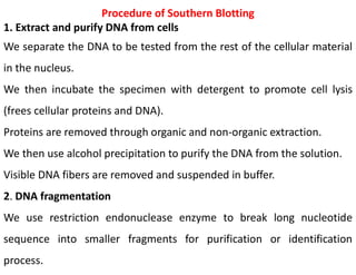
Procedure of Southern Blotting
1. Extract and purify DNA from cells
We separate the DNA to be tested from the rest of the cellular material
in the nucleus.
We then incubate the specimen with detergent to promote cell lysis
(frees cellular proteins and DNA).
Proteins are removed through organic and non-organic extraction.
We then use alcohol precipitation to purify the DNA from the solution.
Visible DNA fibers are removed and suspended in buffer.
2. DNA fragmentation
We use restriction endonuclease enzyme to break long nucleotide
sequence into smaller fragments for purification or identification
process.
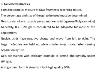
3. Gel electrophoresis
Sorts the complex mixture of DNA fragments according to size.
The percentage and size of the gel to be used must be determined.
Gels consist of microscopic pores and are solid (agarose/Polyacrylmide)
Generally, 0.7 – 2% gel is considered to be adequate for most of the
applications.
Nucleic acids have negative charge and move from left to right. The
large molecules are held up while smaller ones move faster causing
separation by size.
Gels are stained with ethidium bromide to permit photography under
UV light.
A single band form is given to intact high quality DNA.
4. Denature DNA
DNA obtained are double stranded in nature.
Alkalis are used to denature the restriction fragments in the gel that
makes double stranded DNA to become single stranded.
To avoid re-hybridization, we use NaCl so that DNA is neutralized.
5. Blotting
Transfer DNA from the gel to solid support (carrier membrane).
We dry the blot (around 80°C) or use UV radiation to make it
permanent.
6. Washing
Despite using blocking agents, some excess probe binds to the
membrane.
Wash buffers containing NaCl and detergent washes away the excess
probe.
Applications of Southern Blotting
•Identify specific DNA from a DNA sample.
•Identification of viral and bacterial infections.
•Important in the study of gene mutation, deletion and rearrangements.
•DNA fingerprinting (maternity and paternity analysis, forensic studies
and personal identification).
•Diagnosis of neonatal and genetic diseases including cancer.
•Discovery of RFLP (restriction fragment length polymorphism) to map
crucial genomes.
Northern Blotting
•Purpose: Detects specific RNA sequences (and their abundance) within a sample.
•Key Difference: Targets RNA, unlike Southern blotting (DNA) or Western blotting (proteins).
•Steps:
• RNA Isolation: Extract RNA from cells/tissues.
• Gel Electrophoresis: Separate RNA fragments by size using an electric field.
• Transfer: Blot RNA from gel onto a membrane (usually nylon or nitrocellulose).
• Hybridization: Incubate membrane with a labeled probe (DNA or RNA) that's
complementary to the target RNA sequence.
• Detection: Visualize the probe-RNA complex using:
• Radioactive Labeling: Probe emits radiation, detected by X-ray film.
• Non-Radioactive Labeling: Probe linked to an enzyme that produces a colored or
chemiluminescent signal.
Applications:
•Gene Expression Studies: Determine if a gene is active (producing RNA) in a sample.
•RNA Sizing: Estimate the size of specific RNA transcripts.
•RNA Quantification: Compare relative amounts of RNA in different samples.
•Medical Research: Study RNA viruses, gene expression changes in diseases.
Advantages:
•Sensitive: Detects even low levels of RNA.
•Specific: Identifies unique RNA sequences.
•Quantitative: Provides information on RNA abundance.
Blotting & Elisa Techniques Bio-Technology