Bleeding disorder
Dr. Abhishek Singh
CONTENT
 Introduction
 Disorder of coagulation
 Diagnosis of bleeding disorder
 Von willebrand disease
 Bernard soulier syndrome
 Glanzmann’s thrombasthenia
 Hemophilia
INTRODUCTION
 A group of disorders characterized by defective haemostasis with abnormal bleeding.
 These causes are due to :
1. Vessel wall abnormalities
2. Platelet deficiency or dysfunction
3. Derangement of coagulation
DISORDERS OF COAGULATION
HEREDITARY
 Hemophilia A (factor VIII deficiency)
 Hemophilia B(factor IX deficiency)
 Von willebrand disease
 Fibrinolytic disorder
 Bernard-soulier syndrome
 Glanzmann’s thrombasthenia
 Wiscott-Aldrich syndrome
 Chediak-Higashi syndrome
 Hereditary hemorrhagic telengectasia
 Ehlers-Danlos syndrome
 Osteogenesis imperfecta
ACQUIRED
 Disseminated intravascular coagulation
(DIC)
 Liver disease
 Vit K deficiency
 Massive transfusion of stored blood
 Acquired inhibitors of coagulation
 Heparin or oral anticoagulant therapy
 Renal disease
 Idiopathic thrombocytopenic purpura
 Thrombotic thrombocytopenic purpura
 Hemolytic uremic syndrome
 Henoch-schonlein purpura
DIAGNOSIS OF INHERITED BLEEDING
DISORDERS
 HISTORY
 CLINICAL EXAMINATION
 LABORATORY INVESTIGATIONS
CLINICAL HISTORY
 Age of first manifestation
 Family history of bleeding
 Past history
 Spontaneous or after trauma
 Site of bleeding
 Time of manifestation after injury
 H/o defective wound healing
 H/o menorrhagia/PPH
 Drug history
Clinical examination
Platelet disorder Coagulation disorder
Site of bleeding Skin, mucous membranes
(epistaxis, gum, vaginal,
GI tract)
Deep in soft tissues
(joints, muscle)
Petechiae Yes No
Ecchymoses("bruises") Small, Superficial Large, Deep
Hemarthrosis/ Muscle
bleeding
Extremely rare Common
Bleeding after cut &
scratches
Yes No
Bleeding after surgery or
trauma
Immediate, usually mild Delayed(12-24hrs),
often severe
Laboratory approach
PREANALYTICAL VARIABLES
Collection of Sample
 Plastic or polypropylene syringes should be used.
 Venous blood is preferred over capillary blood.
 Don`t use pressure cuff or use for <1 min.
 Blood sample from a indwelling line or catheter should not be used.
 Mix thoroughly with anticoagulant by inverting the container several times.
Anticoagulation
 Most commonly: Trisodium citrate (1:9)
Centrifugation
Preparation of Platelet Poor Plasma(PPP)
 Centrifuged at 2000 rpm for 10 min.
 PPP should be kept at room temp. for PT, lupus anticoagulant & factor VII assay, and at
4 C for other assay.
 Platelet count should be <10,000/mm, it is best achieved by double centrifugation.
End-point detection
 Refers to clot formation.
 Early forming clot is more opaque and easier to detect.
 Slowly forming clot appears as fibrin wisp.
 Different techniques used in Coagulometer :
Electromechanical
Photo optical
Electrochemical etc.
TESTS FOR BLEEDING DISORDER
First line screening Test
 Prothrombin Time(PT)
 Activated Partial Thromboplastin Time(APTT)
 Thrombin Time(TT)
 Clotting Time(CT)
 Platelet count
 Bleeding Time(BT)
Second line specific test for factor abnormalities
 Mixing study
 Factor assay
 Fibrinogen assay
 Clot solubility test
 PFA-100
 D-dimer
PROTHROMBIN TIME(PT)
Significance
 Reflects overall activity of the Extrinsic Pathway.
 Most sensitive to changes in Factor V,VII,X.
 Lesser to Factor I & II.
Principle & Method
Platelet poor plasma(100µl) + Tissue Thromboplastin(200µl)
In Presence of FVII Extrinsic pathway is activated
& clot formed
Normal Range
 11 to 16 seconds
INR- PTtest/PTnormal
Interpretation
Causes of prolonged PT
1. Deficiency of Factor VII,X,V,II,I
2. Vit K deficiency
3. Liver disease esp. Obstructive Jaundice
4. DIC
ACTIVATED PARTIAL THROMBOPLASTIN
TIME (aPTT)
Significance
 Reflects efficiency of Intrinsic Pathway
 Sensitive to changes in Factor VIII,IX,XI,XII
Principle
The test measures the clotting time of plasma after the activation of contact
Factors(Kaolin/Silica) and the addition of CaCl2, but without added tissue
thromboplastin.
Method- Plasma(100µl) + aPTT reagent (100µl)
mix & incubate for 3-5 min
add prewarm Cacl2(100µl) Clot formation
Normal range
26 to 40 seconds (+/-5 of control)
Interpretation
Causes of prolonged APTT
 1. Deficiency of Factor VIII(Hemophilia A)
 2. Deficiency of Factor IX(Hemophilia B)
 3. DIC
 5. Circulating anticoagulants
 6. Liver disease
THROMBIN TIME(TT)
Significance
 Asses the final step of coagulation i.e. conversion of fibrinogen to fibrin in presence of
thrombin.
 Bypasses Extrinsic & Intrinsic pathway.
Principle
Thrombin is added to plasma and the clotting time is measured.
TT is affected by the concentration and reaction of fibrinogen and by the presence of
inhibitory substances including fibrinogen/fibrin degradation products(FDPs) and heparin.
Method – Pt plasma(100µl) + thrombin(200µl) clot formation
Normal range
15–19 sec
A patient’s TT should be within 2 sec of the control
Interpretation
Causes of prolonged TT
 Disorders of fibrinogen-
Afibrinogenaemia.
Hypofibrinogenaemia.
Dysfibrinogenaemia.
 Liver disease
CLOTTING TIME
To measure the time required for a sample of blood to coagulate in vitro
under standard conditions.
Normal value 5-10 minute
The clotting time is prolonged, if there is a marked deficiency in any of
the coagulation factors of the extrinsic/ intrinsic pathway.
CAPILLARY METHOD
PRINCIPLE-
 puncture the skin, blood
is taken to a plain
capillary tube and stop
watch started.
 Formation of fibrin strings is
noted by breaking the capillary
tube at regular intervals.
 The time taken for the first
appearance of the fibrin
string is noted.
OTHER METHOD
whole blood clotting Slide method
Platelet count & Bleeding Time
Platelet count must be done in a suspected bleeding disorder.
PBS must be examined for size & staining properties of plt.
BLEEDING TIME(BT)
 Time required for bleeding to cease following skin puncture
 Assess primary hemostatic defect(vessel wall or platelet)
Methods
Ivy’s- On volar surface of forearm with lancet 2mm deep under venous pressure of 40mmHg
Blood oozing is blotted with filter paper & time taken to stop bleeding is noted.
Template- same as ivy’s with 6-9mm depth of incision
Duke’s – lobes are punctured
Normal Range
Ivy’s method: 2 to 7 mins.
Template method: 2.5 to 9.5 mins.
Interpretation
Causes of prolonged BT
I. THROMBOCYTOPENIA
II. VWD
III. PLATLET FUNCTION DISORDER
IV. DISORDERS OF BLOOD VESSELS
SECOND LINE INVESTIGATIONS
 Relevant second line investigations are carried out with each of the
patterns of abnormalities in first line tests.
Mixing study
CORRECTION TEST USING PT or APTT
PRINCIPLE
 Unexplained prolongation of PT or APTT can be investigated with simple
correction test by mixing the pt`s plasma with normal plasma.
 Correction (should be within few seconds) indicates a possible factor
deficiency, whereas failure to correct suggests the presence of an inhibitor.
.
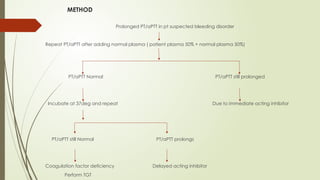
METHOD
Prolonged PT/aPTT in pt suspected bleeding disorder
Repeat PT/aPTT after adding normal plasma ( patient plasma 50% + normal plasma 50%)
PT/aPTT Normal PT/aPTT still prolonged
Incubate at 37deg and repeat Due to immediate acting inhibitor
PT/aPTT still Normal PT/aPTT prolongs
Coagulation factor deficiency Delayed acting inhibitor
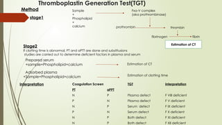
Perform TGT
Thromboplastin Generation Test(TGT)
Sample
+
Phospholipid
+
calcium
Fxa-V complex
(aka prothrombinase)
prothrombin thrombin
fibrinogen fibrin
Estimation of CT
Method
stage1
Stage2
If clotting time is abnormal, PT and aPTT are done and substitusions
studies are carried out to determine deficient factors in plasma and serum
Prepared serum
+sample+Phospholipid+calcium
Adsorbed plasma
+Sample+Phospholipid+calcium
Estimation of CT
Estimation of clotting time
Interpretation Coagulation Screen TGT Interpretation
PT aPPT
N P Plasma defect F VIII deficient
P N Plasma defect F V deficient
N P Serum defect F IX deficient
P N Serum defect F X deficient
N P Both defect F XI deficient
N P Both defect F XII deficient
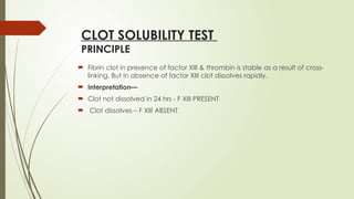
CLOT SOLUBILITY TEST
PRINCIPLE
 Fibrin clot in presence of factor XIII & thrombin is stable as a result of cross-
linking. But in absence of factor XIII clot dissolves rapidly.
 Interpretation—
 Clot not dissolved in 24 hrs - F XIII PRESENT
 Clot dissolves – F XIII ABSENT
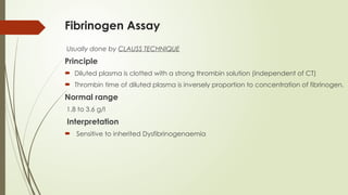
Fibrinogen Assay
Usually done by CLAUSS TECHNIQUE
Principle
 Diluted plasma is clotted with a strong thrombin solution (independent of CT)
 Thrombin time of diluted plasma is inversely proportion to concentration of fibrinogen.
Normal range
1.8 to 3.6 g/l
Interpretation
 Sensitive to inherited Dysfibrinogenaemia
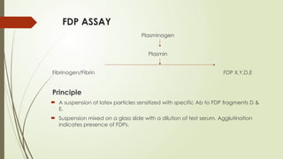
FDP ASSAY
Plasminogen
Plasmin
Fibrinogen/Fibrin FDP X,Y,D,E
Principle
 A suspension of latex particles sensitized with specific Ab to FDP fragments D &
E.
 Suspension mixed on a glass slide with a dilution of test serum. Agglutination
indicates presence of FDPs.
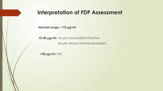
Interpretation of FDP Assessment
Normal range- <10 µg/ml
10-40 µg/ml- Acute myocardial Infarction
Acute venous thromboembolism
>40 µg/ml- DIC
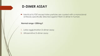
D-DIMER ASSAY
 Identical to FDP except latex particles are coated with a monoclonal
antibody specifically directed against Fibrin D-dimer in human.
Normal range <200mg/l
1. Latex agglutination D-dimer assay
2. Ultrasensitive D-dimer assay
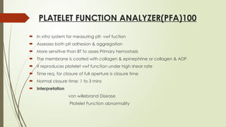
PLATELET FUNCTION ANALYZER(PFA)100
 In vitro system for measuring plt- vwf fuction
 Assesses both plt adhesion & aggregation
 More sensitive than BT to asses Primary hemostasis
 The membrane is coated with collagen & epinephrine or collagen & ADP
 It reproduces platelet vwf function under high shear rate
 Time req. for closure of full aperture is closure time
 Normal closure time: 1 to 3 mins
 Interpretation
von willebrand Disease
Platelet Function abnormality
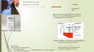
Blood aspirated through
Capillary into machinee collagen
Epinephrin/
ADP
Blood passes throughAperture in machine
Which is coated with platelet Agonist such
as Collagen/epinephrine/ADP
Platelet adhesion, activation and Aggregation
closes the aperture overtime
This time known as closure time is
indicator of platelet function
Abnormals:
Closure time is prolonged in
1. Platelet disorder
2. Von willebrand disease
Not prolonged in vascular disorder
The PFA 100 ANALYSER
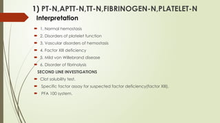
1) PT-N,APTT-N,TT-N,FIBRINOGEN-N,PLATELET-N
Interpretation
 1. Normal hemostasis
 2. Disorders of platelet function
 3. Vascular disorders of hemostasis
 4. Factor XIII deficiency
 5. Mild von Willebrand disease
 6. Disorder of fibrinolysis
SECOND LINE INVESTIGATIONS
 Clot solubility test.
 Specific factor assay for suspected factor deficiency(factor XIII).
 PFA 100 system.
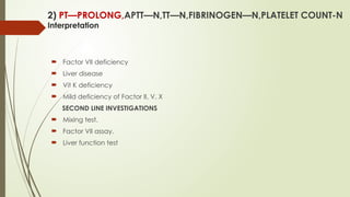
2) PT—PROLONG,APTT—N,TT—N,FIBRINOGEN—N,PLATELET COUNT-N
Interpretation
 Factor VII deficiency
 Liver disease
 Vit K deficiency
 Mild deficiency of Factor II, V, X
SECOND LINE INVESTIGATIONS
 Mixing test.
 Factor VII assay.
 Liver function test
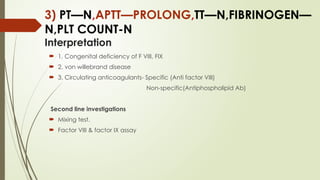
3) PT—N,APTT—PROLONG,TT—N,FIBRINOGEN—
N,PLT COUNT-N
Interpretation
 1. Congenital deficiency of F VIII, FIX
 2. von willebrand disease
 3. Circulating anticoagulants- Specific (Anti factor VIII)
Non-specific(Antiphospholipid Ab)
Second line investigations
 Mixing test.
 Factor VIII & factor IX assay
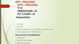
4)PT—PROLONG
APTT—PROLONG
TT-N
FIBRINOGEN—N
PLT COUNT—N
Interpretation
1. Vit k def
2. Liver dis.
3. Rare congenital or acq. deficiency of factor V,X,II
4. Combined factor V+VIII deficiency.
Second line investigations
1. Mixing test.
2. Specific factor assay.
3. Liver function test.
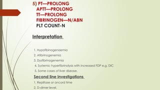
5) PT—PROLONG
APTT—PROLONG
TT—PROLONG
FIBRINOGEN—N/ABN
PLT COUNT-N
Interpretation
1. Hypofibinogenaemia
2. Afibrinogenemia
3. Dysfibrinogenemia
4. Systemic hyperfibrinolysis with increased FDP e.g. DIC
5. Some cases of liver disease.
Second line investigations
1. Replitase or ancord time
2. D-dimer level.
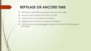
REPTILASE OR ANCORD TIME
 REPTILASE & ANCORD are purified enzymes from snake.
 May be used to replace Thrombin in TT test.
 Snake venom is not inhibited by heparin.
 Normal time for clotting in presence of Heparin.
 Clotting time will be prolonged in presence of raised FDPs/decreased
Fibrinogen.
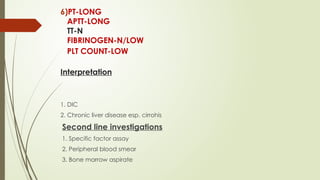
6)PT-LONG
APTT-LONG
TT-N
FIBRINOGEN-N/LOW
PLT COUNT-LOW
Interpretation
1. DIC
2. Chronic liver disease esp. cirrohis
Second line investigations
1. Specific factor assay
2. Peripheral blood smear
3. Bone marrow aspirate
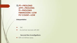
7) PT—PROLONG
APTT—PROLONG
TT—PROLONG
FIBRINOGEN—LOW
PLT COUNT—LOW
Interpretation
 DIC
 Acute liver necrosis with DIC
Second line investigations
 FDP or D-Dimer assay.
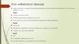
Von willebrand disease
 Most common congenital bleeding disorder characterized by deficiency or functional
defect of vwf.
Type1
 Most common & AD
 Partial quantitative deficiency of vwf
 vwf multimers (small, intermediate & large) are mildly deficient
Type2
 AD & AR
 Qualitative defects in vwf with absence of large vwf multimers
 4 types- 2A, 2B, 2M & 2N(AR)
Type3
 AR
 Complete deficiency of vwf
 Severe deficiency of all forms of vwf multimers
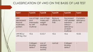
CLASSIFICATION OF vWD ON THE BASIS OF LAB TEST
Type1 Type2A Type2B Type2N Type2M Type3
Mild
decreased/
Normal
multimeric
pattern
Loss of high
and
intermedia
te vWF
multimer
Loss of high
molecular
weight
multimer
Reduce
affinity for
FVIII
Decreased
vWF platelet
adhesion
without
deficiency
of HMW
multimer
Complete
deficiency
of vWF
vWF:RCo/
vWF:Ag >0.6
<0.6 0.5-0.7 <0.6 <0.6 <0.03
- Collagen
binding
defect
Loss of
platelet
binding
activity
normal Collagen
binding
defect
-
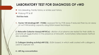
LABORATORY TEST FOR DIAGNOSIS OF vWD
 H/o bleeding, Family history or similar past history
 Prolong PTT & BT
First line tests
1. Factor VIII binding(vWF : FVIIIB): assessed first by FVIII assay if reduced then by an assay
of vwf FVIII binding capacity using ELISA based technique.
2. Ristocetin Cofactor Assay(vWF:RCo) : dilution of pt plasma are tested for their ability to
promote plt agglutination in the presence of ristocetin. Automated, latex beads method
also available.
3. Collagen binding activity(vWF:CB) : ELISA based, in which well coated with collagen is
used to capture vwf.
4. vWF:Ag concentration
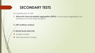
SECONDARY TESTS
for classification of vWF
1. Ristocetin induced platelet aggregation (RIPA) – Inducing plt aggregation by
binding of vwf with GpIb receptor.
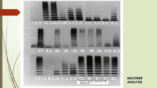
2. vWF multimer analysis
3. MOLECULAR ANALYSIS
 Sanger analysis
 Next generation testing
MULTIMER
ANALYSIS
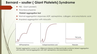
Bernard – soulier ( Giant Platelets) Syndrome
 PBS - Giant platelets
 Thrombocytopenia
Platelet aggregation test
 Normal aggregation responses ADP, epinephrine, collagen, and arachidonic acid
 Impaired aggregation with ristocetin
Platelet aggregation curves in von Willebrand disease and Bernard-Soulier syndrome (absent aggregation
with ristocetin, normal aggregation with ADP, epinephrine, and arachidonic acid)
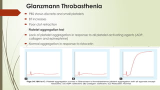
Glanzmann Throbasthenia
 PBS shows discrete and small platelets
 BT increases
 Poor clot retraction
Platelet aggregation test
 Lack of platelet aggregation in response to all platelet-activating agents (ADP,
collagen and epinephrine)
 Normal aggregation in response to ristocetin
HEMOPHILIA A(CLASSICAL)
 Hereditary deficiency of FVIII
 X-linked recessive affecting males
Classification
 Mild- FVIII level >5% , excess bleeding after minor trauma
 Moderate- 1-5%, excess bleeding after mild to moderate trauma
 Severe- <1%, frequent and spontaneous hematomas & hemarthroses
Diagnosis
 Male with h/o spontaneous bleeding in joints
 Positive family history in other male on maternal side
 Excess bleeding following circumcision & tooth eruption
Screening tests
PT-nomal,
APTT- Prolonged,
CBC
Second line test
 Factor VIII assay
 Genetic testing
HEMOPHILIA B (CHRISTMAS DISEASE)
 F IX deficiency
Screening Test
APTT – Prolonged
PT – Normal
TT – Normal
Second line test
 Factor IX Assay
 DNA Analysis
REFERENCES
 Dacie and Lewis practical hematology, twelfth edition
 Shirish M Kawthalkar, essentials of clinical pathology, second edition
 Robbins & Cotran, pathologic basis of disease, tenth edition
BLEEDING DISORDER BY Dr AKS IMS BHU KGMU LKO .pptx