OUTLINE
 DEFINATION
 PRE-OPERATIVE MANAGEMENT
 INDUCTION OF ANAESTHESIA
 INTRAOPERATIVE MANAGEMENT
 SPECIAL PATIENTS CIRCUMSTANCES
 REGIONAL ANAESTHESIA
WHAT IS ANAESTHESIA
 The absence of sensation, especially artificially
induced insensitivity to pain, usually achieved by the
administration of gases or the injection of drugs
 Word has Greek origin: from anaisthesia
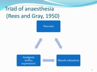
Triad of anaesthesia
(Rees and Gray, 1950)
Narcosis
Muscle relaxation
Analgesia
(reflex
supression)
4
1.0 PRE-OPERATIVE MANAGEMENT
 1.1 PRE-OPERATIVE ASSESSMENT
 1.2 PREPARATION FOR ANAESTHESIA
 1.3 CONCURRENT MEDICAL DISEASE AND
MEDICATION
 1.4 CONCURRENT SURGICAL DISEASE
1.1 PRE OPERATIVE ASSESSMENT
 WHY?
Based on three main aims:
 Reduce risk associated with surgery
 Reduce morbidity
 Reduce mortality
 Screening: History, Examination & Investigations
-General
-Specific
 Evaluation of functional capacity of patient
 Patient optimisation pre-operatively
 Additional specialist input
1.2 PREPARATION FOR
ANAESTHESIA
 A) PREMEDICATION - AIMS
 Anxiolysis
 Smoother induction of anaesthesia
 Reduced requirement for induction agents
 Reduced incidence of awareness
 Reduction in gastric residue
 Raised intragastric pH
 Reduced respiratory secretions
 Prevention of bradycardia
 Reduction in pain from venepuncture
ANXIOLYSIS
 Diazepam 5-20 mg
 Lorazepam 1-4 mg
 Temazepam 10-30 mg
 Midazolam 0.5 mg/kg (oral) 1-5 mg (im/iv)
 Butyrophenones, such as droperidol and haloperidol
and
phenothizines such as chlorpromazine a nd promazine
are much less commonly used
 Do not forget psychological preparation
REDUCING GASTIC VOLUME &
ACIDITY
 Antacids: non-particulate, to reduce pulmonary
damage if inhaled: 0.3M sodium citrate
 H2 receptor blockers: cimetidine, ranitidine,
famotidine
 Proton pump inhibitors: omeprazole, lansoprazole
 Metoclopramide*- promotes gastric emptying
OTHERS
 iv midazolam to reduce awareness and smooth
induction
 Atropine/ hyoscine to reduce respiratory secretions
 Atropine/ hyoscine to reduce incidence of bradycardia
 EMLA or Ametop cream to reduce pain of
venepuncture
B)PRE-OPERATIVE FACTORS
 1. STARVATION
 2. FLUID STATUS
 3. CORRECTION OF FLUID BALANCE
 4. ELECTROLYTE DISTURBANCE
 5. SMOKING
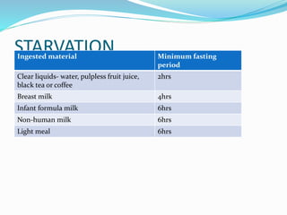
STARVATION
Ingested material Minimum fasting
period
Clear liquids- water, pulpless fruit juice,
black tea or coffee
2hrs
Breast milk 4hrs
Infant formula milk 6hrs
Non-human milk 6hrs
Light meal 6hrs
PERI-OPERATIVE FLUID MX - AIMS
 Assessment of fluid status
 Types of fluids available
 Blood transfusion
 Perioperative requirements
ASSESSMENT BEDSIDE
History:
 Preop NPO
 Intake/output charts
 Preop bowel preparation
 Preop fluid loss (trauma, burns)
ASSESSMENT - BEDSIDE
Examination:
 mental status
 Capillary refil
 blood pressure
 heart rate
 skin colour, temp, turgor
 urinary output
ASSESSMENT ADVANCED
Pressure
 cvp, pulm capillary wedge pressure
Flow
 stroke volume, cardiac output
 Acid-base status
 lactate
CORRECTION OF FLUID STATUS
 Guided by CVP
 Urine output
 Blood pressure
 Heart Rate
 Electrolyte balance
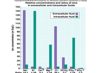
ELECTROLYTE DISTURBANCE
 Disturbances of electrolyte can be due to:
 Underlying disease process
 Drugs( diuretics)
 Iatrogenic
 Caution in correction of electrolyte not to be rapid
SMOKING
 Heavy smoker: 20 cigarettes or more/day
 Problems associated with heavy smoking:
 Hyperactive airways
 Increased sputum production and retention
 Bronchospasms, coughing and atelectasis
 IHD and COPD
 After 12-24hrs following cessation effects of nicotine
and carbon monoxide significantly reduced
 6-8 weeks ciliary and immunological activity restored
1.3 CONCURRENT MEDICAL
DISEASE & MEDICATION
 A) Respiratory
 Infectious: Viral (URTI)
 Obstructive: Asthma, COPD
 B) CVS
 Hypertension, CCF
 IHD
 Valvular disease
 Arrthymias
 C) HAEMATOLOGICAL
 Anaemia
 Sickle cell disease
 Clotting abnormalities
 D) MUSCULOSKELETAL
 Rheumatoid
 E) RENAL & ENDOCRINE
1.4 CONCURRENT SURGICAL
DISEASE
 Intestinal Obstruction
 Acute abdomen
 Head Injury
2.0 INDUCTION OF ANAESTHESIA
 A) METHODS OF INDUCTION
 B) MANAGEMENT OF THE AIRWAY
27
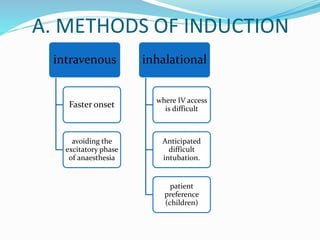
A. METHODS OF INDUCTION
intravenous
Faster onset
avoiding the
excitatory phase
of anaesthesia
inhalational
where IV access
is difficult
Anticipated
difficult
intubation.
patient
preference
(children)
THE IDEAL IV ANAESTHETIC
rapid onset
rapid recovery
analgesia at sub anesthetic concentrations
minimal CVS and resp. depression
no emetic effects
no excitatory or emergence phenomena
no interaction with neuromuscular blocking drugs
no pain on injection
no venous sequelae (thrombosis)
no toxic effects on other organs
no release of histamine
no hypersensitivity reactions
water soluble formulation
long shelf life
no stimulation of porphyria
TYPES OF IV ANAESTHETIC AGENTS
 Barbiturates
 Non barbiturates
• Propofol
• Ketamine
• Etomidate
 Other (adjuvant) agents
• Benzodiazepines
• Opioids
INHALATIONAL AGENTS
 Nitrous oxide
 (Ether)
 Halothane
 (Enflurane)
 Isoflurane
 Sevoflurane
 Desflurane
 (Xenon)
IDEAL INHALATIONAL AGENTS
Physical properties
 Stable to light and heat
 Inert to rubber, metal, sodalime
 Preservative free
 Inflammable
 Non-explosive
 Pleasant odour
 Cheap
 Green
B. MANAGEMENT OF THE AIRWAY
 Different options for airway management:
 Face mask with ‘sniffing morning air position’
 Face mask with guedal airway
 Laryngeal mask airway (LMA)
 Tracheal intubation: Two major indications:
a) ensure airway patency
b) Protect airway from aspiration
3.0 INTRAOPERATIVE
MANAGEMENT
Association of Anaesthetists
(AAGBI) core standards for
monitoring
1. The anaesthetist must be present and care for the
patient throughout the anaesthetic
2. Monitoring devices must be attached before
induction and continued until the patient has
recovered from the anaesthetic
3. These standards also apply to local, regional
anaesthesia and sedation
4. A summary record of information from monitors
should be kept
5. The anaesthetist must ensure that all equipment has
been checked before use. Alarm limits must be set
appropriately and audible alarms enabled
6. If a necessary monitor is unavailable, a note should be
made on the anaesthetic record
7. Additional monitoring may be deemed necessary by the
anaesthetist
8. If the recovery is nearby it is acceptable for a brief
interruption in monitoring, otherwise the patient must
be monitored during transfer
9. The provision, maintenance , calibration and renewal of
equipment is an institutional responsibility
MINIMUM MONITORING
STANDARDS
 ECG
 Pulse oximeter
 Indirect BP
 Capnograph
 Inspired oxygen analyser
 Anaesthetic vapour analyser
RED FLAGS IN ANAESTHESIA
 Maintenance of patient under GA
 Patient position
 Critical incidences
 Failure to breath
4.0 SPECIAL PATIENTS
 1. Full stomach: RSI, minimise gastric pH & volume
 2. Shocked Patient: Prone to CVS collapse following
induction hence optimise
 3. Head Injury: Intubation & ventilation, prevent 2˚
brain injury
 4. Upper airway obstruction: intubation ±
tracheostomy, inhalational
5.0 REGIONAL ANAESTHESIA
 Spinal
 Epidural
 Combined spinal epidural
 Peripheral nerve blocks
 Local infiltration
INDICATIONS & ADVANTAGES
 Full stomach
 Anatomic distortions of upper airway
 TURP surgery
 Obstetrical surgery (T4 Level)
 Decreased post-operative pain
 Continuous infusion
DISTRIBUTION OF SPINAL
ANAESTHESIA
 Factors Effecting Distribution
 Site of injection
 Shape of spinal column
 Patient height
 Angulation of needle
 Volume of CSF
 Characteristics of local anesthetic
 Density
 Specific gravity
 Baracity
 Dose
 Volume
 Patient position (during & after)