2026 Pulmonary Embolism Guidelines: A Practical Clinical Overview
A concise overview of the 2026 Pulmonary Embolism (PE) Guidelines, covering key updates and practical recommendations on the diagnosis, risk stratification, and management of pulmonary embolism. This presentation is intended as a quick, detailed and clinically focused guide for healthcare professionals.
2026 Pulmonary Embolism Guidelines: A Practical Clinical Overview
1.
2026
AHA/ACC/ACCP/ACEP/CHEST/SCAI/SHM/SIR/SVM/S
VN Guideline forthe Evaluation and Management
of Acute Pulmonary Embolism in Adults: A Report
of the American College of Cardiology/American
Heart Association Joint Committee on Clinical
Practice Guidelines
By: Dr. Noshirwan P. Gazder
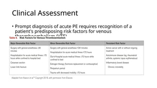
Clinical Assessment
• Promptdiagnosis of acute PE requires recognition of a
patient's predisposing risk factors for venous
thromboembolism (VTE).
4.
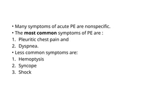
• Many symptomsof acute PE are nonspecific.
• The most common symptoms of PE are :
1. Pleuritic chest pain and
2. Dyspnea.
• Less common symptoms are:
1. Hemoptysis
2. Syncope
3. Shock
5.
• Physical examinationfindings of tachycardia and
hypotension may indicate hemodynamic compromise.
• Tachypnea and hypoxemia may indicate a significant degree
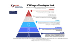
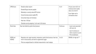
of gas exchange disturbance.
6.
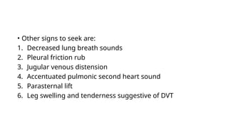
• Other signsto seek are:
1. Decreased lung breath sounds
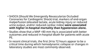
2. Pleural friction rub
3. Jugular venous distension
4. Accentuated pulmonic second heart sound
5. Parasternal lift
6. Leg swelling and tenderness suggestive of DVT
13.
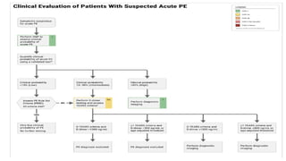
Diagnostic Testing
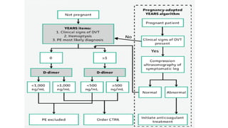
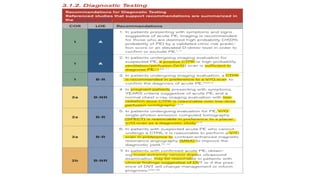
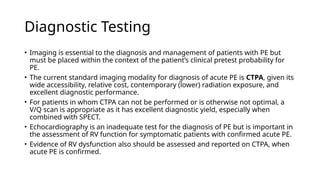
• Imagingis essential to the diagnosis and management of patients with PE but
must be placed within the context of the patient’s clinical pretest probability for
PE.
• The current standard imaging modality for diagnosis of acute PE is CTPA, given its
wide accessibility, relative cost, contemporary (lower) radiation exposure, and
excellent diagnostic performance.
• For patients in whom CTPA can not be performed or is otherwise not optimal, a
V/Q scan is appropriate as it has excellent diagnostic yield, especially when
combined with SPECT.
• Echocardiography is an inadequate test for the diagnosis of PE but is important in
the assessment of RV function for symptomatic patients with confirmed acute PE.
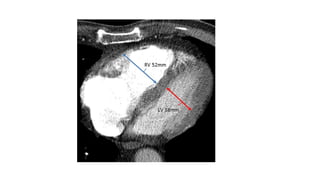
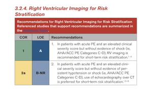
• Evidence of RV dysfunction also should be assessed and reported on CTPA, when
acute PE is confirmed.
14.
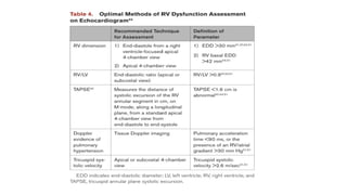
Parameters of RVDysfunction
• RV/ LV end-diastolic ratio
• RV end-diastolic diameter
• Tricuspid Annular Plane Systolic Excursion (TAPSE)
• Estimated right ventricular systolic pressure
• RV free wall hypokinesis with sparing of the apex (McConnell’s
sign)
• Tricuspid systolic velocity
• Paradoxical septal motion
• IVC respirophasic collapse
15.
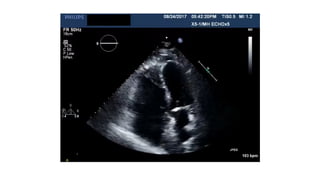
Tricuspid Annular PlaneSystolic
Excursion (TAPSE)
• Echocardiographic measure of right ventricular (RV) systolic
function.
• Measures how far the tricuspid annulus travels.
• It measures how far the tricuspid annulus (D-shaped ring
connecting the right atrium and ventricle) moves toward the
apex during ventricular contraction.
• TAPSE 16 mm Normal RV systolic function
≥ →
• TAPSE <16 mm RV systolic dysfunction
→
16.
Estimated Right VentricularSystolic
Pressure (RVSP)
• Echocardiographic estimate of the pressure generated by
the right ventricle during systole.
• Normal: <35–40 mmHg
• Borderline/Mildly Elevated: 35–40 mmHg or 40–49 mmHg
• Moderately Elevated: 50–59 mmHg
• Severely Elevated: 60 mmHg
• In the absence of significant pulmonic stenosis, RV systolic
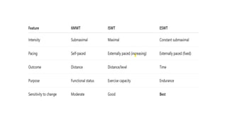
pressure is approximately equal to the pulmonary artery
systolic pressure (PASP).
17.
RV free wallhypokinesis with sparing
of the apex (McConnell’s sign)
• Echocardiographic finding classically associated with acute
Pulmonary Embolism.
• Hypokinesis (reduced movement) of the mid and basal RV
free wall with preserved or hyperdynamic motion of the RV
apex.
19.
Tricuspid Systolic Velocity
•Echocardiographic Doppler measure of right ventricular (RV)
systolic function.
• Measures how fast the annulus moves during systole.
• Measures the speed of longitudinal systolic movement of
the tricuspid annulus toward the apex during RV
contraction.
• S′ 9.5–10 cm/s:
≥ Normal
• S′ < 9.5 cm/s: Suggests RV systolic dysfunction
20.
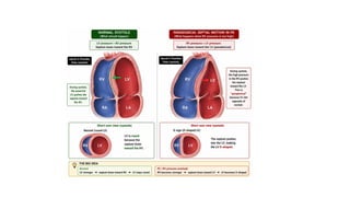
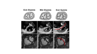
Paradoxical Septal Motion
•Refers to abnormal movement of the interventricular
septum (the wall between the RV and LV), where it moves in
the “wrong” direction during the cardiac cycle.
• Normally: The septum moves toward the left ventricle in a
coordinated fashion during systole.
• In paradoxical motion: Interventricular septum moves
toward the right ventricle during systole, opposite to its
normal motion toward the left ventricle.
23.
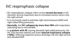
IVC respirophasic collapse
•IVC respirophasic collapse refers to the normal decrease in IVC
diameter during inspiration due to increased venous return into
the right atrium.
• It is commonly used to estimate right atrial pressure (RAP) and
right-sided filling pressures.
• Normally, the IVC collapses by more than 50% with inspiration,
indicating low or normal RAP.
• In a patient with PE causing elevated right-sided pressures, the
IVC may become dilated and show reduced inspiratory collapse
(<50%), reflecting impaired venous drainage into the right atrium
and elevated RAP.
25.
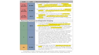
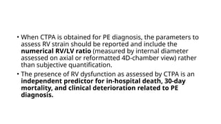
• When CTPAis obtained for PE diagnosis, the parameters to
assess RV strain should be reported and include the
numerical RV/LV ratio (measured by internal diameter
assessed on axial or reformatted 4D-chamber view) rather
than subjective quantification.
• The presence of RV dysfunction as assessed by CTPA is an
independent predictor for in-hospital death, 30-day
mortality, and clinical deterioration related to PE
diagnosis.
26.
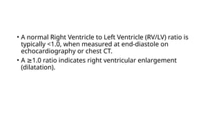
• A normalRight Ventricle to Left Ventricle (RV/LV) ratio is
typically <1.0, when measured at end-diastole on
echocardiography or chest CT.
• A 1.0 ratio indicates right ventricular enlargement
≥
(dilatation).
29.
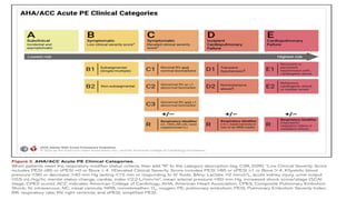
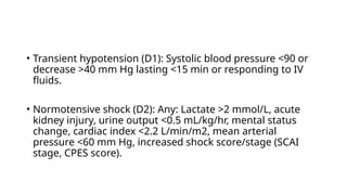
• Transient hypotension(D1): Systolic blood pressure <90 or
decrease >40 mm Hg lasting <15 min or responding to IV
fluids.
• Normotensive shock (D2): Any: Lactate >2 mmol/L, acute
kidney injury, urine output <0.5 mL/kg/hr, mental status
change, cardiac index <2.2 L/min/m2, mean arterial
pressure <60 mm Hg, increased shock score/stage (SCAI
stage, CPES score).
33.
• SHOCK (ShouldWe Emergently Revascularize Occluded
Coronaries for Cardiogenic Shock) trial, markers of end-organ
malperfusion (elevated lactate, acute kidney injury or reduced
urine output, and/or reduced cardiac index) were associated
with higher in-hospital mortality than hypotension alone.
• Studies show that a MAP >80 mm Hg is associated with better
outcomes and reduced in-hospital death for patients with acute
PE.
• In recent clinical trials, the first 24 to 72 hours appear to be a
critical time during which hemodynamic collapse or changes in
laboratory studies are most commonly observed.
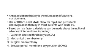
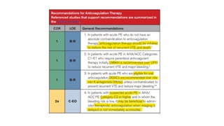
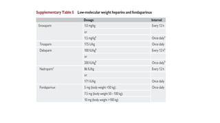
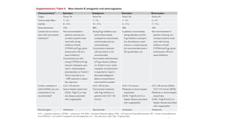
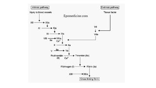
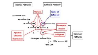
• Anticoagulation therapyis the foundation of acute PE
management.
• Use of DOACs and LMWH allow for rapid and predictable
anticoagulation therapy in most patients with acute PE.
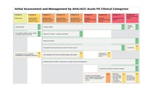
• Based on risk factors, decisions can be made about the utility of
advanced interventions, including:
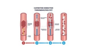
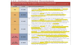
1. Catheter-directed thrombolysis (CDL)
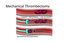
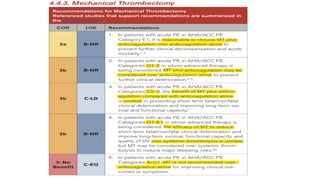
2. Mechanical thrombectomy
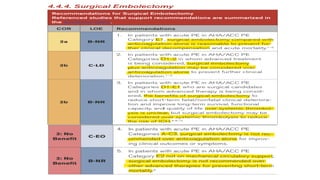
3. Surgical embolectomy
4. Extracorporeal membrane oxygenation (ECMO)
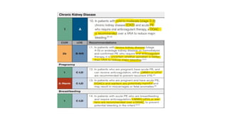
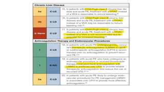
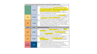
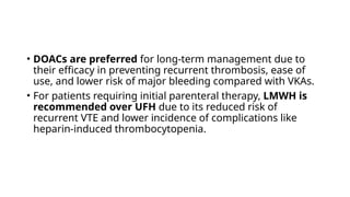
• DOACs arepreferred for long-term management due to
their efficacy in preventing recurrent thrombosis, ease of
use, and lower risk of major bleeding compared with VKAs.
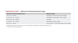
• For patients requiring initial parenteral therapy, LMWH is
recommended over UFH due to its reduced risk of
recurrent VTE and lower incidence of complications like
heparin-induced thrombocytopenia.
59.
• Patients withprofound hypotension due to acute PE may require vasopressor therapy, and selection of
the most appropriate agent depends on the intended effects on the heart and vasculature.
• Based on indirect evidence, norepinephrine (NE) is generally considered the vasopressor of choice.
• NE increases systemic vascular resistance (SVR) and has modest inotropic effects.
• At doses 15 μg/min, NE has little to no effect on PVR. Therefore, it has a favorable net increase in the
≤
SVR/PVR ratio. However, at doses exceeding 15 μg/min, NE may increase PVR.
• Therefore, instead of further increasing the dose of NE, a second vasopressor agent (eg,
vasopressin, phenylephrine) should be added.
• In patients with acute PE who have persistently low cardiac output despite the use of
vasopressors, the addition of an inotropic agent may be indicated.
• A hemodynamic study of 10 patients with acute PE with circulatory failure requiring admission to the
ICU suggests that a continuous infusion of dobutamine (up to 10 μg/ kg/min) may increase cardiac
index at the expense of decreased SVR.
• Thus, dobutamine may be considered as an adjunct to NE in patients with acute PE with low cardiac
output and hypotension (AHA/ACC PE Category E1-2) and may be considered as the initial agent of
choice in patients with normotensive cardiogenic shock (AHA/ACC PE Category D2)
60.
• In acutepulmonary embolism with right ventricular (RV)
dysfunction, fluid management should be very cautious because
the RV is already pressure-overloaded from increased pulmonary
vascular resistance.
• Limited evidence suggests that small fluid boluses ( 500 mL) may
≤
transiently improve cardiac output in selected normotensive
patients with low filling status.
• Larger or repeated fluid administration can worsen RV dilation,
reduce left ventricular preload, and further impair hemodynamics.
Routine or aggressive volume expansion is generally harmful
and should be avoided due to the risk of worsening RV failure.
61.
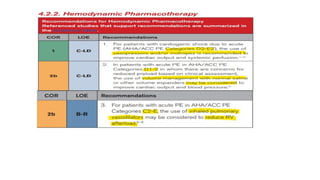
• In acutePE, selective inhaled pulmonary vasodilators may be
useful because they reduce pulmonary vascular resistance and
right ventricular afterload without significantly lowering systemic
blood pressure.
• Evidence from trials of inhaled nitric oxide shows no clear
improvement in primary short-term clinical endpoints but
suggests possible improvement in right ventricular size and
function.
• Systemic agents like sildenafil and epoprostenol have shown little
to no clinical benefit.
• Data is limited.
63.
• Anxiolytic and/oranalgesic drugs, especially sedation for
intubation, can result in catastrophic hemodynamic collapse
in patients with compensated or decompensated RV
dysfunction secondary to acute PE, due to blunting of the
compensatory sympathetic response.
• RV pressure-volume overload associated with acute PE may
cause RV dilation and decreased RV function, resulting in
decreased LV filling and cardiac output.
• Compensatory increases in heart rate and SVR maintain
systemic and myocardial perfusion.
64.
• Anything thatreduces or eliminates these compensatory mechanisms,
can cause or exacerbate hemodynamic decompensation.
• Studies of specific sedation strategies in patients with acute PE are
sparse.
• Any amount of sedation should be administered with caution when
there is evidence or concern for RV dysfunction.
• In addition, deeper sedation and intubation should be avoided unless
there is a strong clinical indication, such as profound hypoxia refractory
to noninvasive oxygenation strategies, or for airway protection, and the
treatment team is readily prepared to support the patient with
vasopressors, inotropes, or VA-ECMO in the event of hemodynamic
collapse.
65.
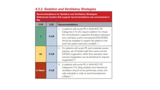
• Acute hypoxemicrespiratory failure is common in patients
with acute PE.
• Modalities for support of oxygenation and ventilation include
conventional nasal cannula, HFNC, noninvasive mechanical
ventilation (NIV), and invasive mechanical ventilation,
• Available evidence from RCTs demonstrate that, compared
with conventional nasal cannula, the use of either HFNC
or NIV is well tolerated, improves oxygenation and work of
breathing, and may reduce the rate of intubation and
mortality.
67.
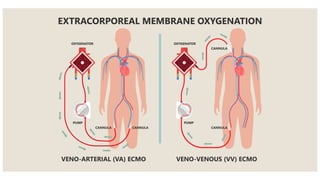
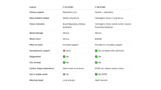
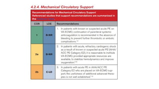
• Mechanical circulatorysupport may be of benefit for patients in
cardiogenic shock as a consequence of known or suspected PE
with evidence of RV dysfunction.
• Use of these supportive measures will depend on local resources
and expertise but most commonly include VA-ECMO.
• VA-ECMO serves to rapidly decrease RV preload while delivering
oxygenated blood to peripheral tissues and end organs, thus
halting the effects of hypoxia and cardiogenic shock.
• Data is limited on the benefit of advanced therapies (eg, MT, CDL)
for patients with acute PE who are being supported with VA-
ECMO
69.
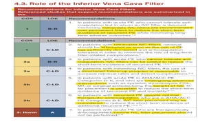
• The PREPICtrial demonstrated that IVC filters led to
significantly higher rates of DVT and IVC thrombosis over
time without improving long-term survival, supporting the
concern that permanent filters increase the risk of chronic
thrombotic complications.
• The PREPIC2 trial, which studied retrievable IVC filters in
patients receiving anticoagulation, found no added benefit
in preventing recurrent PE.
70.
• IVC filtersare associated with a reduction in PE recurrence
( 50%)
∼ , but they also significantly increase the risk of DVT
( 70%)
∼ , with no mortality benefit.
• Permanent filters should be reserved for rare cases in which
retrievable IVC filters are not feasible (eg, mega cava [IVC >30
mm]).
• The FDA issued a safety communication recommending IVC
filter retrieval within 29 and 54 days after placement, once the
risk of PE has subsided, based on evidence of complications
such as device migration, filter fracture, IVC perforation, and
embolization when filters are left in place too long.
71.
• The PREPIC(Prevention du Risque d'Embolie Pulmonaire par
Interruption Cave) trial demonstrated that IVC filter
placement significantly reduced the incidence of PE
recurrence but was counterbalanced by an increased risk of
DVT and no survival benefit over long-term follow-up.
• The PREPIC2 trial found no additional benefit to filter
placement in anticoagulated patients, reinforcing that filters
should be reserved for those in whom anticoagulation is
truly contraindicated.
73.
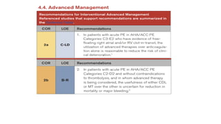
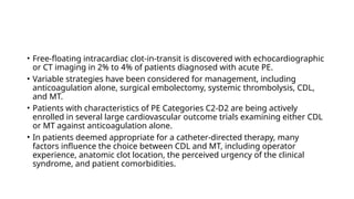
• Free-floating intracardiacclot-in-transit is discovered with echocardiographic
or CT imaging in 2% to 4% of patients diagnosed with acute PE.
• Variable strategies have been considered for management, including
anticoagulation alone, surgical embolectomy, systemic thrombolysis, CDL,
and MT.
• Patients with characteristics of PE Categories C2-D2 are being actively
enrolled in several large cardiovascular outcome trials examining either CDL
or MT against anticoagulation alone.
• In patients deemed appropriate for a catheter-directed therapy, many
factors influence the choice between CDL and MT, including operator
experience, anatomic clot location, the perceived urgency of the clinical
syndrome, and patient comorbidities.
75.
• Four smallRCTs with a total of 224 patients with high risk PE reported that
systemic thrombolysis therapy compared with anticoagulation with heparin
alone leads to improvement in pulmonary obstruction accompanied by a
reduction in RV dilatation on echo.
• The FDA has approved 3 agents for use in PE thrombolysis, including
streptokinase, urokinase, and rt-PA (alteplase).
• The first generation agents (streptokinase and urokinase) are not used in
contemporary practice because they require longer infusion times and are
not readily available.
• Standard dose rt-PA (100 mg in 2 hours) is the most commonly used
thrombolytic agent in patients with PE, although there are no head-to-head
trials of the agents to suggest superiority of a specific thrombolysis agent.
76.
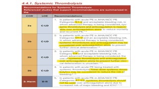
• Meta-analyses ofsystemic thrombolysis trials that included, but were
not limited to, patients with hemodynamically significant PE, defined
primarily as the presence of cardiogenic shock or systolic blood
pressure <90 mm Hg, found that systemic thrombolysis was
associated with a significant reduction in the combined endpoint
of all-cause death or clinical deterioration requiring rescue
treatment.
• The vast majority of patients who present with PE do not have
hypotension at presentation.
• Thrombolysis prevented cardiovascular collapse but increased major
(including intracranial) bleeding, with closely balanced benefits and
harms.
77.
• There isa lack of high-quality evidence regarding dosing of
thrombolytic agents to treat acute PE.
• Evidence is emerging that lower-dose thrombolysis (25-50
mg rt-PA), compared with standard dose (100 mg rt-PA), may
be as efficacious and associated with a reduced risk for
major bleeding.
• The ongoing PEITHO-3 trial assessing the efficacy and safety
of a reduced-dose alteplase regimen with standard heparin
anticoagulation will provide additional insight.
78.
• Systemic thrombolysisis associated with an incremental
increase in major bleeding risk, in particular a greater risk of
ICH.
• Overall, data suggests that patients with the highest risk of
mortality from PE and the lowest risk of bleeding would
obtain the greatest net benefit from thrombolysis, whereas
those with the lowest risk of mortality from PE and the
highest risk of bleeding would obtain the least benefit and
are likely to be harmed.
81.
• Compared withother advanced therapies, the advantages to
percutaneous MT devices include:
1. They do not require concomitant administration of a lytic
medication
2. They do not require an indwelling catheter and post-
procedure ICU stay
3. In some cases an older thrombus can be removed.
• MT has been used as an adjunctive form of treatment in
patients with high-risk acute PE on mechanical circulatory
support, such as ECMO.
83.
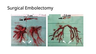
• Modern surgicalpulmonary embolectomy is performed using
cardiopulmonary bypass (CPB), which temporarily replaces heart and lung
function by diverting venous blood through an external pump and
oxygenator before returning it to the arterial system.
• This setup both maintains systemic perfusion and, more importantly,
unloads the failing right ventricle by removing venous return, thereby
reversing pressure and volume overload caused by acute pulmonary
embolism and allowing the RV to recover in a low-stress state. VA-ECMO
provides similar physiological support by decompressing the RV and
sustaining circulation.
• Survival is quite favorable, with mortality rates ranging from 1% to 15%.
• Survival of >97% has been reported among those patients who did not
require CPR.
• 6MWT isa submaximal exercise test that measures the maximum
distance an individual can walk on a flat, hard surface in 6 minutes,
assessing functional capacity for heart and lung conditions.
• ISWT is an externally paced, maximal exercise field test used to assess
functional capacity. Patients walk back and forth along a 10-meter
course, with increasing speeds dictated by audio signals, until they can
no longer maintain the pace or are too exhausted.
• ESWT is an externally paced, sub-maximal field test used to assess
exercise capacity. It requires participants to walk back and forth along
a 10-meter course for as long as possible, keeping pace with an audio
signal (beeps) at a constant speed determined by a previous
incremental shuttle walk test (usually ~85% of ISWT max
87.
• 6MWT “Howfar can patient walk in daily life?”
→
• ISWT “What is their peak exercise capacity?”
→
• ESWT “How long can they sustain exertion?”
→
92.
• Between one-thirdand one-half of the patients who have had a
symptomatic PE will report dyspnea or limitations to physical
activity on follow-up for months to years after a PE.
• The prevalence of shortness of breath is higher after an acute PE
associated with RV dysfunction and can be a presenting symptom
of CTEPD.
• CTEPD with PH complicates 2.3% to 4% of acute PEs.
• The International Consortium for Health Outcomes Measurement
consensus recommendation on PE suggests evaluating the
patients at 3 months, 6 months, 1 year, and annually as long as
the patient is under care.
93.
• In patientswith acute PE, cancer is diagnosed in 4% to 10% during the
first year when no major reversible risk factors for PE are identified.
• Thrombophilia testing is not recommended in patients who had a PE
associated with major reversible risk factors, such as surgery, major
trauma, or immobilization.
• Isolated common genetic mutations (eg, factor V Leiden, prothrombin
gene mutation) are not consistently associated with an increased
risk of recurrence.
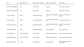
• Deficiencies in natural anti-coagulants (antithrombin, protein C,
protein S), however, are linked to a higher risk of recurrence and
may necessitate extended anticoagulation.
94.
• Coagulation-based testsare often affected by consumption
of factors during the acute phase of a PE and by
administration of anticoagulant medications. Therefore,
coagulation-based tests should not be performed during
the acute phase of a PE, and potential interference with
anticoagulants should be considered to ensure reliable
results.
• Genetic and immunologic testing can be performed at any
time.
98.
• Managing AUBin women on anticoagulants requires a careful
and individualized approach.
• Medical management is often the first line of treatment, with
options such as high-dose progestin-only therapy, combined
hormonal contraceptives (if the patient is on anticoagulation), and
the levonorgestrel intrauterine system.
• In cases where medical management is insufficient, surgical
interventions like endometrial ablation may be considered.
• Interdisciplinary collaboration with gynecology is of paramount
importance to provide an individualized plan for diagnosis and
management.
99.
• Two trialsshowed no increase in VTE recurrence in pre
menopausal or postmenopausal women receiving estrogen-
containing hormonal therapies while on therapeutic
anticoagulation.
• Continuation of hormonal therapy can be considered when
clinically important, as long as the patient is on therapeutic
anticoagulation.