
Recommended
More Related Content
What's hot
What's hot (20)
Similar to Neural tube defect presentation
Similar to Neural tube defect presentation (20)
Recently uploaded
Recently uploaded (20)
Neural tube defect presentation
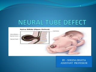
- 1. . BY – SHEENA BHATIA ASSISTANT PROFESSOR
- 2. INTRODUCTION The most common birth defect in India is Neural Tube Defect. The presentation of Neural Tube Defect may vary from Anencepaly, spina bifida occulta and spina bifida cystica. In Year 2015 the birth prevalence of neural tube defects in India was 4.5 per 1000 total birth.
- 3. Neural tube defects are birth defects of the brain, spine, or spinal cord. It happens in the first month of pregnancy, often before a woman even knows that she is pregnant. The two most common neural tube defects are SPINA BIFIDA AND ANENCEPHALY. In Spina bifida, the fetal spinal column doesn't close completely. In Anencephaly defect in brain development occur.
- 4. ETIOLOGY The exact causes of neural tube defects are not known. Genetic Nutritional Factors Environmental factors. Poor intake of folic -acid (also known as folate) Obese Women Intake of anti epileptic medications during pregnancy
- 5. PATHOPHYSIOLOGY THE NEURAL PLATE APPEARS ON THE 17TH DAY OF GESTATION AS A THICKENING OF THE EMBRYONIC ECTODERM . ON DAY 18, THE NEURAL PLATE FOLDS ALONG THE MIDLINE BY THE END OF THE THIRD GESTATIONAL WEEK, THE NEURAL FOLDS FUSE TO FORM NEURAL TUBE. . … . . .
- 6. FUSION BEGINS AT THE HINDBRAIN-CERVICAL JUNCTION FIRST PROCEEDS ROSTRALLY AND THEN CAUDALLY THEN, THE ANTERIOR AND POSTERIOR ENDS (NEUROPORES) CLOSES TO FORM SPINAL CORD DEPENDING ON THE POINT OF INTERUPPTION IN NEURAL TUBE FORMATION DEVELOPS NTDS MAY AFFECT THE BRAIN (ANENCEPHALY)OR SPINAL CORD (SPINA BIFIDA).
- 7. TYPES OF NEURAL TUBE DEFECTS A Spinal cord defects SPINA OCCULTA MENINGOCELE A
- 9. COMMON TYPES OF SPINA BIFIDA Spina bifida occulta which is the mildest form where there is a small gap in the spine but the opening cannot be seen in the back. Brain and spinal cord functions are normal and there is no disabilities. Meningocele is a sac of fluid not involving the spinal cord that comes out through an opening in the back and involve meninges also.
- 10. CONT…………… Myelomeningocele is one of the most common and most severe form of spina bifida . In this the unfused portion of the spinal column allows the spinal cord to protrude through an opening, forming a sac enclosing the spinal elements, such as meninges, cerebrospinal fluid, and parts of the spinal cord and nerve roots.
- 12. SIGN AND SYMPTOMS Loss of bladder or bowel control Partial or complete lack of sensation Partial or complete paralysis of the legs Weakness of the hips, legs, or feet of a newborn Other symptoms may include: Abnormal feet or legs, such as clubfoot Build up of fluid inside the skull (hydrocephalus) Hair present at sacral region Dimpling of the sacral area
- 13. Brain Defects Anencephaly is the absence of a major portion of the brain, skull and scalp that occurs during embryonic development. Infants with this disorder do not survive longer than a few hours or possibly days after their birth.
- 14. CONTD…… Encephalocele, sometimes known as cranium bifidum, is a neural tube defect characterized by sac- like protrusions of the brain and the membranes . Symptoms may include Neurologic problems Hydrocephalus Spastic quadriplegia Microcephaly
- 15. DIAGNOSTIC TESTS Ultrasound Maternal serum Alpha feto protein at 16-20 weeks. Aminiotic Alpha feto protein Aminiotic acetyl cholineterase
- 16. MANAGEMENT Medical Care The patient should be positioned in the prone position to prevent pressure on the defect. The newborn with an open NTD should be kept warm and the defect covered with a sterile wet saline dressing. Intravenous antibiotic should be initiated.
- 17. SURGICAL MANAGEMENT Neurosurgical repair of the defect is considered the mainstay of treatment for open spina bifida. Closed spina bifida does not usually warrant any immediate surgery. The cele closure is typically performed within 1 to 3 days of delivery. Neonates born with severe hydrocephalus should have ventriculoperitoneal shunt placed concurrently.
- 18. NURSING MANAGEMENT ASSESSMENT Depends on the spinal involvement. Visible spinal defect Flaccid paralysis of legs. Altered bowel and bladder pattern. Perform neurological assessment
- 19. NURSING INTERVENTIONS Evaluate the sac and measure lesion . Monitor for increased ICP Measure head circumfrence. Protect sac with non adherent moist dressing Place child in prone position Use aseptic techniques Monitor for early signs of infection Administer Antibiotic. Prepare Family for surgery
- 20. PREVENTION OF NEURAL TUBE DEFECT Experts recommend that all women of childbearing age take a daily supplement of 400 micrograms (mcg) of folic acid. Educate mothers regarding intake of folic acid especially in periconception period and in first trimester as well. Women already had first pregnancy with NTD should take a daily 4mg tablet of folic acid for at least one month before conception and then throughout the first 12 weeks of pregnancy. Genetic Counseling or screening.
- 21. THANK YOU