Scintica's Preclinical Imaging Portfolio
•Download as PPTX, PDF•
0 likes•184 views

This document provides an overview of Scintica's preclinical imaging product portfolio, including their technical capabilities. It describes several of Scintica's imaging systems - the Prospect T1 compact ultrasound system, M-Series compact MRI systems, FIVE2 fluorescence endomicroscopy, Newton 7.0 optical imaging system, and SuperArgus PET/CT systems. Sample images from live demonstrations of the Prospect T1, M-Series, and Newton 7.0 are also presented to showcase their imaging capabilities.

Recommended
More Related Content
Similar to Scintica's Preclinical Imaging Portfolio
Similar to Scintica's Preclinical Imaging Portfolio (20)
More from Scintica Instrumentation
More from Scintica Instrumentation (20)
Recently uploaded
Recently uploaded (20)
Scintica's Preclinical Imaging Portfolio
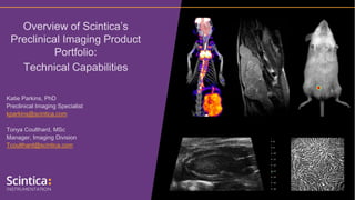
- 1. Katie Parkins, PhD Preclinical Imaging Specialist kparkins@scintica.com Tonya Coulthard, MSc Manager, Imaging Division Tcoulthard@scintica.com Overview of Scintica’s Preclinical Imaging Product Portfolio: Technical Capabilities
- 3. Katie Parkins, PhD Preclinical Imaging Specialist kparkins@scintica.com Tonya Coulthard, MSc Manager, Imaging Division Tcoulthard@scintica.com Overview of Scintica’s Preclinical Imaging Product Portfolio: Technical Capabilities
- 4. • Prospect T1 – high frequency ultrasound • M-Series – compact MRI • FIVE2 – fluorescence in vivo eendomicroscopy • Newton 7.0 – optical imaging • SuperArgus – PET/CT • Understanding the complementary nature of imaging modalities Agenda
- 5. Prospect T1 Compact, High-Frequency, Tablet-based System Small Animal Ultrasound
- 6. • Prospect T1 System Technical Overview • Sample images acquired during the live virtual demo (Oct 1) Topics of Discussion
- 7. Prospect T1 System Technical Overview • System components and standard configuration • Add-on hardware and software components
- 8. Prospect T1 System Components • The Prospect T1 is the first tablet based high-frequency ultrasound system specifically designed for pre-clinical imaging of small animals • System components: • Tablet • Probe • Scanning Platform
- 9. Prospect T1 System Components: Probes • Three single element probes are available: • 20 MHz (user selectable between 15-30 MHz) • Primarily used for rat imaging, as well as harmonic contrast imaging • 40 MHz (user selectable between 30-50 MHz) • Primarily used for mouse imaging, and superficial anatomical targets in larger species like rats • 50 MHz (user selectable between 30-50 MHz) • Primarily used for superficial anatomical targets in both mice and rats
- 10. Prospect T1 System Components: Scanning Platforms • The platform is compact in design, again to limit the footprint of the system • The scanning platform has been designed for ergonomical positioning of the probe • Animal beds have integrated heating, and ECG and respiratory monitoring • Interchangeable beds are available for mice or rats • Animal beds can be precisely adjusted in the X, Y, and Z axis
- 11. Standard System Configuration • Standard system configuration for mouse • B-Mode • M-Mode • Pulsed Wave / Color / Power / Tissue Doppler Mode • Contrast Mode • Comprehensive Measurement and Analysis Tools • Scanning Platform – with mouse bed • 40 MHz probe • Standard system configuration for rat • B-Mode • M-Mode • Pulsed Wave / Color / Power / Tissue Doppler Mode • Contrast Mode • Comprehensive Measurement and Analysis Tools • Scanning Platform – with rat bed • 20 MHz probe
- 12. Add-Ons: 3D Motor • The 3D motor expands the capabilities of the Prospect T1 to acquire 3D B-mode images • Add-on includes the software analysis package to view the 3D images and perform volume calculations
- 13. Add-Ons: Image Guided Needle Injection Mount • The image guided needle injection mount integrates with probe • Injections may be performed with a regular syringe and steel needle, or pulled glass capillary needle • Injections may be made into developing embryos, adult myocardium, or abdominal/muscle targets E15.5 mouse embryo Adult mouse myocardium
- 14. Sample Images • Images acquired during the live virtual demo on Oct 1 • Additional images to show capability not shown in the live demo
- 15. Tumour Imaging – 2D B-Mode Imaging • Tumour identification, and basic linear and area measurements • Investigation of surrounding structures • Longitudinal imaging to monitor tumor progression or therapeutic response Orthotopic Mammary Fat Pad Tumour MDA-MB-231 • Power Doppler (not shown during the demo due to a technical issue with the demo system) can be used to assess tumor vasculature using endogenous signal within the tissue • Contrast Imaging (linear and subharmonic - not shown during the demo as no contrast agent was available) can be used to assess tumor microvasculature using microbubbles
- 16. Tumour Imaging – 2D B-Mode Imaging • The complex nature of tumors can be investigated using B-Mode imaging, as well as surrounding structures IP Injection of Ovarian Tumour Cells SKOV-3 Tumour Stomach IP Injection of Ovarian Tumour Cells SKOV-3 Kidney Splenic Vein TumourIntestine
- 17. Tumour Imaging – 3D B-Mode Imaging • 3D volume measurements • Longitudinal imaging to monitor tumor progression or therapeutic response Volume = 263mm3 Orthotopic Mammary Fat Pad Tumour MDA-MB-231
- 18. Tumour Imaging – 3D B-Mode Imaging • 3D volume measurements • Complex tumor structures can be visualized • Longitudinal imaging to monitor tumor progression or therapeutic response IP Injection of Ovarian Tumour Cells SKOV-3 Tumo ur
- 19. Cardiovascular Imaging – Parasternal Long Axis View of LV B-Mode Frame Rate = 30fps ECG Gated Kilohertz Visualization = 30fps
- 20. Cardiovascular Imaging – Parasternal Short Axis View of LV
- 21. Cardiovascular Imaging – Pulsed Wave Doppler Pulmonary Artery Mitral Valve
- 22. Cardiovascular Imaging – Left Carotid Artery • Color Doppler (not shown during the demo due to a technical issue with the demo system) can be used to identify and assess blood flow velocity and direction within the heart and surrounding vasculature
- 23. M-Series Compact, Self-Shielded, High Performance Permanent Magnet Small Animal MRI
- 24. Topics of Discussion • M-Series System Technical Overview • Sample images acquired during the live virtual demo (Oct 1)
- 25. M-SeriesTM System Technical Overview • M-SeriesTM Compact magnet design • Animal handling system • SimPET insert option for simultaneous PET/MR imaging
- 26. System Components • Compact magnet – select either the M3, M5, or M7 magnet; • Electronics cabinet and User Workstation - is the same for all systems. • Accessories and add-ons • Animal handling system with heating and physiological monitoring • Anesthesia delivery and exhaust gas scavenging • SimPETTM insert – simultaneous PET/MRI (M7 system only)
- 27. M-SeriesTM Compact MRI Systems from Aspect Imaging M3 – Mouse Only M7 – Mouse & Large RatsM5 – Mouse & Small Rats
- 28. M-SeriesTM Compact Magnet Design • Require no special infrastructure • Compact and self shielded with minimal external fringe field • Operate very quietly during image acquisition • Systems are installed with first images being acquired in less than 1 day Installed within an animal facility, or existing laboratory next to other equipment or furnishings
- 29. Animal Handling System • Fully integrated animal handling and coil system includes • Mouse or rat beds to suite varying animal sizes • Anatomy specific coils, with automatic tuning • Heating • Physiological monitoring • Anesthesia delivery and scavenging
- 30. Imaging Coils Type Dimensions Application Inner Diameter Length Mouse Head 23 mm 25 mm Neurological imaging in mice Mouse Body 30 mm 50 mm Extremity, abdominal, and thoracic cavity imaging in mice Mouse Whole Body 30 mm 80 mm Whole body imaging in mice Large Mouse Body 38 mm 50 mm Extremity, abdominal and thoracic cavity imaging in large/obese mice Rat Head 35 mm 40 mm Neurological imaging in rats Rat Body 50/60 mm ellipsoid 90 mm Extremity, abdominal and thoracic cavity imaging in rats Large Rat Body 71 mm 90 mm Extremity, abdominal and thoracic cavity imaging in large rats Imaging coils should fit as tightly as possible to the anatomical target for high quality images
- 31. Most measurements and parameters are functions of time, so we need waveforms
- 32. SimPET insert option for simultaneous PET/MR imaging • The SimPETTM insert expands the capabilities of the M7 system to allow for simultaneous PET/MR imaging • MR images compliment the highly sensitive PET images in detecting functional information, abnormalities, and early disease, providing an anatomical context
- 33. Sample Images • Images acquired during the live virtual demo on Oct 1
- 34. Tumour Imaging – Orthotopic Mammary Fat Pad Tumour • MDA-MB-231 cell line • T1 and T2 weighted image contrast helps to identify anatomy; along with pathological changes, including tumors • 250µm in-plane resolution with 1mm slice thickness • Acquisition was around 4.5 minutes T1 Weighted T2 Weighted
- 35. Tumour Imaging – Orthotopic Mammary Fat Pad Tumour • Tumour volume was found to be 273mm3 T1 Weighted T2 Weighted MDA-MB-231
- 36. Tumour Imaging – IP Injected Ovarian Tumour Cells • SKOV3 cell line, injected IP • These tumors may form anywhere within the peritoneal cavity • Numerous tumors were located throughout the abdomen T2 WeightedT1 WeightedT1 Weighted T2 Weighted
- 37. Tumour Imaging – IP Injected Ovarian Tumour Cells • VivoQuant was used to visualize the acquired images in 3D and to quantify the tumor volumes. • Various manual and semi-automatic tools are available T2 WeightedT1 Weighted Region Of Interest Color Volume (mm³) Upper Tumour red 144 Mid Tumour green 6 Lower Tumour blue 9 Lower Tumour #2 cyan 7 Mid Tumour #2 magenta 64
- 38. Brain Imaging – Normal Brain T2 WeightedT1 Weighted • Various structures can be visualized within the brain • 200µm in-plane resolution, 1mm slice thickness; 7.5-minute acquisition time
- 39. FIVE2 ViewnVivo Fluorescence in vivo endomicroscopy
- 40. Newton 7.0 FT In Vivo Optical Imaging System COURTESY : Matthieu Germain , Nanobiotix/Curadigm
- 41. TOPICS OF DISCUSSION • Applications of optical imaging • Product overview • Images acquired during the live demo
- 42. Main applications of optical imaging Luciferase expressing metastases mCherry expressing cancer cells Fluorescence Imaging Bioluminescence Imaging • Oncology • Neurology • Biodistribution studies • Treatment studies (BLI) • Cell tracking • Ex vivo imaging
- 43. Proprietary CCD Camera 16-bit Scientific CCD Camera Grade 0 – No dead pixels NIST calibrated 4.8 Orders of Magnitude Key Technical Features of the Newton 7.0 FT CAMERA DARQ-9
- 44. Key Technical Features of the Newton 7.0 FT CAMERA RESOLUTION 4.6 MP Newton 7.0 FT / 4.6MP Other systems / >1MP Better Performance in patter recognition
- 45. Key Technical Features of the Newton 7.0 FT LENS APERTURE F/0.70 Newton 7.0 FT = f/0.70 Other systems = f/0.95 More light collection = More sensitivity Wider lens aperture Smaller lens aperture
- 46. Key Technical Features of the Newton 7.0 FT MOTORIZED DARKROOM Infinite imaging Capabilities
- 47. Key Technical Features of the Newton 7.0 FT Green 520nm Green 580nm Red 640nm IR 740nm Blue 480nm NIR 680nm IR 780nm Blue 420nm Full Spectrum Tunability • 8 Excitation channels (400nm > 800nm) • Powerful Laser Class II illumination • Reducing the cross stimulation • Increasing the sensitivity of your images
- 48. Key Technical Features of the Newton 7.0 FT NARROW BAND-PASS FILTERS Emission Filters • 8 Narrow Bandpass emission filters (500nm > 900nm) • Dual Magnetron sputter-coated technology • Ensuring transmission above 90% • Improving spectral separation for multi-spectral imaging
- 49. Key Technical Features of the Newton 7.0 FT CCD camera 3D NIR Camer a 3D NIR Camer a How 3D Optical Tomography is achieved in the Newton 7.0 ? • First, the Topographic image of the mouse is acquired with the help of 3D NIR Cameras • Then, the main CCD camera acquires multiple images of the signal using various emission filters from wavelengths in the spectral emission of the specific probe used. • The signal is reconstructed with the use of an algorithm and repositioned within the topographic image of the animal.
- 50. Key Technical Features of the Newton 7.0 FT Co-registration with digital Organ & Bones Atlas • Choose to add or hide individual organs and bones • Axial, Sagittal and Coronal Views • Quantification of the signal in units of volume
- 51. Images acquired during the live demo Signal Only Image Brightfield Image Overlay Image
- 52. Images acquired during the live demo IP metastasis model (human ovarian cancer) Dual MFP Model (human breast cancer)
- 53. FLI Phantom @ 480nm Multiplex Phantom Images acquired during the live demo Fluorescent beads Fluorescent beads Luminescent beads
- 54. SuperArgus State-of-the-Art Pre-Clinical PET/CT Systems Courtesy of Dr. M. Desco & J.J. Vaquero, UMCE Hospital Gregorio Marañón HGUGM (Madrid, Spain)
- 55. TOPICS OF DISCUSSION • Product Overview • What Makes the Sedecal Systems Unique • Multi-animal Handling System
- 56. • Self-shielded to meet FDA guidelines • no special room preparation, controls, or additional shielding required • Systems can easily be placed within existing laboratory environments, imaging cores, or within the animal facility • Can have the following integrated into animal handling: • Anesthesia – isoflurane with gas scavenging • Heating • Physiological monitoring • Image may be saved as DICOM, Interfile, or JPEG REVIEW OF SUPERARGUS AND COMPACT MODELS
- 57. REVIEW OF SUPERARGUS AND COMPACT MODELS • Compact (Mouse only) Model • PET or CT • Dimensions : 60 x 60 x 40cm • 55 mm bore • Static FOV: 45mm axial; 100mm trans-axial • CT Specifications • 65µm resolution (max.) • 15sec acquisition time (min.) • PET Specifications • 32 tDOI Phoswitch detectors; 4 rings • ≤1.0mm resolution using 3D OSEM reconstruction (≤2 min to perform)
- 58. • Super Argus Models (PET and/or CT) • r models – mouse, rat, marmoset • 90mm bore • FOV – 220mm axial (dynamic); 80mm transaxial • R models – up to 3kg rabbit • 160mm bore • FOV – 350mm axial (dynamic); 120mm transaxial • P models – up to non-human primate, canine, porcine, etc. • 260mm bore • FOV – 650mm axial (dynamic); 210mm transaxial • 2, 4, or 6 PET ring options for a 50mm, 100mm, or 150mm fixed axial FOV • CT is focused on low does, high resolution (15µm) rapid scanning options (15sec) REVIEW OF SUPERARGUS AND COMPACT MODELS
- 59. • Core Phoswich detector technology along with the electronics provide the following best in class specifications: • Highest resolution on the market (≤1.0mm) • Resolution uniformity across the entire FOV, with FOV filling majority of the bore size due to the tDOI technology correcting the parallax error • Highest sensitivity (11%; at 100-700keV) • Real-time imaging (up to 2.5msec frame rate if desired) • Dynamic imaging – possible using time stamp acquisition electronics • Parallel electronics acquisition SUPERARGUS IS CONSIDERED TO BE BEST IN CLASS
- 60. MULTI-ANIMAL HANDLING SYSTEM • Up to 4 mice can be scanned simultaneously in the R model (160mm bore) using the multi-animal handling system • Individually controlled anesthesia flow and heating controls • Separate animal preparation workstation is also included • Additional configurations are available for rats and/or mice depending on specific needs
- 61. MULTI-ANIMAL HANDLING SYSTEM Resolution and sensitivity are maintained throughout the entire FOV due to the unique tDOI Phoswich detectors – correcting for the parallax error
- 62. • Sample images acquired during the live virtual demo (Oct 1) Understanding the Complementar y Nature of Imaging Modalities
- 63. Understanding the Complementar y Nature of Imaging Modalities T1 Weighted • Bioluminescence helps to identify where the tumors may be located, however differentiating tumors from one another, and measuring tumor volume is better done using an anatomical imaging modality such as MRI or Ultrasound Kidney Splenic Vein TumourIntestine IP Injection of SKOV3 Ovarian Tumour Cells
- 64. Understanding the Complementar y Nature of Imaging Modalities T2 Weighted • Bioluminescence helps to confirm viability of the tumor cells, as they express luciferase, approximate volumes may be possible from the BLI signal; anatomical images help to confirm tumor volume - ultrasound (263mm3) or MRI (273mm3) Orthotopic Mammary Fat Pad Tumour (MDA-MB-231)
- 65. Audience Poll
- 66. Katie Parkins, PhD Preclinical Imaging Specialist kparkins@scintica.com Tonya Coulthard, MSc Manager, Imaging Division Tcoulthard@scintica.com Overview of Scintica’s Preclinical Imaging Product Portfolio: Technical Capabilities To ask a question, click the Q&A Button, type your question and click send. Any questions that are not addressed during the live webinar will be answered following the event. Thank you for participating!