Rats Exposed to Libby Amphibole and Amosite Asbestos
•
1 like•656 views

“Subchronic Inhalation Exposure of Rats to Libby Amphibole and Amosite Asbestos: Effects at 18 Months Post Exposure.” Willson GA (presenter), Dodd DA, Roberts KC, Wall HG, Jarabek AM, Gavett SH. The 33rd Society of Toxicologic Pathology Annual Meeting. Washington, DC. June 22-26, 2014. “Subchronic Inhalation Exposure of Rats to Libby Amphibole and Amosite Asbestos: Effects at 18 Months Post Exposure.” Dodd DA (presenter), Willson GA, Roberts KC, Wall HG, Jarabek AM, Gavett SH. The 53rd Annual Society of Toxicology Meeting. Phoenix, AZ. March 23-27, 2014. For full-resolution viewing, please open or save as a PDF.
Recommended

Recommended
More Related Content
Similar to Rats Exposed to Libby Amphibole and Amosite Asbestos
Similar to Rats Exposed to Libby Amphibole and Amosite Asbestos (20)
More from EPL, Inc.
More from EPL, Inc. (9)
Recently uploaded
Recently uploaded (20)
Rats Exposed to Libby Amphibole and Amosite Asbestos
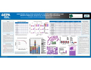
- 1. Innovative Research for a Sustainable FutureInnovative Research for a Sustainable Futurewww.epa.gov/research SUBCHRONIC INHALATION EXPOSURE OF RATS TO LIBBY AMPHIBOLE AND AMOSITE ASBESTOS: EFFECTS AT 18 MONTHS POST EXPOSURE G.A. Willson1, D.E. Dodd2, K.C. Roberts2, H.G. Wall1, S.H. Gavett3. 1Experimental Pathology Laboratories, Inc., RTP, NC, 2The Hamner Institutes for Health Sciences, RTP, NC, 3U.S. EPA, RTP, NC. Abstract Increased asbestosis, lung cancer, and mesothelioma rates are evident after exposures to Libby amphibole (LA). To support dosimetry model development and compare potency, a subchronic nose‐only inhalation study (6 hr/d, 5 d/wk, 13 wk) was conducted in male F344 rats. Rats were exposed to air (control), LA (LO, MED, HI; 1.01, 3.32, 10.06 mg/m3; 159, 693, 1522 fibers/cc), or amosite (AM; 3.34 mg/m3; 230 f/cc). Toxicity endpoints, pathology, and fiber burden evaluation were determined 18 mo post‐exposure. Fiber exposure had no effect on survival. Mononuclear cell leukemia was the main cause of death prior to scheduled necropsy in all groups except the LA 3.3 group. BAL cell numbers, LDH, and protein in AM and LA groups were not statistically different from controls (n=8 rats/group), indicating resolution of earlier inflammation. Histopathology of the left lung, trachea, sternum, pleura, epididymis and testes, and gross tissue lesions was conducted on 50 rats/group. Alveolus inflammation, pleural fibrosis, lung interstitial fibrosis, and foreign bodies were noted in all fiber‐exposed groups. A greater incidence of chronic tracheal inflammation was noted in the LA groups. Alveolar bronchiolar adenoma occurred in 2 rats in each of the AM, MED LA, and HI LA groups, and 1 alveolar bronchiolar carcinoma was observed in the HI LA group. No pleural mesotheliomas were observed in any group. In conclusion, both AM and LA induced dose‐related lung fibrotic responses; tumor incidences were apparently increased but not beyond historical control ranges. (This abstract does not represent US EPA policy.) Exposure Fiber Characterization • Overall, LA fiber is slightly longer and has a slightly greater aspect ratio compared to UICC Amosite. • UICC amosite had a higher proportion of particles (L/D <3) compared to LA. • Other exposure atmosphere characteristics (T, %RH, airflow) were equivalent. Air Control UICC Amosite LA low LA medium LA high Target Concentration (mg/m3) 0.0 3.30 1.00 3.30 10.0 Actual Concentration (mg/m3) 0.0 3.32 1.01 3.34 10.0 APS: Count Median Aero‐ dynamic Diameter (m) 1.41 0.93 1.05 1.02 1.13 APS: g 2.01 1.40 1.51 1.46 1.52 APS: Particles Mean #/cc 1.2 404 43 171 280 SEM: All Objects Mean #/cc ‐ 3101 1158 3728 6718 SEM: Mean Fiber Concentration (f/cc) (L/D> 3) ‐ 1627 663 2753 5093 SEM: Mean Fiber Concentration (f/cc) (L/D > 3; L > 5) ‐ 230 159 693 1522 SEM: Mean Fiber Length (µm) (L/D > 3) ‐ 3.05 3.94 4.16 4.63 SEM: Mean Fiber Diameter (µm) (L/D > 3) ‐ 0.33 0.39 0.41 0.42 Experimental Design • Samples: LA obtained from the USGS collection in 2007. Amosite (Amo, UICC) obtained from Dr. Phil Cook (EPA, Duluth, MN). • Exposure, analysis: Samples lofted and aerosolized by rotary brush aerosol generators were analyzed by SEM (JEOL JSM‐840A) and Image‐Pro Plus (v. 5.0.1.11) to count, measure particle and fiber size. • Exposure (nose‐only) of male 10‐wk old F344 rats (89‐98/group) for 6 hr/d, 5 d/week, 13 weeks, at target concentration levels: • Air control • Amosite: 3.3 mg/m3 • LA Low: 1.0 mg/m3, LA Med: 3.3 mg/m3, LA High:10 mg/m3 • Major endpoints at necropsy (1‐day, 1‐mo, and 3‐mo post‐exposure, n=7‐8/group) were bronchoalveolar lavage (BAL) fluid cytology and biochemistry, and lung histopathology. At 18‐mo post‐exposure, necropsy included up to 50/group for lung, trachea, sternum, pleura, epididymis, testes, and relevant gross tissue lesions histopathology, n=8 for BAL, and n=3‐4/tissue/groupfor fiber burden analysis. • Data not reported here: Tissue fiber burden analyses are being determined for individual or combined lung lobes, trachea/larynx, lung lymph nodes, upper respiratory tract, and pleura at each time point. Background • Vermiculite ore from Libby, Montana contains several types of amphibole asbestos, including winchite, richterite, tremolite, and magnesioriebeckite, with crystalline forms ranging from asbestiform to acicular or prismatic (1). • Occupational exposure to Libby amphibole (LA) is associated with significant increases in asbestosis, lung cancer, and mesothelioma compared to the rest of the U.S. population (2‐4). • The overall goal of this research is to inform the scientific basis for the risk assessment of LA‐contaminated communities by conducting animal toxicology studies to help define key determinants of internal dose and provide additional insight on key health or pathologic endpoints. • The specific aims of the study are to determine the biological potency of inhaled LA fibers over the near‐life span of the rat compared to the potency of inhaled UICC amosite, a known fibrogenic amphibole asbestos fiber, and to develop fiber burden data to use in a dosimetry model of amphibole fiber deposition, clearance and retention in the respiratory tract. Summary and Conclusions • SEM results of exposure atmospheres indicate that the LA fiber is slightly longer and has a greater aspect ratio compared to the AM fiber. • At 18 months post‐exposure, the mean survival percentages were similar. The main cause of early animal deaths/animals euthanized moribund was mononuclear cell leukemia (MCL), but group incidences of MCL (6‐14%) were below NTP’s historical control average of 36% for male F344 rats. • Results of BAL cytology and enzymology (data not shown) at 18 months were unremarkable, indicating a reversal of inflammatory effects observed at earlier time points. • Mesothelioma was not detected in the thoracic cavity of any rats, including the AM group. Primary thoracic mesotheliomas are rare in NTP studies (0.037% of 66941 control and treated rats, Willson, Soc. Toxicol. Pathol., 2003). • Fibers could be identified in the majority of animals from each fiber exposure group, indicating a persistence of the fiber throughout 18 months post‐exposure. The fibers continued to be sequestered in macrophages and sometimes syncytial giant cells were observed. • Throughout the 18 months post‐exposure, the most significant histopathological findings were observed in the lungs of rats exposed to LA HI (10 mg/m3). • Lung effects at 1‐day, 1‐mo, 3‐mo, and 18‐mo post‐exposure were similar between the AM and LA groups exposed to the same fiber concentration of 3.3 mg/m3. • Dosimetry model predictions of various internal dose metrics may provide further insight on factors determining this pathogenesis. Air Control UICC Amosite LA Low LA Medium LA High 1 D 1 Mo 3 Mo 18 Mo 1 D 1 Mo 3 Mo 18 Mo 1 D 1 Mo 3 Mo 18 Mo 1 D 1 Mo 3 Mo 18 Mo 1 D 1 Mo 3 Mo 18 Mo Bronchiole Epithelial Hyperplasia 0 0 0 0 0 1 (0.1) 0 0 0 0 0 0 0 0 0at this 0 8 (1.0) 8 (1.0) 0 0 Alveolar Epithelial Hyperplasia 0 0 0 1 (0.1) 0 0 0 9 (0.3) 0 0 0 6 (0.2) 0 0 0 6 (0.2) 0 1 (0.1) 1 (0.3) 6 (0.3) Alveolus Inflammation 0 0 0 0 8 (1.0) 8 (1.1) 8 (1.0) 45 (1.0) 8 (1.0) 8 (1.0) 8 (1.0) 40 (0.9) 8 (1.0) 8 (1.0) 8 (1.1) 47 (1.1) 8 (2.0) 8 (2.0) 8 (1.4) 48 (1.1) Interstitial Fibrosis 0 0 0 0 8 (1.0) 8 (1.0) 8 (1.0) 42 (0.8) 8 (1.0) 7 (0.9) 8 (1.0) 29 (0.6) 8 (1.0) 8 (1.0) 8 (1.0) 43 (0.9) 8 (1.0) 8 (1.0) 8 (1.0) 42 (0.9) Pleural Fibrosis ‐ ‐ ‐ 0 ‐ ‐ ‐ 25 (0.5) ‐ ‐ ‐ 16 (0.3) ‐ ‐ ‐ 19 (0.4) ‐ ‐ ‐ 22 (0.4) Foreign Body (Fibers; not graded) 0 0 0 0 8 8 8 47 8 8 8 40 8 8 8 48 8 8 8 49 Bronchiolization 0 0 0 0 8 (1.0) 7 (0.9) 8 (1.0) 5 (0.1) 7 (0.9) 7 (0.9) 8 (1.0) 5 (0.1) 8 (1.0) 7 (0.9) 8 (1.0) 0 8 (1.0) 8 (1.4) 8 (1.0) 0 Alveolar Bronchiolar Adenoma 0 0 0 0 0 0 0 2 0 0 0 0 0 0 0 2 0 0 0 2 Alveolar Bronchiolar Carcinoma 0 0 0 0 0 0 0 0 0 0 0 0 0 0 0 0 0 0 0 1 This research was supported under the US EPA’s ORD Libby Action Plan and via EPA Contract #EP‐W‐08‐051 to The Hamner Institutes for Health Sciences. The views expressed in this poster do not necessarily represent the views or policies of the U.S. Environmental Protection Agency. References available upon request. Values shown are incidence, number animals with a finding, n=8 or 50 (18‐mo) animals examined per group, and mean severity score (in parentheses). Severity score: 1=minimal, 2=mild, 3=moderate, 4=marked, 5=severe. Survival Curves Post‐Exposure 50% 60% 70% 80% 90% 100% 0 20 40 60 80 Survival Weeks Post-Exposure Air AM 3.3 LA 1.0 LA 3.3 LA10.0 mcl mcl mcl mcl mcl oms oms oms oms pa pa pa pa pm pgc ne ne un un un pm os os sgc sf ms tfc labc mf me acc tfc ss Severity Score Severity Score 18 Months Bronchoalveolar Lavage Cells Significant Differences (p < 0.05): † vs. all others * vs. Air # vs. LA 1.0 § vs. LA 3.3 ‡ vs. Amo 3.3 Incidence, n = 50 LA 10.0 Alveolar Epithelial Hyperplasia, Inflammation, Interstitial Fibrosis Causes of Early Death/Moribund Necropsy Interstitial Fibrosis Pleural Fibrosis Alveolar Bronchiolar Adenoma (AM 3.3) Tracheal Inflammation Incidence, n = 50 Major Histopathology Findings (18 Months) • Alveolus inflammation, interstitial fibrosis, foreign bodies (fibers), and pleural fibrosis were observed in the lungs of all fiber exposure groups. Pleural fibrosis was not observed at earlier post‐exposure time points. • Alveolar epithelial hyperplasia was diagnosed in all fiber exposure groups. Except in the LA HI group, this lesion had not been observed at earlier post‐ exposure time points. • Alveolar bronchiolar adenomas were observed in the AM, LA MED, and LA HI groups and a single alveolar bronchiolar carcinoma was diagnosed in the LA HI group. • All rats with mesotheliomas also had tumors in the testes/epididymides; the tumors are thought to originate in the tunica vaginalis. • Pulmonary neoplasms were evident only in fiber exposure groups at this 21 month time, while NTP’s historical control incidence range for 24 months is 0‐8%.