5. organ support techniques
•
6 likes•2,824 views

critical care

Recommended
More Related Content
What's hot
What's hot (20)
Viewers also liked
Viewers also liked (20)
Similar to 5. organ support techniques
Similar to 5. organ support techniques (20)
More from BP KOIRALA INSTITUTE OF HELATH SCIENCS,, NEPAL
More from BP KOIRALA INSTITUTE OF HELATH SCIENCS,, NEPAL (20)
Recently uploaded
Recently uploaded (20)
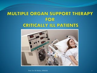
5. organ support techniques
- 1. 1Prof. Dr. RS Mehta, BPKIHS
- 2. Introduction The intensive care unit (ICU) is the hospital facility within which the highest level of continuous patient care and treatment care are provided. ICU cases include a variety of severe cases due to major surgical interventions, trauma, hemodynamic instability, sepsis and so on. All of these factors can easily lead to MODS (multiple organ dysfunction syndromes). 2Prof. Dr. RS Mehta, BPKIHS
- 3. Introduction … Multiple organ dysfunction syndromes are the leading cause of mortality in critically ill patients and is responsible for a large amount of healthcare expenditure. Since the probability of death is directly correlated to the number of failing organs beyond the kidney and the degree of physiological derangement, a clinically sensible approach is to broaden the spectrum of physiological endpoints targeted by extracorporeal therapy. 3Prof. Dr. RS Mehta, BPKIHS
- 4. Introduction … Blood is the vital element that regulates all body systems from cellular to organ level. A multiple organ support therapy is a logical and should be the goal of extracorporeal blood purification in the intensive care unit. 4Prof. Dr. RS Mehta, BPKIHS
- 5. Indication of ICU admission Patients requiring, likely require, advanced respiratory support alone. The patient requiring support of two or more organ systems. Patients with co-morbidity who require support for an acute reversible failure of another organ system 5Prof. Dr. RS Mehta, BPKIHS
- 6. 6Prof. Dr. RS Mehta, BPKIHS
- 7. Clinical feature of patient requiring organ system support Confusion Decrease GCS Shortness of breath Rapid or irregular heart beat Rapid, shallow breathing 7Prof. Dr. RS Mehta, BPKIHS
- 8. Clinical feature of patient requiring organ system support… Grunting sounds Flaring of the nostrils Decrease urine output= (<400ml/24 hours oliguria or 50ml/12hours anuria) 8Prof. Dr. RS Mehta, BPKIHS
- 9. Investigations Echocardiography-two-dimensional with Doppler flow studies may show ventricular hypertrophy, dilation of chambers, and abnormal wall motion. ECG (resting and exercise) may show ventricular hypertrophy and ischemia. Chest X-ray may show cardiomegaly, pleural effusion, and vascular congestion. Cardiac catheterization to rule out CAD ABG studies may show hypoxemia due to pulmonary vascular congestion. Liver function studies may be altered because of hepatic congestion. 9Prof. Dr. RS Mehta, BPKIHS
- 10. Investigations… Renal function test may be altered because of renal congestion. Imaging Chest X-ray may show cardiomegaly, pleural effusion, and vascular congestion. Computed tomography (CT) scan is a structural imaging study that uses a computer-based X-ray to provide a cross-sectional image of the brain. MRI is a noninvasive structural imaging procedure that uses powerful magnetic field and radio frequency waves to create an image of brain and others organ. Positron-emission tomography (PET): A computer-based functional imaging technique that permits study of the brain's metabolism, blood flow, and chemical processes. 10Prof. Dr. RS Mehta, BPKIHS
- 11. Objective of multiple organ support therapy Protect the organs before organ failure Restrict tissue hypoxia Reduce an excessive inflammatory response Protect against oxidant damage if multiple organ failure is already established, the cells might need to be rested. 11Prof. Dr. RS Mehta, BPKIHS
- 12. Categories of organ system support therapy Respiratory support therapy Circulatory support therapy Renal support therapy Hemodynamic monitoring or support therapy Neurological monitoring or support 12Prof. Dr. RS Mehta, BPKIHS
- 13. Respiratory support therapy a. Advanced respiratory support therapy b. Basic respiratory support therapy 13Prof. Dr. RS Mehta, BPKIHS
- 14. Advanced respiratory support Immediate tracheal intubation and mechanical ventilation support (excluding mask continuous positive airway pressure, CPAP) or non- invasive ventilation. 14Prof. Dr. RS Mehta, BPKIHS
- 15. Indication Upper and Lower airway obstruction as a result of blockage caused by blood or pus or bronchospasm and edema. Neuromuscular disorders as in Myasthenia gravis, Poliomyelitis, Guillain-Barré syndrome, Snake bite and inadequate reversal of anesthesia. Lung diseases which prevent proper exchange of O2 and CO2 as in chest injuries pneumothorax, lung infections, COPD, Adult Respiratory Diseases Syndrome (ARDS). 15Prof. Dr. RS Mehta, BPKIHS
- 16. Indication … Post-operative cardiac surgery, any other surgery, shock & trauma. Respiratory arrest Acute respiratory acidosis with partial pressure of carbon dioxide (pCO2) > 50 mmHg (normal : 35- 45) and pH < 7.25. Hypoxemia. 16Prof. Dr. RS Mehta, BPKIHS
- 17. Basic respiratory support Assisting in coughing, deep breathing and alveolar recruitment techniques ( e.g., CPAP) Chest percussion positioning (e.g. fowlers position) Bronchodilators Suctioning 17Prof. Dr. RS Mehta, BPKIHS
- 18. Basic respiratory support… Tracheostomy care. Physiotherapy to clear secretions at least 2 hourly Use of supplemental oxygen (restricted to certain situations like COPD) Use of an incentive spirometer to increase inhaled lung volume and eliminate mucous and saliva 18Prof. Dr. RS Mehta, BPKIHS
- 19. Basic respiratory support… Inspiratory muscle training to help strengthen diaphragm muscles Nebulization Feeding modifications to reduce aspiration risks 19Prof. Dr. RS Mehta, BPKIHS
- 20. Indications The possibility of progressive deterioration to the point of needing advanced respiratory support. Patients in whom the tracheal tube has been removed recently after prolonged period of intubation and mechanical ventilation. The need for mask CPAP or non-invasive ventilation. Patients whose trachea is intubated to protect the airway but who do not need mechanical ventilation. Bed ridden patients or prolonged immobility. 20Prof. Dr. RS Mehta, BPKIHS
- 21. Nursing consideration Assess respiratory rate and depth; Inspect thorax for symmetry of movement. Assess the patient for oxygenation such as oxygen saturation, signs and symptoms of hypoxia (tachypnea, nail beds, ABG analysis, auscultation for air entry). Observe for tube misplacement. Prevent accidental extubation by taping tube securely, checking q.2h. Maintain ventilator settings as ordered. Elevate head of bed 60-90 degrees. 21Prof. Dr. RS Mehta, BPKIHS
- 22. Circulatory support therapy Mechanical circulatory support Use of Intra-aortic balloon pump with a ventricular assist device (VAD). Medical therapy including use of angiotensin converting enzyme inhibitors, beta blockers, and aldosterone antagonists. 22Prof. Dr. RS Mehta, BPKIHS
- 23. Intra-aortic balloon pump A ventricular assist device 23Prof. Dr. RS Mehta, BPKIHS
- 24. Indications Cardiogenic shock resulting from acute myocardial infarction (AMI). Postsurgical myocardial dysfunction Acute cardiac failure from myocarditis Decompensated chronic heart failure Dilated cardiomayopathy 24Prof. Dr. RS Mehta, BPKIHS
- 25. Nursing consideration The nurse plays a critical role in caring for the patient receiving intra-aortic balloon counterpulsation . The nurse makes ongoing timing adjustments of the balloon pump to maximize its effectiveness by synchronizing it with the cardiac cycle. The patient is at great risk for circulatory compromise to the leg on the side where the catheter for the balloon has been placed; therefore, the nurse must frequently check the neurovascular status of the lower extremities. Auscultate heart sounds frequently and monitor cardiac rhythm. 25Prof. Dr. RS Mehta, BPKIHS
- 26. Renal support therapy Acute renal replacement therapy: is a term used to encompass life- supporting treatments for renal failure. it includes: hemodialysis, peritoneal dialysis. Hemodialysis: Hemodialysis is a process of cleansing the blood of accumulated waste products. In haemodialysis the blood flows through a dialysis machine that filters away the waste products. 26Prof. Dr. RS Mehta, BPKIHS
- 27. Renal support therapy… Peritoneal dialysis: Peritoneal dialysis involves the repeated cycles of instilling dialysate into the peritoneal cavity , allowing time for substance exchange , and then removing the dialysate. PD is typically used for client with severe cardiovascular disease 27Prof. Dr. RS Mehta, BPKIHS
- 28. Indication of dialysis Oliguria (urine output <200 mL/12 h) Anuria/extreme oliguria (urine output <50 mL/12 h) Hyperkalemia (K >6.5 mEq/L) Severe acidemia (pH <7.1) Azotemia (urea >30 mg/dL) Pulmonary edema 28Prof. Dr. RS Mehta, BPKIHS
- 29. Nursing consideration Assess for bleeding at the access site or elsewhere. Use standard precautions at all times. Renal failure and heparinization during dialysis increase the risk for bleeding. Assess for dialysis disequilibrium syndrome, with headache, nausea and vomiting, altered level of consciousness; and hypertension. Assess for other adverse responses to dialysis, such as dehydration, nausea and vomiting, muscle cramps, or seizure activity. Assess and document vital signs, weight, and vascular access site condition. Monitor BUN, serum creatinine, serum electrolyte, and Hematocrit levels between dialysis treatments. 29Prof. Dr. RS Mehta, BPKIHS
- 30. Liver support therapy Liver failure is defined as an insufficiency of any facet of liver function to a degree that this insufficiency leads to secondary organ failures and creates a life threatening situation if untreated. Artificial extracorporeal liver support is a term that is used to describe measures that are used to carry out liver function and are outside of the body. The Molecular Adsorbent Recirculation System (MARS) is an example of artificial extracorporeal liver support. The ultimate goal of extracorporeal liver support is to prolong the survival time of patients with liver failure by preventing progression of secondary organ failure. 30Prof. Dr. RS Mehta, BPKIHS
- 31. The Molecular Adsorbent Recirculation System (MARS) The MARS system combines the efficacy of sorbents to remove albumin-bound toxins with the high selectivity of highly biocompatible dialysis membranes. In this way, common dialysis or CRRT machines can be expanded into a modern system for liver support therapy. 31Prof. Dr. RS Mehta, BPKIHS
- 32. Nursing consideration During MARS therapy, there is potentially chance to occur bleeding complications and mortality so observe feature of bleeding. Monitor BUN, serum creatinine, serum electrolyte, ammonia, albumin, AST, ALT between dialysis treatments. 32Prof. Dr. RS Mehta, BPKIHS
- 33. Hemodynamic support Hemodynamics are the forces which circulate blood through the body. Specifically, hemodynamics is the term used to describe the intravascular pressure and flow that occurs when the heart muscle contracts and pumps blood throughout the body. 33Prof. Dr. RS Mehta, BPKIHS
- 34. Hemodynamic support… Hemodynamic monitoring refers to measurement of pressure, flow and oxygenation of blood within the cardiovascular system. Hemodynamic support includes: Fluid resuscitation/Blood transfusion Use of vasoactive drugs like nitroglycerine, amlodipine, nitric oxide, hydralazine. 34Prof. Dr. RS Mehta, BPKIHS
- 35. Indications Decreased urine output from dehydration Hemorrhage G.I bleed Burns or surgery All types of shock; cardiogenic shock, neurogenic shock or anaphylactic shock. Any deficits or loss of cardiac function: such as myocardial infarction, congestive heart failure, cardiomyopathy 35Prof. Dr. RS Mehta, BPKIHS
- 36. Nursing consideration Administer fluid as prescription. Assess the sign of cardiac overload like dyspnea, increase CVP, edema, weight gain, crackles (rales) and bounding pulse etc. Measure intake and output. 36Prof. Dr. RS Mehta, BPKIHS
- 37. Neurological monitoring or support Central venous system depression sufficient to compromise the airway and impair protective reflexes. Invasive neurological monitoring: A common complication of many serious neurologic conditions is an elevation of the pressure within the skull, the intracranial pressure or ICP. In adults, the average ICP ranges from <10-15 mm Hg . 20 mm Hg is considered to be the maximal upper limit of desirable ICP and pressures exceeding 40 mm Hg are considered extremely elevated. 37Prof. Dr. RS Mehta, BPKIHS
- 38. Neurological monitoring or support Whatever the underlying cause an increase in intracranial pressure is extremely dangerous. The type of monitor used is dependent on a number of clinical factors, not the least of which is the neurologic disease causing the pressure increase. The following devices commonly used to monitor and treat intracranial pressure: Intraventricular catheter 38Prof. Dr. RS Mehta, BPKIHS
- 39. Intraventricular catheter 39Prof. Dr. RS Mehta, BPKIHS
- 40. Nursing consideration Proper positioning helps to reduce ICP. The head is kept in a neutral (midline) position, maintained with the use of a cervical collar if necessary, to promote venous drainage. Elevation of the head is maintained at 30 degrees to aid in venous drainage unless otherwise prescribed. Assess the level of consciousness( GCS). Pupillary reaction. Maintain aseptic technique while measuring ICP. 40Prof. Dr. RS Mehta, BPKIHS
- 41. 41Prof. Dr. RS Mehta, BPKIHS