SlideShare a Scribd company logo
1 of 186
Download to read offline
Structure-function relationship of clinically important Peptides
Dr. Rohini C Sane
Protein structure-function relationship
• The overall conformation of a protein , the particular position of amino acid
side chains in three-dimensional space determines the function of the
protein.
• The diversity of protein structure and its correlation with function can be
explained by the following clinically important proteins.
Protein Function
Carbonic anhydrase Formation and degradation of carbonic acid
Myoglobin(Mb) Stores oxygen in muscle cells
Hemoglobin (Hb) Transports oxygen from lungs to the tissue
Collagen Providestensile strengthtomanytissueinthebody
Elastin(withrubberlikeproperties) Occur in distensible structure
Quaternary structure of enzyme regulates its functions
Structural conformation of the active site the
enzyme precisely oriented for the substrate binding
Binding of enzyme to the substrate .
enzyme catalysis →product + enzyme set free for
substrate binding again
Disordered region of enzyme and its Importance
Schematic diagram
Many Disordered region become ordered region when a specific ligand is bound .
Disordered region gives flexibility and performs a vital biological role.
Disordered regions of
enzyme- catalytic site
ordered regions of
enzyme
Substrate added to
enzyme catalyzed
reaction mixture
Enzyme with
Disordered regions
set free at the end of
reaction
Product
Enzyme-substrate complex –induced fit
+
QuaternarystructureofenzymeCarbonicanhydraseregulatesitsfunctions
❖Function of Carbonic anhydrase : catalyzes the reversible hydration of carbon
dioxide.
H2O + CO2  H2CO3(Carbonic acid)
1.H2O binds to Zinc ion (located in deep cleft of Carbonic anhydrase and is
coordinated to Histidine residue present at its active site).
Binding of CO2 to histidine residue is close to Zinc ion.
2.H2O→ H+ + OH- (Ionization of water to hydroxyl ion)
3.OH- +CO2(located proximally to OH- ions)→HCO-
3
Carbonic anhydrase facilitates the precise positioning of CO2 molecule and hydroxyl
ion OH- for the formation of bicarbonate ion HCO-
3.
4.HCO-
3 + H+ → H2CO3(Carbonic acid)
Thus , two substrates CO2 and H2O are brought in close proximity for reaction to
proceed and to facilitate the function of Carbonic anhydrase .
QuaternarystructureofenzymeCarbonicanhydrase
QuaternarystructureofenzymeCarbonicanhydraseregulatesitsfunctions
StepsofenzymecatalysisbyCarbonicanhydrase
1.H2O binds to Zinc ion (located in a deep cleft of Carbonic anhydrase and is coordinated to
Histidine residue present at an active site of enzyme )
2.H2O→ H+ + OH- (Ionization of water)
3. Binding of CO2 toHistidine residue close to Zinc ion of Carbonic anhydrase
4.OH- + CO2( located proximally to OH- ions)→HCO-
3
5.HCO-
3 + H+ →H2CO3(Carbonic acid)
Structure of Myoglobin
❖StructureofMyoglobin:
❖ Oxygenbindingcytosolicmetalloprotein functionalwithinskeletal&cardiacmuscle.
• hasasinglepolypeptidechainwith153aminoacidsanditcontainsonehemegroup(iron
containingporphyrinringsooneFe2+ ionwhichimpartsredcolortomyoglobin–
chromoprotein).Itisaglobularproteinwithlowmolecularweight(16700Dalton).
• 80%ofitspolypeptidechain isfoldedinto8-helices,whicharelabeledas AtoH.
-helicalregionsareterminatedbypresenceofProline(5-memberedringofProcannot
beaccommodatedinalpha-helix→Proishelixdestabilizingaminoacid).Restofthe
polypeptidechainformsturnsandloopsbetweenhelices.
• Thehelicescreatehydrophobicoxygen(O2)bindingpocketcontainingtightlybound
hemewithanironatom(Fe2+)initscenter.WithinthepocketofMyoglobin,O2 binds
directlytotheFe2+ ionoftheporphyrinring.
• hasnobetasheets.Thisstructureisunusual fortheglobularproteins.
Myoglobincontentofmuscles
Type of muscle Myoglobincontentofmusclesin gm/100gmof muscles
Skeletal 2.5
Cardiac 1.4
Smooth 0.3
Structure of Myoglobin
Structure of Myoglobin(Mb) :hasasinglepolypeptidechainwith153aminoacidsand
containsonehemegroup(ironcontainingporphyrinringsooneFe2+ ion-impartingred
colortoMyoglobin).Itis a globularproteinwithmolecularweight(16700Dalton) andhas
cytosoliclocation.ThisOxygenbindingmetalloproteinproteinfunctionalwithinskeletal&
cardiacmuscles.Onemoleculeof(Mb)containsoneoxygenmolecule.
JohnKendrewandMaxPerutz(Noble1962):determinedthestructureofMyoglobinby
highresolutionX-crystallography.
Tertiary Structure of Myoglobin(Mb)
Myoglobin:PrimarystructuresimilartosinglemonomericunitofHemoglobin withasinglepolypeptidechainhaving
153aminoacids(molecularweight16700Dalton).It haseightalpha–helices(AtoH)andonehemegroup(iron
containing porphyrin)tofacilitateitsfunctionofoxygenstorageincardiacandskeletalmusclesinhumanbody,Whales
andSeals.
Tertiary Structure of Hemoglobin (Hb)
Hemoglobin:Tetramericwith4hemegroups.Eachpolypeptidechainhassimilarstructureto single
polypeptidechainofMyoglobin.Ithasaloweraffinity foroxygenthanMyoglobin.FoursubunitsofHb
functioncooperatively.TetramericstructureofhemoglobinfacilitatessaturationwithO2inthelungand
releaseofoxygenasittravelsthroughthecapillarybed.
Functions of Myoglobin
❖Functions of Myoglobin(Mb): acts as storage and transport protein for oxygen
in skeletal&cardiacmuscles. In resting muscle cells ,myoglobin binds oxygen
(O2)that has been released by Hemoglobin(Hb) under the conditions of
oxygen deprivation (in severe exercise for use O2 by muscle).
❖HbO2 (oxyhemoglobin) → → → Mb(Myoglobin) →MbO2(oxymyoglobin)→
tissue → Respiration
Oxygen (O2) transport
Oxygen dissociation curve of Myoglobin and hemoglobin
When the amount oxygen (O2)bound to the
Hemoglobin and Myoglobin is plotted
against the partial pressure of oxygen (pO2),
a hyperbolic curve is obtained for
Myoglobin whereas that for Hemoglobin is
sigmoidal.
When the (pO2) is high both Myoglobin and
Hemoglobin are saturated with oxygen.
At lower levels of (pO2), however ,
Myoglobin contains more oxygen O2 than
Hemoglobin .
Bohr’s effect ,co-operative effect and 2,3
BPG effect are absent for Myoglobin .
Mb has higher affinity for Oxygen (O2) than Hb.
At PO2 in tissue (30mmHg): Mb is 90% saturated, Hb is 50%
saturated . Physical exercise: PO2 in tissue is (5mmHg), when
Mb releases all bound Oxygen (O2).
Clinical aspects associated with Myoglobin
❖Myoglobin : under physiological conditions , Myoglobin( being a small
molecular weight protein)is filtered and excreted in urine.
• is a sensitive maker for muscleinjury,makingitpotential cardiacmarkerfor
myocardialinfarctioninpatientswithchestpain.
• rises rapidly after the chest pain at about the same rate as CK-MB.
• is non-specific ,since it is raised following any form of muscle damage.
• Estimation ofMyoglobin–usingmonoclonalantibodyforbyRIA/ELIZA/
chemiluminescence
• Cost of the analysis has prevented its widespread use.
• TemporalpatternofserumMyoglobin&Creatinekinase-2inpatientswithmyocardial
infarctionisdepictedinadiagram.
SerumMyoglobin&Creatinekinase-2(CK-MB)inpatientswithmyocardialinfarction
• The cellular release Myoglobin is often associated with an increase in Creatine
kinase (CPK), Aldolase , Lactate dehydrogenase(LDH) , Serum Glutamate
Pyruvate Transaminase(SGPT) in patients with myocardial infarction.
Myoglobinuria
❖Myoglobinuria : under physiological conditions , Myoglobin being low
molecular weight protein, it is filtered and excreted in urine. Presence of
excessive Myoglobin in the urine usually associated with muscle destruction
or rhabdomyolysis . Urine color becomes dark brown.
❖Damage to muscle( crush injury), releases Myoglobin to the circulation and
filtered by the kidney. If too much of Myoglobin is released into circulation ,
Myoglobin precipitate and obstruct the renal filtration system results in
acute renal failure.
❖Myoglobin is released from myocardium during Myocardial infarction (MI)
and is elevated in serum(cardiac marker)followed by its excretion in urine.
ClinicalconditionassociatedwithMyoglobinuria
❖ClinicalconditionassociatedwithMyoglobinuriainadultinclude:
1. Extremeexercise(mostcommoncauseofrhabdomyolysisinadolescents):excessive
physicalactivitycanproduceimbalancebetweenenergyconsumptionand
productionresultinginmuscledestruction(duetooxidativestress)
2. Trauma/Vascularproblems
3. Prolongedethanol/Alcoholconsumption
4. Drugabuse
5. ToxicityofVenoms
Myoglobinuria
Rhabdomyolysis
Quaternary structure of Hemoglobin
Hemoglobin (HbA1)has
4 Polypeptides chains
( tetramer) associated by
non-covalent bonds :
2 Alpha() chains
+
2 Beta( )chains
It possesses Quaternary
structure(oligomeric).
Inthis, Rgroupcontactsare
presentbetweensimilarside
chainsandthereisverylittle
contactbetweendissimilar
side chains.
HbA:Tetramer22
Each chain/subunit has one heme
group and so one Fe2+ ion similar
to that of myoglobin.
Hb is found
exclusively in RBC
Structure of normal Hemoglobin
Hemoglobin variant Structure of normal Hemoglobin Abbreviation
Hemoglobin (HbA 1) 2 Alpha() chains and 2 Beta( )chains 22
Hemoglobin (HbA 2) 2 Alpha() chains and 2 delta( )chains 22
Fetal Hemoglobin (HbF) 2 Alpha() chains and 2 gamma ( )chains 22
Functions of Hemoglobin
❖Functions of Hemoglobin:
• Hemoglobin is found exclusively in RBC .It transports four molecules
oxygen(O2) from lungs(high pO2) to the tissues (low pO2) . Myoglobin is still
very saturated with oxygen at the (pO2) of tissue. It can transport H + and
carbon dioxide (CO2) from peripheral tissues to lungs for its excretion in air.
oxygenation
Deoxyhemoglobin + 4O2 oxyhemoglobin (in lungs)
Deoxygenation is the reverse process by which O2 is liberated.
deoxygenation
Oxyhemoglobin 4O2+ Deoxyhemoglobin
• Hemoglobin is rich in amino acid Histidine ,helps to maintain pH of blood (acts
as buffer).
• Transports oxygen(O2)by Hb is regulated by 2-3-bisphosphate
glycerate(2,3BPG).
Functions of Hemoglobin as a transporter of oxygen(O2) from lungs(high pO2) to the tissues
(low pO2)
Quaternary structure of Hemoglobin favors its functions
❖ Hemoglobin found exclusively in red cells and functions as transporter of
oxygen(O2) from lungs to tissue . Each molecule carry four
oxygen(O2)molecules from lungs to the cells .This function is favored by
presence of one heme unit one each in monomer (4 heme→4 oxygen(O2)/
hemoglobin).
➢Binding of oxygen(O2) to one heme unit of tetramer facilitates oxygen
binding by other subunits. Each subunit has a heme binding pocket similar to
that of myoglobin .
➢It can transport H+ and CO2 from the tissue to the lung.
➢Binding of H+ and CO2 promotes release of oxygen(O2) from hemoglobin .
➢This allosteric interaction is physiologically important and as Bohr’s effect .
➢Even a single amino acid substitution alters the structure and thereby
functions.
Co-operative binding of oxygen to hemoglobin
• Binding of oxygen to Heme will increase binding of oxygen to other heme
• Affinity of oxygen for hemoglobin
• Last oxygen binds with affinity 100 time greater than first oxygen
• Heme –Heme interaction ( co-operative binding of oxygen to Heme )
• Release oxygen from one Heme will release oxygen from other
• As there is communication between Heme groups of hemoglobin
• Myoglobin is reservoir (transient )& supplier of oxygen
Lung Tissue
Oxygen concentration high Oxygen concentration low
Oxygen binds to
hemoglobin
Oxygen is released to tissue
Structural changes in hemoglobin on oxygen binding
• Studied using x ray crystallographic
• Homotropic effect→ binding of oxygen to hemoglobin
• Heterotrophic effect → binding of 2,3BPG to hemoglobin
• Distance between two Beta-chain decreases oxygenation from 4nm to 2nm
• Increasing affinity for oxygen with addition every molecule of oxygen
• On oxygenation iron moves in plane of Heme
• Decrease in diameter of iron (movement of iron accompanied by pulling of
proximal Histidine
• affinity of hemoglobin for last oxygen > first oxygen ( 100 times greater )
• Cooperative binding of oxygen to hemoglobin or Heme →Heme interaction
• Release of oxygen from one Heme → Release of oxygen from other Heme
❖Therefore communication between Heme groups of hemoglobin.
On oxygenation iron moves in plane of Heme → decrease in diameter of Iron
→movement of Fe Is accompanied by pulling of proximal site →primary event of Heme
–Heme interaction of Hemoglobin
Structural changes in Hemoglobin on its Oxygenation and deoxygenation
Deoxy –Hb Hb O₂ Hb O₄ Hb O₆ Hb O₈
T form ↓ ↓ ↓ ↓ ↓
↑
R form
Oxygenation of hemoglobin
Quaternary structure of Hemoglobin favors its functions :Binding of oxygen(O2) to
one heme unit facilitates oxygen binding by other subunits.
Structural changes in Hb on oxygen binding
• Structural change in one subunit of hemoglobin on oxygenation is
communicated to other subunits
• Binding of oxygen to one Heme distorts globin chain to which it is
attached → distortion in neighboring chain →oxygen binds more
easily.
Effect of 2,3 BPG on oxygen affinity of Hb
• 2,3 BPG :Most abundant phosphate in RBC
• Molar concentration of 2,3 BPG = Molar concentration of Hb
• Synthesis ( synthesis through Rapport Leu Bering cycle)
2, 3 BPG mutase ( Glycolysis )
1,3 BPG 2,3 BPG
• Reinhold's & Ruth Benesch’s (1967 )→ 2,3 BPG decreases affinity of Oxygen to
Hemoglobin
• 2,3 BPG regulates the binding of oxygen
• 1mole of 2,3 BPG binds to 1mole of Deoxy Hb not to oxy –Hb
• Molecular concentration 2,3 BPG = Molecular concentration of hemoglobin
• At partial pressure of oxygen(O₂) in tissue: HbO₂+ 2,3 BPG→Hb 2,3 BPG +O₂
(release of O₂)
(Oxy–Hb) (De-oxy Hb)
• In tissue → 2,3 BPG shift curve towards right
1mole of 2,3 BPG binds to 1mole of Deoxy Hb not to oxy –Hb
Regulation of oxygen binding by 2,3 BPG
Effect of 2,3 BPG on oxygen affinity of Hb
2,3 BPG decreases affinity of Oxygen to Hemoglobin. In tissue → 2,3 BPG shift curve towards right
At partial pressure of O₂ in tissue: HbO₂+ 2,3 BPG→ Hb 2,3 BPG +O₂ (release of O₂)
(Oxy –Hb) (De-oxy Hb)
2,3 BPG shift oxygen dissociation curve of hemoglobin towards right to release of oxygen (O₂)
in hypoxic conditions ,anemia ,hyperthermia.
Effect of 2,3 BPG shift oxygen dissociation curve of hemoglobin
Clinical significance of 2,3 BPG:1
❖Function of 2,3BPG : Release of oxygen to tissue ( supply of oxygen to tissue )
to cope with oxygen demand → varied concentration of 2,3 BPG
1. Hypoxia : concentration of 2,3BPG in RBC increases during chronic hypoxic
conditions e.g.
a. Adaptation to high altitude
b. Obstruction to pulmonary odema ( air flow in bronchial blocked )
2. Anemia : concentration of 2,3 BPG in RBC increases in chronic anemic
conditions → to cope with oxygen (O₂) demand of body even at low Hb
concentration.
Clinical significance of 2,3 BPG:2
3. Blood Transfusion : storage of blood in acid citrate dextrose → decrease in
concentration of 2,3, BPG ( O₂ remains bound to Hb )
• Blood stored in ACD fails to supply O₂ to tissue→ with 24-48 hr. until 2,3 BPG
restored
• O₂ supply /tissue O₂ demand met adequately after 24-48 hrs.
• Blood with (ACD )+ Inosine ( Hypoxanthine Ribose )→ prevent decrease in 2,3, BPG
• Inosine→ phosphorylation of tissue→ entry into HMP shunt → get converted to
2,3BPG →increase in Conc in 2,3 BPG → release of oxygen
4.Fetal hemoglobin(HbF) : the binding of 2,3BPG to fetal hemoglobin is very weak.
Therefore, HbF has higher affinity for oxygen compared to adult hemoglobin (HbA).
This is needed for transfer of oxygen from the maternal blood to fetus.
Protein structure-function relationship in
Adult Hemoglobin(HbA1) and Fetal Hemoglobin (HbF)
• Function of Fetal Hemoglobin (HbF) : The transfer of oxygen from the
mother to the fetus.
• This function of Fetal Hemoglobin (HbF) is facilitated by structural
differences in between the hemoglobin molecule of the mother and that of
fetus (HbF).
• Adult Hemoglobin(HbA1) : 2 Alpha() chains and 2 Beta()chains (22)
• Fetal Hemoglobin (HbF) : consist of 2 Alpha() chains and 2gamma ()
chains- 22
• The difference in amino acid composition between the beta chain of HbA1
and gamma ( )chains of (HbF) results in structural changes that causes
HbF to have lower affinity for 2,3- biphosphoglycerate (2,3 BPG) than HbA1
and thus greater affinity for oxygen.
➢Therefore , the oxygen released from the mother’s HbA1 is readily bound by
HbF in the fetus.
Protein structure-function relationship in Myoglobin and adult Hemoglobin(HbA1):1
Criteria Myoglobin Hemoglobin(HbA1)
Primarystructure SimilartosinglemonomericunitofHemoglobin
withasinglepolypeptidechainhaving 153
aminoacids(molecularweight16700)andone
hemegroup(ironcontaining porphyrin).
Tetramericwith4hemegroups.Each
polypeptidechainhassimilarstructure
to singlepolypeptidechainof
Myoglobin.
Affinityforoxygenof
oxygenbinding
protein
Hasahigheraffinity foroxygenthan
hemoglobin.ThebindingofO2 tohemegroup
ofeachmoleculeisindependentofanother
moleculebecauseitcontains onlyoneheme
group(onepolypeptidechain).
Hasaloweraffinity foroxygenthan
myoglobin.Foursubunits of Hb
functioncooperatively.
Function of
metalloprotein
Astoragereserve foroxygen,releasesoxygen
theboundoxygenforcellularusewhenoxygen
supplyisreduced.Storeofoxygenindeep-
divingmammals.
Allowsefficienttransferofoxygenfrom
lungtotissue.Tetramericstructureof
hemoglobinfacilitatessaturationwith
O2inthelungandreleaseofoxygenas
ittravelsthroughthecapillarybed.
Structure of Myoglobin and adult Hemoglobin(HbA1)
PrimarystructureofMyoglobin:SimilartosinglemonomericunitofHemoglobin withasinglepolypeptide
chainhaving 153aminoacids(molecularweight16700)andonehemegroup(ironcontaining porphyrin).
PrimarystructureofadultHemoglobin(HbA1):Tetramericwith4hemegroups.Eachpolypeptidechainhas
similarstructureto singlepolypeptidechainofMyoglobin.
Functions ofMyoglobin and adult Hemoglobin(HbA1)
Functionofmyoglobin:Astoragereserve foroxygen,releasesoxygentheboundoxygenforcellularuse
whenoxygensupplyisreduced.Storeofoxygenindeep-divingmammals.
Functionofadulthemoglobin(hba1):allowsefficienttransferofoxygenfromlungtotissue.Tetrameric
structureofhemoglobinfacilitatessaturationwith O2inthelungandreleaseofoxygenasittravelsthrough
Protein structure-function relationship of Myoglobin and adult Hemoglobin(HbA1):2
Condition Myoglobin Hemoglobin(HbA1)
Highpartialpressureof
oxygen(pO2)
Saturated saturated
Lowpartialpressureof
oxygen(pO2 )
ContainsmoreO2thanhemoglobin Containslesser O2thanmyoglobin
Agraphplottedwith the
amountofO2 boundtothe
proteinagainstthepartial
pressureofoxygen(pO2 )
Hyperboliccurve Sigmoidcurve
Application Stillverysaturatedwith oxygenatthe
lowpartialpressureofoxygen(pO2)
intissue facilitatingstoragefunction
intissue.
Effectivetransporterof oxygen,binding
withoxygeninthe lungswhere(pO2)is
highandreleasingitintissuewhere(pO2)
islow .
OxygenbindingaffinityofMyoglobinandHemoglobin
Hyperboliccurve of oxygenbinding forMyoglobin
Sigmoidcurveof oxygenbinding forHemoglobin
At Highpartialpressureofoxygen(pO2)both myoglobin and hemoglobinaresaturatedwithoxygen.
Application : Myoglobin stillverysaturatedwith oxygenatthelowpartialpressureofoxygen(pO2)in
tissue facilitatingstoragefunctionintissue.Hemoglobinis aeffectivetransporterof oxygen,bindingwith
oxygeninthe lungswhere(pO2)ishighandreleasingitintissuewhere(pO2)islow .
Transport of oxygen by hemoglobin
PO₂ (mm) of Hg % saturation
Inspired air 158
Alveolar air 100
lung 90 97%
Capillary bed 40 60%
37% - 40% O ₂ release of oxygen at tissue level
Structural modification of hemoglobin by glycosylation and its application
• Glycosylated hemoglobin (Hb A1C): is formed by non –enzymatic reaction of
the aldehyde group of Glucose with amino terminal (N –terminal residue)
Valine of beta chains of HbA when blood Glucose enters the erythrocytes.
• Normal concentration of Glycosylated hemoglobin (Hb A1C): low ( to the
extent of 5% of total hemoglobin
• Formation of (Hb A1C) is proportional to blood glucose concentration.
• Glycosylated hemoglobin (Hb A1C) increases in Diabetes Mellitus : 12% or
more of total hemoglobin as serum glucose is high
• Since RBC have life span of 120 days , the content of Hb A1C is an indicator of
how effectively blood glucose levels have been regulated over the previous
2 or 3 months.
• Application of Glycosylated hemoglobin (Hb A1C) estimation : to follow the
effectiveness of treatment (management)for Diabetes Mellitus .
Glycosylated hemoglobin (Hb A1C)
Glycosylated hemoglobin (Hb
A1C): is formed by non- enzymatic
reaction of HbA with Glucose when
blood Glucose enters the
erythrocytes.
Application of Glycosylated
hemoglobin (Hb A1c) estimation :
to follow the effectiveness of
treatment for Diabetes Mellitus
Primary structure of Normal protein determines biological functions
Unique amino acid sequence specified by genes in a Normal protein
Specific amino acid sequence→ confers specific 3 dimensional structure
(conformation )
Specific Function arises from conformation
Normal physiologically active protein e.g. Normal hemoglobin
Consequences of Altered primary Structure in Abnormal protein
Mutation→ altered genetic constitution ( base sequence of DNA)
Altered amino acid sequence→ Altered 3 dimensional structure(conformation)
Altered or loss of functions arises from Altered conformation of protein
Abnormalorphysiologicallyinactiveprotein→diseaseconditione.g.hemoglobinopathy
Structure function relationship of proteins
Normal protein
• Unique amino acid sequence
specified by genes
• Specific amino acid sequence→
confers specific 3 dimensional
structure ( conformation )
• Specific Function arises from
conformation
• Normal physiologically active
protein e.g. Normal hemoglobin
Abnormal protein
• Mutation→ altered genetic
constitution / base sequence of
DNA
• Altered amino acid sequence
→Altered 3 dimensional structure
( conformation )
• Altered or loss of Function arises
from Altered conformation
• e.g. abnormal or physiologically
inactive protein→ disease
condition e.g. hemoglobinopathy
Altered primary Structure of Abnormal hemoglobin
Abnormalhemoglobin→
hemoglobinopathy
Altered primary Structure leading anemia in
early life
Sickle cell hemoglobin ( Hb S) no
change in amino acid sequence of 
chain
6 th amino acid in beta chain of HbA1 (Glutamic acid
replaced by Valine in HbS )
HbM (Methemoglobinemia ) Substitution of Tyrosine with Histidine
Hb Chesapeake Arginine is replaced by Leucine at 92th amino acid in
alpha chain of Hb A
Alpha(  ) thalassemia ( normally the
rate of synthesis  and  chains
identical )
Deficiency or absence of alpha chains → HbA of fetus
has tetramer  4 or 4 chains ( enlargement of live
and spleen)
Beta ( )thalassemia ( more common
inherited disease than Alpha
thalassemia )
Deficiency or absence of beta chains →hemoglobin
found in RBC are HbA2 (  2 2) and HbF (  2 2 )
Deoxygenated HbS
• This substitution generates ‘A stick patch’ on the surface of the beta chain of
both oxygenated and deoxygenated HbS .
• On the surface of deoxygenated normal HbA and deoxygenated HbS , there
exists a complement to the sticky patch.
• When HbS is deoxygenated ,its sticky patches can bind to the complementary
patches on another deoxygenated HbS .
• Binding of large number of deoxygenated HbS causes polymerization of
deoxygenated HbS forming long fibrous precipitate that mechanically disturbs
the red cell→ sickle shaped→ casing lysis and anemia.
• This polymerization will not take place when HbS is in oxygenated form as in
arterial blood .
Molecular basis of Sickle cell anemia
Linus Pauling ( 1954 Noble prize ) reported abnormal electrophoretic mobility & peptide
mapping Glutamic acid ( sixth position on beta globin chain ) replaced by Valine (Recessive
Mutation ) Hb A & Hb F prevent sickling.
sixth amino acid in beta() chain of HbA1 (Glutamic acid )replaced by Valine in HbS
Altered primary Structure leading anemia in early life
Sickle cell disease
Binding of large number of deoxygenated HbS causes polymerization of deoxygenated HbS
forming long fibrous precipitate that mechanically disturbs the red cell→ sickle shaped→
casing lysis and anemia.
HbM (Methemoglobinemia )
❖HbM (Methemoglobinemia) :
Substitution of Tyrosine with Histidine on hemoglobin result in
oxidation of ferrous is to ferric in heme . Hemoglobin in blood is
oxidized to methemoglobin. This condition leads to
Methemoglobinemia .
HbM (Methemoglobinemia) :Substitution of Tyrosine with Histidine
 Substitution of Tyrosine with Histidine
oxidation of ferrous is to ferric in heme→
Hb Chesapeake
❖Hb Chesapeake
➢Substitution of Arginine by Leucine at 92th amino acid in alpha chain
of Hb A results in increased affinity of hemoglobin for oxygen and
does not release as much oxygen to the peripheral tissue as does
normal hemoglobin HbA .
➢This leads to tissue hypoxia and polycythemia ( increased number of
RBC per unit volume) in ode to meet the oxygen needs.
Quaternary structure of Aspartate trans carbamylase
Aspartate trans carbamylase : An allosteric enzyme with 2 subunits : catalytic
subunit (C) and the regulatory subunit (R)
Quaternary structure of Lactate dehydrogenase (LDH )
❖Quaternary structure of Lactate dehydrogenase (LDH ):
• Tetramer
• two types of polypeptide chains :H and M type
• five isoenzymes :
1. LDH 1 →H4
2. LDH2 → H3M
3. LDH3→ H2M2
4. LDH 4→ HM3
5. LDH 5 →M4
Isoenzymes of Lactate dehydrogenase
Subunit composition of LDH1
isoenzymes in heart cells
favors conversion of Lactate
to Pyruvate
Subunit composition of LDH5
in muscle cells favors
conversion of Pyruvate to
Lactate
Quaternary structure of Lactate dehydrogenase (LDH ): Tetramer with 2 types of
polypeptide chains →H and M type
Structure of Human Insulin
In 1953 , Frederick Sanger determined primary structure of Insulin ( a pancreatic protein
hormone ) and showed for the first time that a protein has a precisely defined amino acid
sequence ( primary structure.)
Insulin and Glucagon(Polypeptide Hormones)
Insulin
Hormone secreted by  pancreatic cells
Polypeptide and a Dimer with 51 amino acids
regulates glucose metabolism and Induces
hypoglycemia
Amino acid sequence varies in different
mammalian species
Glucagon
Hormone secreted by  pancreatic cells
Polypeptide and monomer with 29 amino acids
regulates glucose metabolism and Induces
hyperglycemia
Amino acid sequence is same in all mammalian
species
Structure of Human Insulin
❖Structure of Human Insulin : described by Sanger(Noble-1955 )
❖Dipeptide (2 polypeptide chains)of insulin has 51 amino acids
A polypeptide chain : 21 amino acids
B polypeptide chain : 30 amino acids
❖ Dipeptideofinsulinarerequiredforbiologicalactivityandheldby
➢inter chain disulphide bonds:
a. between cysteine residues ( 7th amino acid of A chain and 7th
amino acid of B chain )
b. between cysteine residues (20 th amino acid of A chain and 19 th
amino acid of B chain)
➢intra chain disulphide bond : between cysteine residues (6 th
amino acid of A chain with 11 th amino acid of B chain)
Structure of Human Insulin
Carboxy terminal end
A chain : Asparagine
B chain : Threonine
Amino terminal end
A chain : Glycine
B chain : Phenylalanine
A polypeptide chain :
21 amino acids
B polypeptide chain :
30 amino acids
intra chain disulphide bond : between
cysteine residues( 6 th amino acid of A
chain with 11 th amino acid of B chain)
inter chain disulphide bonds 


Primary Structure of human Insulin
S S
A chain H2N Gly Cys Cys Cys Cys Asn
1 6 7 11 20 21
S S
S S
B chain H2N Phe Cys Cys Thr
1 7 19 30
Pig insulin differs from human insulin in only one position ,30th amino acid is alanine instead of Threonine.
Insulin from other animals like cattle ,sheep, horse etc. differ from human insulin in having a different
sequence of amino acids in the positions 8-9-10 in A chain .
This minor altered sequence does not result inappreciable change biological activity.
Amino acid substitution in Primary Structure of human Insulin
8 9 10 Achainof insulin
ofspecies
Thr Ser Ile Human
Ala Ser Val Bovine
Thr Ser Ile Pig
Ala Gly Val Sheep
Thr Gly Ile Horse
Aminoacidcompositionat30th amino
acidinBchainof insulinofspecies
Thr Human
Ala Bovine
Ala Pig
Porcineandhumaninsulinare
similar(homologous)exceptC-terminal
aminoacidinBchain(Thr→Ala).Itmay
produceantibodiesinhumanafter repeated
injections.Dealaninatedporcineinsulinwill
notproduceanyantibodiesindiabetic
patientsevenafterlongtermuse.
Aminoacidsequencehasbeenconservedto
thegreatextentduringevolution.Human
insulinrequiredforreplacementtherapy,is
nowsynthesizedbyrecombinantDNA
technology.
Site of Insulin Biosynthesis
❖Amino acids form primary structure → definite function
❖Peptide < 10 amino acids
❖Polypeptide > 10 amino acids
Preproinsulin (singlepolypeptidechainwith108aminoacids,molwt.11500)
↓
Proinsulin(single polypeptide chain with 86 amino acids, molwt.9000)
↓
Human Insulin ( dipeptide 51 amino acids : polypeptide chain A →21
amino acid and polypeptide chain B →30 amino acids held together b
interchain disulfide bridges molwt.5734 )
➢The gene for insulin synthesis : located on chromosome 11 in beta cells
of pancreatic cells.
Removal of signal sequence in endoplasmic reticulum
Removal of C-peptide in Golgi apparatus
Pre proinsulin ( single polypeptide chain ):
108 amino acids ,mol wt. 11500
Proinsulin ( single polypeptide
chain):86 amino acids ,mol wt. 9000
Insulin ( dipeptide chain ):51
amino acids ,mol wt. 5734
Biosynthesis of Insulin from Preproinsulin:1
Removal of
signal
sequence in
endoplasmic
reticulum
Removal
of C-
peptide in
Golgi
apparatus
The gene for insulin synthesis : located on chromosome 11 in beta cells of pancreatic cells.
Biosynthesis of Insulin from Preproinsulin:2
In the beta pancreatic cells , insulin and proinsulin combines with Zinc to form complexes.
In this form it is stored in the granules of the cytosol which is released in response to
various stimuli by exocytosis.
Pre proinsulin ( single polypeptide chain ) :108 amino acids ,mol wt. 11500
Proinsulin ( single polypeptide chain):86 amino acids ,mol wt. 9000
Insulin (dipeptide
chain):51 amino
acids, mol wt. 5734
Structure function relationship of C–peptide and insulin
C – peptide
has no
biological
activity .
Single polypeptide chain with 86 amino acids

Beta cells of panaceas synthesize insulin as a prohormone – Proinsulin . Biologically active
insulin (a dipeptide) is formed by removal of C – peptide ( the central potion ) of proinsulin .
C–peptide and insulin are synthesized in equimolar concentration. It useful index for the
endogenous production of insulin.
BiochemicalandclinicalaspectsofCollagen
Epidermolysis bullosa
Scurvy
Ehlers-Danlos syndrome
Biochemical aspects of Collagen
❖Biochemical aspects of Collagen:
1. Most abundant fibrous protein(major macromolecules) in human body :70 kg
body weight→ 12-14 kg of total protein→5kg of Collagen (1/3 of total protein)
2. Main Component of : connective tissue, skin(70%) ,bone (90%) tendon(85%),
cartilage ,teeth and liver(4%)
3. Synthesized by fibroblast in connective tissue and osteoblasts in bone
4. Made up of small fibrils → tropocollagen( fundamental units ) containing 3
polypeptide chains each of them in left-handed helix with 3 amino acid per turn.
5. rich in Glycine and rare amino acids like hydroxyproline, hydroxylysine
6. Cysteine and Tryptophan absent
7. have a triple helical secondary structure and rich in helix destabilizing amino
acids (Glycine ,Proline and Hydroxyproline). These amino acids prevent the
formation of the usual - helical and - pleated structure. Instead it forms a triple
helical secondary structure.
Triple stranded helix Structure of Collagen
Made up of small fibrils → Tropocollagen( fundamental units ) containing 3
polypeptide chains each of them in left-handed helix with 3 amino acid per
turn.
Triple stranded helix Structure of Collagen
• Collagenhas3 polypeptidechainswoundarounditself. Eachpolypeptidechainsubunitis
calledalpha-chains. EachofAlpha-chain is twistedintoleft-handedhelix ofthreeresidues
perturncomparedwith 3.6forright-handedalpha-helix.Threeoftheseleft–handedhelices
arethenwoundtoright-handedsuperhelixtoformastiffrodlike molecule(Triplehelical
secondarystructure).
• Itisrich in helixdestabilizingaminoacids(Glycine ,ProlineandHydroxyproline).Theseamino
acids preventtheformationoftheusual- helicaland-pleatedstructure.Instead,it formsa
triplehelicalsecondarystructure.
• Every3rdresidueis Glycineandtheonlyaminoacidthatcanfitintothetriplestrandedhelix.
• QuarterstaggeredtriplestrandedhelixofCollagenis stabilizedbythestericrepulsionof
ringshydroxyproline andhydrogenbondsbetweenthem.
• Triplehelicalsecondarystructureimpartsthetensilestrengthofsteeltocollagen(has
unusualstrength)
❖Types arrangementofcollagenfibril:
a. Parallelbundles:in tendons,cartilage
b. Sheets:layered atmanyanglesin skin
Arrangements of collagen fibers in cartilage of bone
Types of Collagen
❖19 different Types of Collagen , composed of 30 distinct polypeptide
chains encoded by separate genes.
❖ Numbering for Types of Collagen: Roman numerals I, II, III….XIX
❖Structure of collagen types : in principle , all types of collagen are
triple helical structures . The triple helix may occur throughout the
molecule or only a part of the molecule.
❖Each one suited to performed specialized function in tissue
❖e.g. Collagen Type I →skin, Collagen Type II → bone
Most abundant types of collagen found in human tissue and their
distribution
Type of collagen Distribution Composition of triple helix
I Skin , bone, tendon , cornea 2 -1(I), -2(I)
II Articular cartilage, intervertebral disc,
vitreous body
3-1(II)
III Fetal skin ,cardiovascular system,
reticular fibers
3-1(III)
IV Basement membrane 2 -1(IV), -2(IV)
V Placenta, Skin 2 -1(V), -2(V)
Structure of collagen Type 1
❖ Structure of collagen Type 1:
1. Triple stranded helical structure present throughout the collagen
molecule
2. Shape : rod-like molecule → 1.4 nm diameter and 300 nm length
3. Number of Amino acid residues : 1000 per for each polypeptide
chain (3000 /molecule)
4. Amino acid contribution : 1/3 rd of amino acids are Glycine (every
third amino acid in collagen is Glycine.
5. Repetitive amino acid sequence : (Gly – X –Y )n ,where X and Y
represent other amino acids
6. Proline and hydroxyproline : 100 per for each polypeptide chain
7. Function of Proline and hydroxyproline : confer rigidity to the
collagen molecule
8. Collagen Fibril formation : Triple helical molecule of collagen
assemble to form elongated fibrils . It occurs by a quarter staggered
alignment i.e. each triple helix is displaced longitudinally from its
neighbor collagen molecule by about one-quarter of its length
9. Collagen Fiber formation : Collagen Fibrils assemble to form rod like
fibers .
10. Strength of Collagen Fiber : contributed by covalent cross linking of
formed between Lysine and hydroxylysine and also between Proline
and hydroxyproline.
Collagen molecules in Collagen fibers
Triple helical molecule of
collagen assemble to
form elongated fibrils .
Triple stranded helical structure
present throughout the collagen
molecule
Collagen Fibrils
assemble to form
rod like fibers .
Repetitive amino acid sequence (Gly – X –Y )n
Proline and hydroxyproline confer rigidity to the collagen molecule
Arrangement of Tropocollagen molecules in collagen fibril
Heads of Tropocollagen molecules 64 nm Cross striations
Sections of Tropocollagen moleculeCollagen Fibril formation :
Triple helical molecule of collagen
assemble to form elongated fibrils .
It occurs by a quarter staggered
alignment i.e. each triple helix
is displaced longitudinally from
its neighbor collagen molecule
by about one-quarter of its length.
Tropocollagen molecule
❖Tropocollagen : Subunits of Collagen
• Shape : rod shaped
• Length : 300nm
• Thickness : 1.4 nm
• Molecular weight : 300,000
• Constituent polypeptides: three helically interwind polypeptides
of equal length (each with 1000 amino acid residues)
• Primary structure of collagen : all 3 or two out of three chains
have identical in amino acid sequence. Rich in Glycine (35%)and
Alanine(11%) , Gly-Pro-X or Gly-Hpr-X or
• Repetitive amino acid sequence : (Gly – X –Y )n ,where X and Y
represent other amino acids
• Secondary structure of collagen : Each of three polypeptide
chains of tropocollagen is itself -helix. Proline and
hydroxyproline form bends in polypeptide chains that they are
not compatible with -helix structure.
Collagen fibrils
❖Collagen fibril :
• Triple helical molecules are associated into Collagen fibrils.
• It consists of recurring polypeptide subunits called tropocollagen, arranged
head to tail in parallel bundles . The heads of the tropocollagen molecules are
staggered along the length of fibers ,accounting for the characteristic 64 nm
spacing of the cross striations in most collagens .
• A section of tropocollagen molecule shows the backbone of triple helix . Each
of three polypeptide chains of tropocollagen is itself -helix whose pitch and
spacing is determined by the rigid R group of the numerous Proline and
hydroxyproline residue .
• The gap between the end of one triple helix and the beginning of the next
where there is the deposition of hydroxyapatite crystals in bone formation.
Constituent amino acids of triple stranded helix Structure of Collagen
• ScvConstituent amino
acids of Collagen
% of total amino acids
Glycine 33
Proline and hydroxy
proline
21
Lysine and hydroxy
Lysine
3
Alanine 11
Arginine 5
Cysteine and
Tryptophan
absent
Scurvy:vitaminCdeficiency→failure ofhydroxylationofProlineandLysineleadstoreducedhydrogen
bonding→weaknessofcollagen→Brittlebonedisease:mutation→replacementofcentralGlycine
Triple helical secondary structure of Collagen
Forces stabilizing Triple helical secondary structure of Collagen
❖Forces stabilizing Triple helical secondary structure of Collagen:
1. Hydrogen bonds : three left-handed helices are bound together by
interchain hydrogen bonds.
2. Lysinonorleucine bond: covalent cross links both within and
between triple helical units further stabilize Collagen fibers.
3. Electrostatic interactions
4. Hydrophobic interactions
Covalent cross-links in Collagen fibers
• Strength of Collagen Fiber : contributed by covalent cross linking formed
between Lysine and hydroxylysine and also between Proline and
hydroxyproline.
• Covalent cross links are formed both within and between triple helical units
further stabilize Collagen fibers.
• The degree of covalent cross-linking in Collagen molecule increases with age .
• In Elder individuals : skin, blood vessels (Collagen containing tissue) become
less elastic and more stiff → health complications
Skin :Collagen containing tissue
In Elder individuals , skin, blood vessels (Collagen containing tissue) become less elastic and
more stiff → health complications
Collagen and calcific aortic valve stenosis(CAVS)
Biosynthesis of collagen
❖Biosynthesis of collagen: collagen is an extracellular protein but synthesized as an
intracellular precursor molecule before becoming a mature collagen fibril.
• Site : fibroblast ,osteoblasts in bones , chondroblasts in cartilage, odontoblasts in teeth
• Cellular location : ribosomes in endoplasmic reticulum (ER)
• Precursor : preprocollagen (a single polypeptide chain) with leader peptide at amino
terminal 20000 MW and carboxy terminal 35000MW.Both are not present in mature
collagen.
• Function of preprocollagen: contains a signal peptide which directs the protein to each
endoplasmic reticulum (ER)
• Synthesis of procollagen : from preprocollagen in (ER) after cleavage of a signal peptide
• Post transcriptional modification of procollagen : hydroxylation, glycosylation and
disulfide formation . Followed by its secretion in extracellular medium by the way of Golgi
complex .
• Synthesis of collagen in extracellular medium : from preprocollagen after action of
aminopeptidase and carboxypeptidase to remove terminal amino acids. This followed by
a spontaneous assembly of polypeptide chains to form triple helical structure (with 1000
amino acids each) of collagen .
Types of cross links in collagen
Lysine
Lysil oxidase
Allolysine
Allysine Lysine
H2O H2O
Aldol condensation Schiff base
Reduction
Lysinonoleucine
Synthesis of Collagen
Structural modification of Collagen during its Synthesis
Procollagen
Tropocollagen
Collagen
Glycosylationloss of peptide potion from N-terminal and C-terminal
Each of the 3 chains is in a left handed
helix with 3 amino acids per turn.
3 Chains are further twisted in right handed way to give
cable like structure.
Hydroxylation of Proline and Lysine by Lysyl hydroxylase
and Proline hydroxylase in presence of vitamin C→
Cross linking of hydroxy proline and hydroxy lysine
Since vitamin C is required for collagen synthesis ,a connective tissue , there is a delay in
wound healing process in vitamin C deficiency.
Intracellular and extracellular alterations of Collagen during
post-translational processing
Intracellular alterations of Collagen Extracellular alterations of Collagen
Hydroxylation of Proline and some
Lysine residues
Formation of intra and interchain
crosslinks
Glycosylation of some of the
hydroxylysine residue
Oxidative deamination of epsilon amino
groups of Lysine and hydroxylysine
residues
Formation of intrachain and interchain
disulphide bonds ,mainly in the carboxy
and amino terminal ends
Cleavage of 25-35 kD portions at both
carboxy and amino terminal ends
Formation of triple helix Formation of quarter staggered
alignment
Functions of Collagen
❖Functions of Collagen : triple helical molecules are associated into fibrils. There is
gap between the end of one triple helix and the beginning of the next where there
is deposition of hydroxyapatite crystals in bone formation.
1. Gives tensile strength, support and shape to tissue . To break a collagen fiber of 1
mm in diameter, a load of 10-40 kg is needed. In disease status tensile strength is
reduced.
2. Contributes to proper alignment of cells ,which in turn help in cell proliferation
and their differentiation to different tissue and organs .
3. Collagen which is exposed in blood vessels contributes to thrombus formation.
❖Collagen can be converted to
a. gelatin by boiling by splitting off some amino acids .Gelatin is highly soluble and
easily digestible. It forms gel on cooling and is provided as diet for convalescents
and invalids. But it lacks essential amino acid Tryptophan.
b. a tough hard substance on treatment with tannic acid (tannic process)
Genetic aspects of Collagen Synthesis
❖Genetic aspects of Collagen Synthesis :
1. Complex process
2. Involves at least 30 genes in human
3. about 8 post –transcriptional modifications
4. Inherited diseases due to gene mutations linked with collagen synthesis:
a. Ehlers-Danlos syndrome
b. Alport syndrome
c. Osteogenesis imperfecta
d. Epidermolysis bullosa
Abnormalities associated with collagen synthesis
Disease Abnormalities associated with collagen synthesis
Ehlers-Danlos
syndrome
Inherited disorders characterized by hyperextensibility of skin and
abnormal tissue fragility , hypermobile and lax joints
Alport syndrome Defect in formation of type IV collagen fibers found in the basement
membrane of renal glomeruli→ hematuria and renal disease
Osteogenesis
imperfecta
Characterized by abnormal bone fragility due to deceased synthesis of
collagen
Epidermolysis bullosa due to alteration in in the structure of type VII collagen fibers→ skin
breaks and blister formation even with minor trauma
Scurvy Deficiency of vitamin C→ defective post translational modification of
collagen→ bleeding gums ,poor wound healing, subcutaneous
hemorrhage
Lathyrism (disease of
bone deformities )
CausedbyconsumptionofLathyrussativa(kesaridal)containingtoxiccompound
BetaOxalylAminoAlanine(BOAA).BOAAinhibitsenzymeLysyloxidaseand
interfereswiththecrosslinkingoflysineaminoacidresiduesincollagen.
Types of Ehlers-Danlos syndrome
❖Types of Ehlers Danlos syndrome :
• Ehlers-Danlos syndrome type V: inherited deficiency of Lysyl oxidase(copper
requiring enzymes)→prevents cross-linking of collagen→ arterio-vascular and
skeletal changes.
• Ehlers-Danlos syndrome type VI: inherited deficiency of Lysyl hydroxylase
→abnormalities of the eye ,severe scoliosis (abnormal vertebral curvature)
and hyperextensibility of skin and joints.
• Ehlers-Danlos syndrome type VII: non-serving of procollagen as a substrate for
the procollagen amino protease →hip dislocation , increased skin elasticity
and short stature.
Ehlers-Danlos syndrome : Clinical manifestations
Hyperextensibility of skin and joints
severescoliosis
(abnormalvertebralcurvature)
Ehlers-Danlos syndrome
Alport syndrome
Alport syndrome :Defectinformationof typeIVcollagenfibersfoundinthebasementmembraneof
renalglomeruli→ hematuriaandrenaldisease
Alport syndrome :Clinical manifestations
Visual abnormality Deafness
Glomerular Nephritis
Epidermolysis bullosa
Epidermolysisbullosa:duetoalterationininthestructureoftypeVIIcollagenfibers→skinbreaksandblister
formationevenwithminortrauma
Epidermolysis bullosa
Osteogenesis imperfecta : Clinical manifestations
Osteogenesisimperfecta:Characterizedby abnormalbonefragilityduetodeceasedsynthesisofcollagen
Marfan's syndrome
❖An autosomal dominant trait.
❖Molecular basis : defect in the gene coding for fibrillin -1 located on
chromosome 15 → deficient of deposition of fibrillin -1 and elastin which are
components of microfibrils or defect in the gene coding for fibrillin -2 located
on chromosome 5 → deficient of deposition of fibrillin -2→congenital
contractual Arachnodactyly.
❖Clinical manifestations of Marfan's syndrome :
a. Arachnodactyly (long digits)
b. Ectopia lentis(dislocation of lenses)
c. Hyperextension of joints
d. Aortic aneurism
Molecular basis of Marfan's syndrome
Molecular basis : defect in the gene coding for
fibrillin -1 located on chromosome 15 → deficient of
deposition of fibrillin -1 and elastin which are
components of microfibrils or defect in the gene
coding for fibrillin -2 located on chromosome 5 →
deficient of deposition of fibrillin -2→congenital
contractual Arachnodactyly.
An autosomal dominant trait
Clinical manifestations of Marfan's syndrome
RoleofLysyloxidasecrosslinkingoflysineaminoacidresiduesincollagen
Hyperhomocysteinemia
Accumulation of Homocysteine
Reaction of Homocysteine with Lysyl aldehyde formed by Lysyl oxidase
Prevention of cross- linking of Lysine residues in connective tissue
Skeletal deformities ,vascular and ocular defects
Hyperhomocysteinemia
❖Normal Homocysteine levels (blood): 5-15 micromoles/L
❖Hyperhomocysteinemia : homocysteine levels (blood) increased 50-100 times→
increased risk of coronary artery diseases ,urinary excretion of homocysteine
increases( >300 mg/24 hr.).
❖Causes of Hyperhomocysteinemia:
a. Vitamin B6 and/or B12 deficiency
b. Hypothyroidism
c. Tobacco smokers
d. Alcoholics →chronic pancreatitis
e. Congenital diseases
f. Pre –eclampsia of Pregnancy
g. Elderly persons
Congenital Hyperhomocysteinemia
❖Congenital Hyperhomocysteinemia : due Cystathionine beta-synthase deficiency
❖Clinical Signs and symptoms of Hyperhomocysteinemia :
a. Mental retardation
b. Charley Chaplin gait
c. Skeletal deformities
d. ocular defects: glaucoma myopia , Ectopia lentis(dislocation/ subluxation of
lenses)
e. vascular defects: intravascular thrombosis
❖Molecular basis /changes : increased Homocysteine→ activation of Hageman’s
factor→ increased platelet adhesiveness → intravascular thrombosis→ life
threatening
❖Biochemical changes : increased serum Homocysteine and Methionine levels,
increased urinary excretion of Homocysteine( > 300mg/24 hr.), reduced plasma
cysteine levels
Clinical manifestations of Congenital Hyperhomocysteinemia
Management of Hyperhomocysteinemia
❖Management of Hyperhomocysteinemia :
a. Dietary supplementation of Vitamin B6( 500mg /per day) and/or B12
b. Diet Low in Methionine and rich in Cysteine supplemented
❖Cyanide – nitroprusside Biochemical test for diagnosis (in urine): positive
➢Other diseases associated with Hyperhomocysteinemia: neurological
disorders
Protein structure-function relationship of Menke’s kinky hair syndrome
❖Menke’s kinky hair syndrome :
• An x-linked defect (affects only male child).
• Molecular basis :absence of an intracellular copper binding ATPase protein(mutation
in ATP7 A gene)→dietary copper absorbed from GI tract; but cannot be transported
to the blood .
• Copper that has entered into intestinal cells is not able to get out of the cell and so it
gets accumulated there . Therefore , Copper(a constituent of Lysyl oxidase)is not
available for metabolism ,resulting in defective cross linking in collagen molecule of
connective tissue.
• Defective Vascular( weakening of walls of major blood vessels including aorta
→aneurysm→ fatal rupture of aorta→ cardiac failure) and connective tissues.
• Child dies in infancy.
➢Copper binding ATPase protein present in intestinal cells are different from that
present in liver and extrahepatic tissues . Therefore , Clinical manifestations of
Wilson’s disease and Menke’s disease are different .
Menke’s disease
Absence of an intracellular copper binding ATP ase protein( mutation in ATP7 A gene)
Accumulation of Copper in intestinal cells (Copper that has entered into is not able to get
out of the cell and so it accumulated gets there)
Unavailability of Copper for metabolism and function of Lysyl oxidase
Defective cross-linking in collagen molecule of connective tissue
Defective formation of Vascular and connective tissues
Death of the Child in infancy
Menke’s kinky hair syndrome
Scurvy
Scurvy: Deficiency of vitamin C→ defective post translational modification( hydroxylation )of
collagen → fragility of blood vessels → bleeding gums ,poor wound healing, subcutaneous
hemorrhage
Scurvy
Lathyrism
Lathyrism :CausedbyconsumptionofLathyrussativa(kesaridal)containingtoxiccompoundBetaOxalyl
AminoAlanine(BOAA).BOAAinhibitsenzymeLysyloxidaseandinterfereswiththecrosslinkingoflysine
aminoacidresiduesincollagen.
Lathyrussativa
Lathyrism
Degradation of collagen by Collagenase
Collagen
peptides
Amino acids
Collagenase
❖ Clinical applications of degradation
of Collagen by collagenase:
• Reabsorption of bone and cartilage
• Osteoporosis
• Postpartum involution of uterus
• Rickets
• Paget’s disease
• Osteoarthritis
• Rheumatoid arthritis
• Scurvy
• Gas gangrene
• Tumor metastasis
➢ Adult human tissue do not have
significant amount of collagenase
activity.
➢ Tissue collagenase is active in animals
whose tissue undergo a degree of
remolding e.g. tadpoles
Peptidase
❖ Gas gangrene:
• Collagenase produced by
Clostridium histolyticum splits
each polypeptide chain at the
site indicated ( X-Gly-Pro-Y)
• Connective tissue barriers
destroyed by bacterium
→invasiveness
✓ Collagen: a protein resistant to
action of by ordinary
proteolytic enzymes.
Clinical applications of degradation of Collagen structure by Collagenase
Gas gangrene : degradation of collagen
structure by collagenase by bacterium
Clostridium Histolyticum
Osteoarthritis and Rheumatoid Arthritis
Connective tissue proteins
Connective
tissue
proteins
Foundinlarge
quantities
Functionsof
proteins
Abnormities associatedwithprotein
Elastin Lungs,elasticligaments,
arterialbloodvessels
Extensibilityand
elasticityoftissue
Williams'ssyndrome:impairmentofelastinsynthesis
duetogenemutation→defectiveconnectivetissue
andcentralnervoussystem,pulmonaryemphysema
Fibrillin Myofibrilsfoundinvarious
tissue
extensibilityofmuscles Marfansyndrome:impairmentoffibrillin
synthesisduetogenemutation→hyperextensibilityof
jointsandskeletalsystem→longdigitsandtallness,
Cardiovascularcomplications(e.g.AbrahamLincoln)
Fibronectin Connectivetissue InvolvedinInteraction
ofcellswithextracellular
matrix,celladhesion
,cellmigration
Tumorcellmetastasis: impairmentoffibronectin
synthesisduetogenemutation→lackofcelladhesion
amongtumorcells→cellmigration→metastasis
Laminin Basallaminaofglomerular
membraneofrenalcells,
Extracellularprotein
Involvedinneuronal
growthandnerve
degeneration
Alzheimer'sdisease→excessivefibronectinsynthesis
duetogenemutation→highconcentrationofLaminin
Elastin
❖Elastin:
1. A Connective tissue protein imparting high tensile strength
2. Occurrence : the major component in yellow elastic fibers of connective
tissue →lungs, elastic ligament , arterial blood vessels( especially large
vessels like aorta ,tendons
3. Formed in large amount in uterus during pregnancy
4. Are hydrolyzed by pancreatic elastase enzyme
Amino acid composition of Elastin
❖Tropoelastin(the basis subunit of elastin fibrils):
• contains about 800 amino acid residues
• Rich in non-polar amino acids such as Alanine, Leucine, Valine , Isoleucine
and Proline .
• Contain high amounts of Glycine, Proline(like collagen)
• One-third the residues are Glycine but No repeat sequence of (Gly-X-Y)n
(unlike collagen)
• Less hydroxyproline
• Do not contain Cysteine, Methionine , Histidine , 5-hydroxylysine,
glycosylated hydroxylysine.
• No triple helix
Biosynthesis of Elastin
Biosynthesis : is synthesized as Tropoelastin
Tropoelastin
Post-translational modifications
formation of hydroxyproline
Elastin
➢Collagen has aldol cross links, while elastin has Desmosine cross links.
➢When elastin matures , Desmosine cross links are formed from lysine
residues.
➢Once elastin matures , elastin is very stable ,turn overrate is very low due to
different crosslinks .
Desmosine: Cross links of Elastin
❖Cross links of elastin:
• More complex than those in collagen.
• the major cross links in elastin are Desmosine.
• are formed from 4 Lysine residues. Some Lysine residue of Tropoelastin get
oxidized by lysine oxidase( copper containing enzyme) to aldehyde
derivative of lysine called Allysine.
3Allysine + unmodified Lysine →lysinonoleucine cross links of
Desmosine (by condensation)
• permit the elastin to stretch in two dimensions and subsequently recoil
during the performance of its physiologic functions.
• are destroyed by elastase .
✓Deficiency of alpha-trypsin (an inhibitor elastase)can result in Emphysema.
Cross links of elastin
Emphysema: a clinical condition related to loss of Elastin function
Deficiency of alpha-trypsin (an inhibitor elastase)can result in emphysema.
Comparison of primary structure of Collagen and Elastin
Collagen Elastin
Many different genetic type One genetic type
It has no capacity to stretch It has capacity to stretch and subsequently to
recoil
Primarystructurehasrepeating(Gly-X-Y)sequences Primarystructurehasnorepeating(Gly-X-Y)sequences
Formation of triple helix secondary structure No triple helix secondary structure
Presence of Hydroxylysine absence of Hydroxylysine
Presence of Glycosylated hydroxylysine absence of Glycosylated hydroxylysine
Formation of intramolecular aldol cross links Formation of intramolecular Desmosine cross
links
Abnormalities associated with elastin biosynthesis
Disease Molecularbasis ClinicalManifestation
William-Beurensyndrome Deletionofgeneforelastinonchromosome7 Severedevelopmentalabnormalitiesin
connectivetissuealloverthebody.
Defectiveconnectivetissueandcentral
nervoussystem,pulmonary
emphysema
Pseudoxanthomaelasticum Inheriteddefectinformationofelastin Inheriteddisorderscharacterizedby
hyperextensibilityofskinandabnormal
tissuefragility,hypermobileandlax
joints(similartoEhlers-Danlos
syndrome)
Copperdeficiency Blockstheformationofaldehydeswhichare
essentialforcross-linking.Somelysineresidues
areoxidizedbycoppercontainingLysyloxidase
andresultingaldehydederivativewhichcondense
withunmodifiedlysinetoformLysinonorleucine.
Reducedcrosslinkageofcollagen
Human fibrinogen
❖Human fibrinogen ( factor I):
1. Soluble glycoprotein
2. 2-3 % plasma protein(plasma fibrinogen
concentration: 0.3g/dl)
3. Consist of 6 polypeptide chains → two A  ,two B
, two 
4. Structural formula= (A )2 (B )2  2
Structure –function relationship of fibrinogen
Fibrinogen
Fibrin monomer formation
Fibrin monomers stick together to form hard clot formation
Stabilization of clot formation by cross linking between Glutamine and Lysine
Red cells get entangled in fibrin clot → red color of clot
Proteolytic cleavage catalyzed Thrombin (IIa)→ Release of fibrinopeptides A and B
Prothrombin→ Thrombin(IIa)
Fibrinogen
Schematic diagram of fibrin clot formation from fibrinogen
Fibrin monomer
Proteolytic cleavagecatalyzed Thrombin(IIa) → Releaseof fibrinopeptidesAandB
Fibrin clot
Prothrombin→ Thrombin(IIa)
Stabilizationofclotformation bycrosslinkingbetween
GlutamineandLysine
Redcellsgetentangledinfibrinclot→redcolorofclot
Biochemistry of Albumin
❖Biochemistry of Albumin:
• Plasma concentration : 3.5 - 5.5 gm/dl(60% of plasma proteins )
• Molecular weight : 69000
• Structure : a single polypeptide with 585 amino acids with 17 disulfide
bridges
• Site of synthesis : liver ( 12gm/day→25% of total hepatic protein)
• Half life : 20 days
• Application of Measurement of Plasma concentration→ Liver function test
Functions of Albumin (Globular proteins)
❖ Functions of Albumin :
1.Nutritive(serum albumin ,ovalbumin , Lactalbumin):
serves as source of amino acids for protein synthesis
particularly in nutritional deprivation of amino acids.
2. Transport :binds and transports plasma free fatty acids
,bilirubin, steroid hormones ,Calcium and Copper in
circulation
3. Buffering function : among the plasma proteins,
albumin has maximum buffering capacity(lower than
bicarbonate buffer system).
4.Osmotic function : due to high concentration and low
molecular weight .It plays predominant role in maintaining
blood/plasma volume and body fluid distribution. It
contributes to 75-80% of the total plasma osmotic
pressure(25 mm Hg) .
Hypoalbuminemia:Lowplasmaalbumin<2g/dl(e.g.kwashiorkor,
nephroticsyndrome,cirrhosis )→edema
TherapeuticuseAlbumin:treatmentof
burns,hemorrhageandkwashiorkor
Protein misfolding and diseases
Four orders of protein structure
Protein misfolding
• The process of Protein folding is complex.
❖Causes of Protein misfolding :
1. Spontaneous
2. Gene mutations
❖Consequences of Protein misfolding : misfolded protein usually get
degraded . However ,as the individual age progresses , misfolded protein
get accumulated and cause number of diseases.
❖Group of diseases due to Protein misfolding :
a. Prion diseases
b. Amyloidosis
Prion diseases due to Protein misfolding
❖Prion: represents proteinous infectious agents.
❖Prions protein(PrP):
1. the altered forms of normal proteins.
2. No difference in the primary structure (amino acid sequence) and post-
translational modifications observed.
3. Certain changes in three –dimensional structure.
4. Major alterations is the replacement of alpha-helices by beta-sheets in PrP which
confers resistance to proteolytic digestion of Prions protein.
5. highly infectious agents and can act as template to convert non-infectious
proteins with alpha-helices to infectious form .
6. The process continues in exponential manner to accumulate a large number of
prion proteins in tissue.
Prion: represents proteinous infectious agents
Apolipoprotein E 2 (APO-E2) : responsible for production of chaperons of Tau protein→
the risk for Alzheimer's disease.
A model for the formation of infectious prions
Alpha-helix of a protein(non-infectious) Infectious prion (with beta –sheets}
Two molecules of Infectious prions (with beta-sheets)
These two molecules of Infectious prions separate and convent another two non-
infectious proteins to Infectious prions with beta –sheets.
Exponential increase in Infectious prions
interaction
A model for the formation of infectious prions
OXPHOS(Oxidative phosphorylation)Diseases
• Defects in mitochondrial genome will lead to myopathies. Leber’s Hereditary
Neuropathy (LHON)is caused by a single base mutation which alters one
Arginine to Histidine in NADH Coenzyme Q reductase.
OXPHOS(Oxidative phosphorylation)Disease Clinical features
Leber’s Hereditary Neuropathy (LHON) Complex I defect, blindness ,cardiac
conduction defects
Leigh’s syndrome Complex I defect , NDUFS gene defect,
movement disorders
Myoclonic epilepsy ragged red fiber disease
(MERRF)
Myoclonic epilepsy, myopathy , dementia
Mitochondrial encephalopathy lactic acidosis
stroke like episodes ( MELAS)
Complex I defect , lactic acidosis, stroke ,
myopathy, seizures, dementia
Amyloidosis
❖Amyloidosis: refers to the altered proteins with beta sheets that accumulate in the body
particularly in the nervous system.
❖Amyloids :
1. Extracellular Proteins found in tissue and body fluids resembling starch.
2. pathological deposit formed by protein misfolding or due to gene mutations associated
with a group of disorders collectively called amyloidosis.
3. pathological deposit exert pressure on the vital organs and eventually cause their death.
4. at least 15 different proteins found in a amyloidosis
5. are not infectious agents as prion proteins.
6. accumulate as the age advances (aging).
7. implicated in degenerative diseases ( e.g. Alzheimer'sdisease) and multiple myeloma.
8. Secondary amyloidosis associated with inflammatory or infectious diseases.
9. Familial amyloidosis: inherited genetic mutations
10. Diagnosis / Detection of amyloidosis: Amyloids + Congo red + polarized light→ Apple
green fluorescence
Pathogenesis of Amyloidosis
Protein misfolding in Amyloidosis
Diseases associated with Amyloidosis due to Protein misfolding
Diseases Abnormal misfolded Protein
Alzheimer’s disease Beta amyloid
Cystic fibrosis CFTR
Parkinson’s disease Alpha synuclein
Huntington’s disease Huntingtin
Creutzfeldt Jakob disease Prion
❖Chaperones :
• Three dimensional conformation of proteins important for biological
functions.
• some proteins can generate the functionally active conformation
spontaneously e.g. ribonuclease.
• Majority of attain correct conformation ,through assistance of certain
proteins called Chaperones.
Role of Chaperones in protein folding
Functions of Chaperones
❖Functions of Chaperones :
1. are heat shock proteins.
2. facilitate and favor interactions on the polypeptide surfaces to
finally give the specific conformation of a protein.
3. can reversibly bind to hydrophobic regions of unfolded proteins and
folding intermediates.
4. can stabilize intermediates and prevent the formation of incorrect
intermediates.
5. prevent undesirable interactions with other proteins.
6. these activities of chaperones help the protein to attain compact
and biologically active conformation.
Functions of Chaperones
Chaperones facilitate and favor interactions
on the polypeptide surfaces to finally give
the specific conformation of a protein.
Chaperones can reversibly bind to
hydrophobic regions of unfolded proteins
and folding intermediates.
Types of chaperones
❖Types of chaperones :
1. heat shock protein (Hsp)system : consist of 70 kDa heat shock
protein (Hsp 70) and 40 kDa heat shock protein (Hsp 40) . These
proteins can bind individually to the substrate protein and help in
formation of protein folding.
2. Chaperonin system : a large oligomeric assembly which forms a
structure into which the folded proteins are inserted. It mainly
consist of Hsp 60 and Hsp 10 i.e. 60 kDa and 10 kDa Hsp. They are
required at later part of protein folding process and work in
association with Hsp 70 system.
Chaperonin system : a Type of Chaperone
It is a large oligomeric assembly which forms a
structure into which the folded proteins are
inserted
It is required at later part of protein folding
process and work in association with Hsp 70
system.
Heat shock protein (Hsp)system: a Type of Chaperone
Heat shock protein (Hsp)system : consist of 70 kDa heat shock protein (Hsp
70) and 40 kDa heat shock protein (Hsp 40) . These proteins can bind
individually to the substrate protein and help in formation of protein folding.
Protein misfolding and diseases
❖Protein misfolding and diseases :
The failure of a protein to fold properly and generally leads to its rapid
degradation. Prions (proteinous degraded infectious agents) are aggregates
of misfolded proteins or their partially degraded products . Prions exhibit
the characteristics of viral and microbial pathogens.
❖Protein misfolding and Cystic fibrosis (CF):
1. A common autosomal recessive disease
2. with mutations that result in abnormal protein cystic fibrosis
transmembrane conductance regulator (CFTR)
3. Mutated CFTR cannot fold properly ,not being able to get glycosylated
and transported . Therefore ,CTFR gets degraded.
❖Protein misfolding and neurological diseases :
Prion are implicated in Alzheimer’s disease , mad cow disease ,
Huntington’s disease, Creutzfeldt – Jacob disease
Cystic fibrosis (CF) and Alzheimer'sdiseasearedueProtein misfolding
The failure of a protein to fold properly and generally leads to its rapid
degradation.
Alzheimer’s disease and chaperones
PrionsareimplicatedinAlzheimer’sdisease.Alzheimer'sdiseaseis duecellular
accumulationofaggregatesofmisfoldedproteins(withbetasheets)ortheir partially
degradedproductsinthebodyparticularlyinthenervoussystem.
Alzheimer'sdisease
❖Alzheimer'sdisease was reportedbyAloysiusAlzheimerin1906.
❖Alzheimer'sdisease:
1. aneurodegenerativedisease→seriouspsychologicalproblem.
2. affects5-10%ofthepeopleabove60yearsofage.
3. asthediseaseprogresses,thepatientmayentervegetativestate(affectentirefamily).
4. maydieafter10yearsafterthefirstonsetofthediseasesymptoms/manifestation.
❖Molecularbasis:ApolipoproteinEpromotestheconformationchangeofalphaamyloid
tobetaamyloid.Followedbyselfaggregationofbetaamyloidsinthenervoussystem.
ClinicalmanifestationsofAlzheimer'sdisease
❖ClinicalmanifestationsofAlzheimer'sdisease:
a. Memoryloss/dementia
b. Confusion
c. Hallucinations
d. Personalitychangeswithabnormalbehavior→patients entervegetativestatewithno
comprehensiveoftheoutsideworld.
e. Requireroundtheclockcareandprotection
f. Shakespeare’sKingLear(whoislosinghismemoryandbecomingdisoriented)isawell
–knownexample.
Genes associated with Alzheimer's disease
Genes associated with Alzheimer's disease Located onChromosome
number
Amyloidprecursorprotein(APP) 21
Presenilin-1 14
Presenilin-2 1
AD-3 14
AD-4 1
APO E4(Apolipoprotein E4) 19
Gene S 182 14
PathologicalhallmarksofAlzheimer'sdisease
❖PathologicalhallmarksofAlzheimer'sdiseaseinclude:
• Cerebralamyloiddeposition-Amyloid precursor protein (APP)
• NeurofibriltanglesinCNS(Tauproteins)
• Senileneuroticplaques
➢Inflammationwithinthebrainplaysa roleindevelopmentAlzheimer'sdiseaseandlong
termuseofanti-inflammatorydrugwasfoundtoreducetheincidenceofdisease.
➢Aluminumtoxicity:Alpha-helicesofAPPundergoconformationchangetobeta-pleated
sheetsinpresenceofAluminum.ThisAPPproteinwithabnormalconformationbeing
insolublegetsdepositedinCNSleadingtoDementia.
Alpha()secretase( proteolytic enzyme)
❖Alpha ()secretase:
1. Expressed and present on cell surfaces(trans-membrane region).
2. A proteolytic enzyme.
3. Are members of ADAM(a Disintegrins and metalloprotease domains).
➢Secretase complex is a prime target for pharmacological interventions of AD.
Amyloid precursor protein(APP- a transmembrane protein )
Alpha ()secretase Beta ()secretase Gamma( )secretase
Soluble Alpha protein
(sAPP)into the
extracellular environment
insolubleproteins withAlpha()andbeta ()regions–toxic
conformation→getdeposited
Structure-functionrelationshipofAmyloid precursor protein(APP) in Alzheimer's
disease
Criteria Normal Amyloid precursor protein( APP) Amyloid precursor protein
(APP) in Alzheimer's disease
Genetic
aspect
Itiscodedbyagenelocatedonalongarmof
chromosome21.Normalconstituentofserum
andalsofoundintransmembraneregion.
Mutation of a gene coding for
APP→ substitution of Valine by
Isoleucine
Solubility Soluble Insoluble(cannot be degraded by
Cathepsins and are deposited in
neurons)→neurotic plaque
Substrate for Alpha ()secretase( proteolytic enzyme) Beta ()secretase( proteolytic
enzyme)
Product of
secretase
catalysis
Soluble protein(sAPP) Insoluble beta- protein with 40-42
amino acids gets deposited →
neurotoxic effect
→dementia(Alzheimer's disease)
Selfaggregationofbeta-amyloidsinthenervoussysteminAlzheimer'sdisease
Action of beta and gamma secretase on the Amyloid precursor protein(APP) can result in toxic
conformation , called alpha-beta peptide (40-42amino acids )with neurotoxic activity→
dementia of Alzheimer'sdisease.
Structure-functionrelationshipofBeta-Amyloid precursor protein(APP) in Down syndrome
Trisomyofchromosome21/mutationofagenecodingfor Amyloidprecursor
protein(APP)
Substitution of Valine by Isoleucine
Increased rate of biosynthesis of Beta-APP
Deposition of insoluble Amyloid plaques
Dementia
sAPP and Alzheimer's disease
❖ sAPP : soluble form of Amyloid precursor protein results from its proteolytic cleavage
by -Secretase. It is released from neurons in response to Electrical activity.
❖Functions of sAPP in Modulation of :
1. Synaptic plasticity
2. Synaptogenesis
3. Neuronal excitability
4. Neurite outgrowth
5. Neuronal Cell survival
sAPP
C-Gmp (guanosine 3’,5’ –cyclic monophosphate )
Modulation of activities of K-channels
Signaling pathway
Beta APP
sAPP : a soluble neuroprotective protein APP with C-terminal end of truncated (s-APP ) +amyloid beta-
peptide(A –beta) with neurotoxic activity
-Secretase - secretase
Alzheimer's disease: is associated with mutation in secretase leading to decreased levels of sAPP protein and or
elevated levels of A-beta( APP )protein→ Dementia
- secretase
amyloid beta peptide with neurotoxic activity
Regulation of Beta-Amyloid precursor protein(APP) synthesis by 25- hydroxy
cholesterol
➢27- hydroxy cholesterol(27OHC)→ regulate a number of key enzymes within
the brain→ regulate synthesis of Beta- Amyloid precursor protein(APP)→
prevent dementia/ Alzheimer's disease .
➢27- hydroxy cholesterol→ suppresses expression of gene responsible for
synthesis of Arc( a cytoskeletal-associated protein responsible for memory
consolidation).
➢Alzheimer's disease is associated with low levels of Arc.
Toxic effects of Beta-Amyloid precursor protein(APP) on neurofibrils
❖Toxic effects of Beta -Amyloid precursor protein(APP) on neurofibrils:
1. Beta -Amyloid precursor protein(APP) cause oxidative injury and changes
2. Changes in intracellular calcium homeostasis
3. Cytoskeletal recognition
➢The gene coding beta-APP is located on chromosome 21 .Therefore , in
Trisomy 21(Down syndrome) the rate of production of Beta-Amyloid precursor
protein(APP) is increased leading to early onset of Alzheimer’s in patients with
Down syndrome.
Tauproteins
❖Tauproteins:
• Microtubuleassociatedproteinsthatareabundantinneuronsinthecentralnervous
system.Itenhancespolymerizationoftubulin.
• PhosphorylationofTauisregulatedbyahostofkinasese.g.PKN(Serine/Threonine
kinases)
ActivationofPKN(Serine/Threoninekinases)
PhosphorylationofTau
Disruptionofmicrotubuleorganization
Pathogenesis of Alzheimer's disease and other Tauopathies
Self –assembly of tangles of paired helical
filaments and straight filaments
HyperphosphorylationofTau(Tauinclusions)
Enhanced activity of protein kinases and
diminished activity of phosphatases
Structure-functionrelationshipofTauproteinsinAlzheimer's disease
Criteria Normal Tau proteins Tau proteins in Alzheimer's disease
Solubility Soluble and
catabolized easily
Insoluble(cannot be degraded by
Cathepsins and are deposited around
neurons)
Function Required for
Stabilizing axonal
microtubules and
facilitating the
communication
channels in nerve
fibers.
Loss stability of microtubules ,hyper
polymerization of tubulins in neurons
→deceased synthesis of Acetyl CoA→
dementia .
Diabetes mellitus ,hypertension are
risk factors for Alzheimer's disease.
BiochemicalchangesduetoTauproteinsinAlzheimer's disease
Tauopathies:Biochemical changes duetoTau proteins in Alzheimer's disease : 1. Threonine
/Serine kinases catalyze phosphorylation of Tau proteins → self-assembly of paired helical
filaments of Tau proteins → tangles of Tau→ disruption of microtubules in neurons of CNS →
Dementia 2.Changes in intracellular Calcium homeostasis .
Dementia :due to Mutation of a gene coding for
Transmembrane protein Presenilin-1 and Presenilin-2
Mutation of a gene coding for Transmembrane proteins
Presenilin-1 and Presenilin-2
Excessive production of Beta- Amyloid precursor protein(APP)
Deposition of insoluble Amyloid plaques in neurofibrils
Dementia
Apolipoprotein E4 associated with Familial Alzheimer's disease
❖Apolipoprotein E : Arginine rich protein. It is present in chylomicrons, LDL, and VLDL . It is a ligand
for hepatic uptake. Normal Blood levels Apo E 2 mg/dL. Gene for Apolipoprotein E is polymeric and
located on chromosome 19 . It has has 3 alleles and therefore 6 possible combinations .
1. ABC
2. ACB
3. BAC
4. BCA
5. CAB
6. CBA
❖Liver and also Astrocytes make Apo-E. It is involved in cellular transport of lipids in CNS. Apo-E is
associated with lipoprotein glomerulopathy. Apo-E has four isoforms viz. ApoE- I, II, III, IV.
❖Two of these isoforms i.e. Apolipoprotein E- II and Apolipoprotein E- IV, increase the risk for
Alzheimer's disease by eight folds.
❖Apo E- IV is implicated in the development of senile dementia and Familial Alzheimer's disease
(AD).
❖Familial Alzheimer's disease: 30% cases of (AD) with genetic background are associated with
Apolipoprotein E- IV .
➢Apolipoprotein E- II (Apo E -II) : responsible for production of chaperons of Tau protein→ the risk
for Alzheimer's disease.
CerebralamyloiddepositionbyApolipoproteinE4inAlzheimer'sdisease
Alzheimer'sdisease:ApolipoproteinE4promotestheconformationchangeofalphaamyloidtobetaamyloid
Zinc deficiency and Alzheimer's disease
❖Zinc binds to beta amyloid to form plaque →dementia → Alzheimer's disease
Glutamate transporters in Alzheimer's disease
• L –Glutamate : an excitatory neurotransmitter in mammalian CNS.
• EAAT : high affinity glutamate transporters (5 types viz EAAT 1-5)
• Neural EAATs : play specialized roles at neural synapses.
• The transporters EAAT1 and EAAT2 present in glial neural cells and are
responsible for majority of Glutamate uptake.
• Dysfunction of EAAT is implicated in pathology of neurogenerative conditions
including Alzheimer's disease( other conditions : Huntington’s disease
,epilepsy , ischemic stroke, amyotrophic lateral sclerosis).
• Trafficking ,splicing, and post-transcriptional modifications of EAAT are
exploited in treatments of these conditions.
Clinical aspects of N-acetyl –D-aspartate receptors and Transcription factor NFKB
❖Genetic mutations or age-related changes in N-acetyl –D-aspartate receptors
and Transcription factor NFKB may promote neuronal degeneration indicated
in Alzheimer's disease by increasing production of A-beta and or decreasing
levels of neuroprotective s-APP-alpha .
Role of Glutathione in prevention of Alzheimer's disease
Alzheimer's disease: is due cellular accumulation of aggregates of misfolded proteins or
their partially degraded products . The term Prion ( proteinous infectious agents ) is coined
to collectively represent them. Reduced Glutathione prevents amyloid formation by
activating enzyme Glyoxalase and thus restores memory.
Diagnostic tests for Alzheimer's disease
❖Diagnostic tests for Alzheimer's disease
1. Elevated levels of Tau proteins in CSF
2. Elevated levels of soluble -Amyloid precursor protein Beta in CSF (sAPP)
3. Decreased levels of (s-APP)
4. Serum DHEA
➢APP-beta mutations are linked to some inherited forms of Alzheimer's
disease.
➢ s-APP is approximately 100-fold more potent than (s-APP) in protecting
hippocampal neurons against excitotoxicity.
Management of Alzheimer's disease
1. N –methyl aspartate an antagonist → slows the progression of disease
2. Omega-3-fatty acids → prevention disease
3. Long term anti-inflammatory drugs→ reduce incidence of disease
4. Beta() and Gamma ()secretase inhibitors
➢ Limitation of Gamma ()secretase inhibitor therapy : Gamma ()secretase is
required by variety of physiological substrates therefore ,there is a need to
develop substrate specific compounds.
Alpha-synuclein and Parkinson’s disease
❖Parkinson’s disease:
• A degenerative disease –affecting muscular coordination.
• Two genes are associated with this disease
1. Gene for Alpha-synuclein is mutated in Parkinson’s disease. Alpha-
synuclein is found in Lewy bodies(=inclusion bodies) found in many regions
of the brain in this disease . Lewy bodies are responsible for neuronal
degeneration of Parkinson’s and Alzheimer’s disease.
2. Gene Codes for protein- Parkin which is associated with juvenile form of
Parkinson’s disease.
Cystic Fibrosis
❖Cystic Fibrosis (CF) is a multisystem disease that presents
• in neonates ,with failure to pass the first feces containing bile,
intestinal debris and mucus ( meconium ileus )
• in early childhood with respiratory infections
• in the adults
❖Inheritance of Cystic Fibrosis (CF) : autosomal recessive disorder
Pathogenesis of Cystic Fibrosis
❖Cystic Fibrosis arises due to mutations in gene located on
chromosome 7 encoding the Cystic Fibrosis transmembrane regulator
(CFTR ) protein that regulates transmembrane chloride transport .
❖ The most common mutation is F 508 mutation which refers to
deletion of three base pairs , resulting in the absence of Phenylalanine at
position 508 in amino acid sequence of CFTR .
Structure-functionrelationshipofCystic Fibrosis transmembrane regulator (CFTR )
❑Absence of Cystic Fibrosis transmembrane regulator (CFTR ) /chloride
channel leads to following Consequences :
• Exocrine pancreatic insufficiency with impaired secretion of sodium,
bicarbonates and water resulting in increased viscosity (mucoviscoidosis)
,obstruction of pancreatic duct , pancreatic fibrosis and obstruction of
pancreatic tissue.
• Chronic airways infection that affects mucus secretion in the bronchi with
recurrent respiratory infections ,bronchiectasis and chronic lung disease
• Malabsorption ,cirrhosis of liver and cholelithiasis due to defective secretion
of chloride and water
• abnormal sweat gland function due to excessive excretion of sodium and
chloride in sweat
• Abnormal urogenital functions
Diagnosis of Cystic Fibrosis
❖Diagnosis of Cystic Fibrosis is based on:
1. Clinical symptoms
2. Measurement of pilocarpine induced sweat electrolyte concentration:
3. Na ⁺ and Cl⁻ in sweat ( 70 mmols /L or mequ /L )
4. Neonatal screening test : increased plasma immunoreactive trypsin
5. Prenatal screening test : for F 508 mutation
Diagnosis of Cystic Fibrosis
Sweat test for Diagnosis of Cystic Fibrosis
Structure function relationship of clinically important peptides

More Related Content

What's hot

porphyrin metabolism
porphyrin metabolismporphyrin metabolism
porphyrin metabolismjyoti arora
 
Protein structure, Protein unfolding and misfolding
Protein structure, Protein unfolding and misfoldingProtein structure, Protein unfolding and misfolding
Protein structure, Protein unfolding and misfoldingNamrata Chhabra
 
BIOSYNTHESIS OF PURINE NUCLEOTIDES
BIOSYNTHESIS OF PURINE NUCLEOTIDESBIOSYNTHESIS OF PURINE NUCLEOTIDES
BIOSYNTHESIS OF PURINE NUCLEOTIDESYESANNA
 
Nucleic acid chemistry and structure
Nucleic acid chemistry and structureNucleic acid chemistry and structure
Nucleic acid chemistry and structureSalman Ali
 
Ch21 conversion-of-amino-acids-to-specialized-products 1
Ch21 conversion-of-amino-acids-to-specialized-products 1Ch21 conversion-of-amino-acids-to-specialized-products 1
Ch21 conversion-of-amino-acids-to-specialized-products 1BASALINGAPPA GUTTEDAR
 
Structure of proteins
Structure of proteinsStructure of proteins
Structure of proteinsDevyani Joshi
 
HEME CHEMISTRY
HEME CHEMISTRYHEME CHEMISTRY
HEME CHEMISTRYYESANNA
 
Urea Cycle Disorders
Urea Cycle DisordersUrea Cycle Disorders
Urea Cycle DisordersCSN Vittal
 
Protein Folding Mechanism
Protein Folding MechanismProtein Folding Mechanism
Protein Folding MechanismSabahat Ali
 
Purine catabolism
Purine catabolism Purine catabolism
Purine catabolism ranjani n
 
Urea cycle and disorder
Urea cycle and disorderUrea cycle and disorder
Urea cycle and disorderfarranajwa
 
Protein folding slids
Protein folding slidsProtein folding slids
Protein folding slidsanam tariq
 

What's hot (20)

Purine & pyrimidine metabolism and disorders
Purine & pyrimidine metabolism and disordersPurine & pyrimidine metabolism and disorders
Purine & pyrimidine metabolism and disorders
 
Protein dna interaction
Protein dna interactionProtein dna interaction
Protein dna interaction
 
Biosynthesis of amino acids
Biosynthesis of amino acidsBiosynthesis of amino acids
Biosynthesis of amino acids
 
porphyrin metabolism
porphyrin metabolismporphyrin metabolism
porphyrin metabolism
 
Protein structure, Protein unfolding and misfolding
Protein structure, Protein unfolding and misfoldingProtein structure, Protein unfolding and misfolding
Protein structure, Protein unfolding and misfolding
 
BIOSYNTHESIS OF PURINE NUCLEOTIDES
BIOSYNTHESIS OF PURINE NUCLEOTIDESBIOSYNTHESIS OF PURINE NUCLEOTIDES
BIOSYNTHESIS OF PURINE NUCLEOTIDES
 
Nucleic acid chemistry and structure
Nucleic acid chemistry and structureNucleic acid chemistry and structure
Nucleic acid chemistry and structure
 
Ch21 conversion-of-amino-acids-to-specialized-products 1
Ch21 conversion-of-amino-acids-to-specialized-products 1Ch21 conversion-of-amino-acids-to-specialized-products 1
Ch21 conversion-of-amino-acids-to-specialized-products 1
 
Structure of proteins
Structure of proteinsStructure of proteins
Structure of proteins
 
HEME CHEMISTRY
HEME CHEMISTRYHEME CHEMISTRY
HEME CHEMISTRY
 
Urea Cycle Disorders
Urea Cycle DisordersUrea Cycle Disorders
Urea Cycle Disorders
 
Protein Folding Mechanism
Protein Folding MechanismProtein Folding Mechanism
Protein Folding Mechanism
 
Purine catabolism
Purine catabolism Purine catabolism
Purine catabolism
 
Urea cycle and disorder
Urea cycle and disorderUrea cycle and disorder
Urea cycle and disorder
 
Regulation of enzyme activity
Regulation of enzyme activity Regulation of enzyme activity
Regulation of enzyme activity
 
Nucleotides metabolism
Nucleotides metabolismNucleotides metabolism
Nucleotides metabolism
 
Glycoproteins
Glycoproteins Glycoproteins
Glycoproteins
 
Lysosome mechanism
Lysosome mechanismLysosome mechanism
Lysosome mechanism
 
Bpt 114. purine and pyrimidine metabolism
Bpt 114. purine and pyrimidine metabolismBpt 114. purine and pyrimidine metabolism
Bpt 114. purine and pyrimidine metabolism
 
Protein folding slids
Protein folding slidsProtein folding slids
Protein folding slids
 

Similar to Structure function relationship of clinically important peptides

slides conjugate proteins (1).pptx
slides conjugate proteins (1).pptxslides conjugate proteins (1).pptx
slides conjugate proteins (1).pptxAlhuurulayniyyah3714
 
Protein Function - General Biology 2 Lesson
Protein Function - General Biology 2 LessonProtein Function - General Biology 2 Lesson
Protein Function - General Biology 2 LessonCyrusEsguerra6
 
Hemoglobin and myoglobin
Hemoglobin and myoglobinHemoglobin and myoglobin
Hemoglobin and myoglobinUsama Aamir
 
Hemoglobin and myoglobin(new)
Hemoglobin and myoglobin(new)Hemoglobin and myoglobin(new)
Hemoglobin and myoglobin(new)BUITEMS
 
Oxygen Binding by Myoglobin and Hemoglobin
Oxygen Binding by Myoglobin and HemoglobinOxygen Binding by Myoglobin and Hemoglobin
Oxygen Binding by Myoglobin and HemoglobinAlecks Madrona
 
hemoglobin and myoglobin (1).pptx
hemoglobin and myoglobin (1).pptxhemoglobin and myoglobin (1).pptx
hemoglobin and myoglobin (1).pptxDrThangarajMD
 
hemoglobin-lab-presentation.pptx
hemoglobin-lab-presentation.pptxhemoglobin-lab-presentation.pptx
hemoglobin-lab-presentation.pptxAshrafiAli
 
Protein ligand interaction by KK Sahu sir
Protein ligand interaction by KK Sahu sirProtein ligand interaction by KK Sahu sir
Protein ligand interaction by KK Sahu sirKAUSHAL SAHU
 
Protein ligand interaction
Protein ligand interactionProtein ligand interaction
Protein ligand interactionNikita Dewangan
 
protein ligend interaction by kk sahu
protein ligend interaction by kk sahuprotein ligend interaction by kk sahu
protein ligend interaction by kk sahuKAUSHAL SAHU
 
Hemoglobin and myoglobin
Hemoglobin and myoglobinHemoglobin and myoglobin
Hemoglobin and myoglobinVarinderKhepar
 
Hemoglobin and myoglobin are two important proteins involved in the transport...
Hemoglobin and myoglobin are two important proteins involved in the transport...Hemoglobin and myoglobin are two important proteins involved in the transport...
Hemoglobin and myoglobin are two important proteins involved in the transport...tekalignpawulose09
 
HAEMOGLOBIN & MYOGLOBIN STRUCTURE.pptx
HAEMOGLOBIN & MYOGLOBIN STRUCTURE.pptxHAEMOGLOBIN & MYOGLOBIN STRUCTURE.pptx
HAEMOGLOBIN & MYOGLOBIN STRUCTURE.pptxNiranjanShakya1
 
hemoglobin and iron metabolism.pptx
hemoglobin and iron metabolism.pptxhemoglobin and iron metabolism.pptx
hemoglobin and iron metabolism.pptxSimran942930
 

Similar to Structure function relationship of clinically important peptides (20)

Hemoglobin
HemoglobinHemoglobin
Hemoglobin
 
slides conjugate proteins (1).pptx
slides conjugate proteins (1).pptxslides conjugate proteins (1).pptx
slides conjugate proteins (1).pptx
 
Protein Function - General Biology 2 Lesson
Protein Function - General Biology 2 LessonProtein Function - General Biology 2 Lesson
Protein Function - General Biology 2 Lesson
 
GLOBULAR PROTEINS
GLOBULAR PROTEINSGLOBULAR PROTEINS
GLOBULAR PROTEINS
 
Hemoglobin and myoglobin
Hemoglobin and myoglobinHemoglobin and myoglobin
Hemoglobin and myoglobin
 
Hemoglobin and myoglobin(new)
Hemoglobin and myoglobin(new)Hemoglobin and myoglobin(new)
Hemoglobin and myoglobin(new)
 
Oxygen Binding by Myoglobin and Hemoglobin
Oxygen Binding by Myoglobin and HemoglobinOxygen Binding by Myoglobin and Hemoglobin
Oxygen Binding by Myoglobin and Hemoglobin
 
hemoglobin and myoglobin (1).pptx
hemoglobin and myoglobin (1).pptxhemoglobin and myoglobin (1).pptx
hemoglobin and myoglobin (1).pptx
 
hemoglobin-lab-presentation.pptx
hemoglobin-lab-presentation.pptxhemoglobin-lab-presentation.pptx
hemoglobin-lab-presentation.pptx
 
Protein ligand interaction by KK Sahu sir
Protein ligand interaction by KK Sahu sirProtein ligand interaction by KK Sahu sir
Protein ligand interaction by KK Sahu sir
 
Protein ligand interaction
Protein ligand interactionProtein ligand interaction
Protein ligand interaction
 
protein ligend interaction by kk sahu
protein ligend interaction by kk sahuprotein ligend interaction by kk sahu
protein ligend interaction by kk sahu
 
Lecture 4
Lecture 4Lecture 4
Lecture 4
 
Globular proteins
Globular proteinsGlobular proteins
Globular proteins
 
Hemoglobin and myoglobin
Hemoglobin and myoglobinHemoglobin and myoglobin
Hemoglobin and myoglobin
 
Hemoglobin and myoglobin are two important proteins involved in the transport...
Hemoglobin and myoglobin are two important proteins involved in the transport...Hemoglobin and myoglobin are two important proteins involved in the transport...
Hemoglobin and myoglobin are two important proteins involved in the transport...
 
Royal Mirage (ZHCET-AMU)
Royal Mirage (ZHCET-AMU)Royal Mirage (ZHCET-AMU)
Royal Mirage (ZHCET-AMU)
 
Haemoglobin
HaemoglobinHaemoglobin
Haemoglobin
 
HAEMOGLOBIN & MYOGLOBIN STRUCTURE.pptx
HAEMOGLOBIN & MYOGLOBIN STRUCTURE.pptxHAEMOGLOBIN & MYOGLOBIN STRUCTURE.pptx
HAEMOGLOBIN & MYOGLOBIN STRUCTURE.pptx
 
hemoglobin and iron metabolism.pptx
hemoglobin and iron metabolism.pptxhemoglobin and iron metabolism.pptx
hemoglobin and iron metabolism.pptx
 

More from rohini sane

Biotin (vitamin b7) biological functions, clinical indications and its techn...
Biotin (vitamin b7)  biological functions, clinical indications and its techn...Biotin (vitamin b7)  biological functions, clinical indications and its techn...
Biotin (vitamin b7) biological functions, clinical indications and its techn...rohini sane
 
Scurvy (deficiency of vitamin C)
Scurvy (deficiency of vitamin C)Scurvy (deficiency of vitamin C)
Scurvy (deficiency of vitamin C)rohini sane
 
Vitamin C (Ascorbic acid)
Vitamin C (Ascorbic acid)Vitamin C (Ascorbic acid)
Vitamin C (Ascorbic acid)rohini sane
 
Microscopic examination of urine
Microscopic examination of urineMicroscopic examination of urine
Microscopic examination of urinerohini sane
 
Urinalysis for detection of abnormal constituents
Urinalysis for detection of abnormal constituentsUrinalysis for detection of abnormal constituents
Urinalysis for detection of abnormal constituentsrohini sane
 
Urinalysis for detection of normal inorganic and organic constituents
Urinalysis for detection of normal  inorganic and organic constituentsUrinalysis for detection of normal  inorganic and organic constituents
Urinalysis for detection of normal inorganic and organic constituentsrohini sane
 
Biochemical kidney function tests with their clinical applications
Biochemical kidney function tests with their clinical applicationsBiochemical kidney function tests with their clinical applications
Biochemical kidney function tests with their clinical applicationsrohini sane
 
Total parenteral nutrition ( TPN )
Total parenteral nutrition ( TPN )Total parenteral nutrition ( TPN )
Total parenteral nutrition ( TPN )rohini sane
 
Nutritional management of clinical disorders
Nutritional management of clinical disordersNutritional management of clinical disorders
Nutritional management of clinical disordersrohini sane
 
Prescription of diet
Prescription of dietPrescription of diet
Prescription of dietrohini sane
 
Nutritional importance of vitamins and minerals
Nutritional importance of vitamins and mineralsNutritional importance of vitamins and minerals
Nutritional importance of vitamins and mineralsrohini sane
 
Nutritional importance of proteins
Nutritional importance of proteinsNutritional importance of proteins
Nutritional importance of proteinsrohini sane
 
Biochemical aspects of obesity
Biochemical aspects of obesityBiochemical aspects of obesity
Biochemical aspects of obesityrohini sane
 
Nutritional aspects of lipids
Nutritional aspects of lipidsNutritional aspects of lipids
Nutritional aspects of lipidsrohini sane
 
Basal metabolic rate(bmr)and nutrition
Basal metabolic rate(bmr)and nutrition Basal metabolic rate(bmr)and nutrition
Basal metabolic rate(bmr)and nutrition rohini sane
 
Nutritional importance of carbohydrates
Nutritional importance of carbohydratesNutritional importance of carbohydrates
Nutritional importance of carbohydratesrohini sane
 
Physical activity of the human body and nutrition
Physical activity of the human body and nutritionPhysical activity of the human body and nutrition
Physical activity of the human body and nutritionrohini sane
 
Specific dynamic action and nutrition
Specific dynamic action and nutritionSpecific dynamic action and nutrition
Specific dynamic action and nutritionrohini sane
 
Respiratory quotient of nutrients
Respiratory quotient of nutrientsRespiratory quotient of nutrients
Respiratory quotient of nutrientsrohini sane
 

More from rohini sane (20)

Biotin (vitamin b7) biological functions, clinical indications and its techn...
Biotin (vitamin b7)  biological functions, clinical indications and its techn...Biotin (vitamin b7)  biological functions, clinical indications and its techn...
Biotin (vitamin b7) biological functions, clinical indications and its techn...
 
Scurvy (deficiency of vitamin C)
Scurvy (deficiency of vitamin C)Scurvy (deficiency of vitamin C)
Scurvy (deficiency of vitamin C)
 
Vitamin C (Ascorbic acid)
Vitamin C (Ascorbic acid)Vitamin C (Ascorbic acid)
Vitamin C (Ascorbic acid)
 
Microscopic examination of urine
Microscopic examination of urineMicroscopic examination of urine
Microscopic examination of urine
 
Urinalysis for detection of abnormal constituents
Urinalysis for detection of abnormal constituentsUrinalysis for detection of abnormal constituents
Urinalysis for detection of abnormal constituents
 
Urinalysis for detection of normal inorganic and organic constituents
Urinalysis for detection of normal  inorganic and organic constituentsUrinalysis for detection of normal  inorganic and organic constituents
Urinalysis for detection of normal inorganic and organic constituents
 
Biochemical kidney function tests with their clinical applications
Biochemical kidney function tests with their clinical applicationsBiochemical kidney function tests with their clinical applications
Biochemical kidney function tests with their clinical applications
 
Total parenteral nutrition ( TPN )
Total parenteral nutrition ( TPN )Total parenteral nutrition ( TPN )
Total parenteral nutrition ( TPN )
 
Food toxicants
Food toxicantsFood toxicants
Food toxicants
 
Nutritional management of clinical disorders
Nutritional management of clinical disordersNutritional management of clinical disorders
Nutritional management of clinical disorders
 
Prescription of diet
Prescription of dietPrescription of diet
Prescription of diet
 
Nutritional importance of vitamins and minerals
Nutritional importance of vitamins and mineralsNutritional importance of vitamins and minerals
Nutritional importance of vitamins and minerals
 
Nutritional importance of proteins
Nutritional importance of proteinsNutritional importance of proteins
Nutritional importance of proteins
 
Biochemical aspects of obesity
Biochemical aspects of obesityBiochemical aspects of obesity
Biochemical aspects of obesity
 
Nutritional aspects of lipids
Nutritional aspects of lipidsNutritional aspects of lipids
Nutritional aspects of lipids
 
Basal metabolic rate(bmr)and nutrition
Basal metabolic rate(bmr)and nutrition Basal metabolic rate(bmr)and nutrition
Basal metabolic rate(bmr)and nutrition
 
Nutritional importance of carbohydrates
Nutritional importance of carbohydratesNutritional importance of carbohydrates
Nutritional importance of carbohydrates
 
Physical activity of the human body and nutrition
Physical activity of the human body and nutritionPhysical activity of the human body and nutrition
Physical activity of the human body and nutrition
 
Specific dynamic action and nutrition
Specific dynamic action and nutritionSpecific dynamic action and nutrition
Specific dynamic action and nutrition
 
Respiratory quotient of nutrients
Respiratory quotient of nutrientsRespiratory quotient of nutrients
Respiratory quotient of nutrients
 

Recently uploaded

Call Girls Viman Nagar 7001305949 All Area Service COD available Any Time
Call Girls Viman Nagar 7001305949 All Area Service COD available Any TimeCall Girls Viman Nagar 7001305949 All Area Service COD available Any Time
Call Girls Viman Nagar 7001305949 All Area Service COD available Any Timevijaych2041
 
Book Call Girls in Yelahanka - For 7001305949 Cheap & Best with original Photos
Book Call Girls in Yelahanka - For 7001305949 Cheap & Best with original PhotosBook Call Girls in Yelahanka - For 7001305949 Cheap & Best with original Photos
Book Call Girls in Yelahanka - For 7001305949 Cheap & Best with original Photosnarwatsonia7
 
VIP Call Girls Lucknow Nandini 7001305949 Independent Escort Service Lucknow
VIP Call Girls Lucknow Nandini 7001305949 Independent Escort Service LucknowVIP Call Girls Lucknow Nandini 7001305949 Independent Escort Service Lucknow
VIP Call Girls Lucknow Nandini 7001305949 Independent Escort Service Lucknownarwatsonia7
 
See the 2,456 pharmacies on the National E-Pharmacy Platform
See the 2,456 pharmacies on the National E-Pharmacy PlatformSee the 2,456 pharmacies on the National E-Pharmacy Platform
See the 2,456 pharmacies on the National E-Pharmacy PlatformKweku Zurek
 
9873777170 Full Enjoy @24/7 Call Girls In North Avenue Delhi Ncr
9873777170 Full Enjoy @24/7 Call Girls In North Avenue Delhi Ncr9873777170 Full Enjoy @24/7 Call Girls In North Avenue Delhi Ncr
9873777170 Full Enjoy @24/7 Call Girls In North Avenue Delhi NcrDelhi Call Girls
 
Call Girls Jayanagar Just Call 7001305949 Top Class Call Girl Service Available
Call Girls Jayanagar Just Call 7001305949 Top Class Call Girl Service AvailableCall Girls Jayanagar Just Call 7001305949 Top Class Call Girl Service Available
Call Girls Jayanagar Just Call 7001305949 Top Class Call Girl Service Availablenarwatsonia7
 
97111 47426 Call Girls In Delhi MUNIRKAA
97111 47426 Call Girls In Delhi MUNIRKAA97111 47426 Call Girls In Delhi MUNIRKAA
97111 47426 Call Girls In Delhi MUNIRKAAjennyeacort
 
Call Girls Hebbal Just Call 7001305949 Top Class Call Girl Service Available
Call Girls Hebbal Just Call 7001305949 Top Class Call Girl Service AvailableCall Girls Hebbal Just Call 7001305949 Top Class Call Girl Service Available
Call Girls Hebbal Just Call 7001305949 Top Class Call Girl Service Availablenarwatsonia7
 
Case Report Peripartum Cardiomyopathy.pptx
Case Report Peripartum Cardiomyopathy.pptxCase Report Peripartum Cardiomyopathy.pptx
Case Report Peripartum Cardiomyopathy.pptxNiranjan Chavan
 
Statistical modeling in pharmaceutical research and development.
Statistical modeling in pharmaceutical research and development.Statistical modeling in pharmaceutical research and development.
Statistical modeling in pharmaceutical research and development.ANJALI
 
Russian Call Girls Gunjur Mugalur Road : 7001305949 High Profile Model Escort...
Russian Call Girls Gunjur Mugalur Road : 7001305949 High Profile Model Escort...Russian Call Girls Gunjur Mugalur Road : 7001305949 High Profile Model Escort...
Russian Call Girls Gunjur Mugalur Road : 7001305949 High Profile Model Escort...narwatsonia7
 
Call Girls Service in Virugambakkam - 7001305949 | 24x7 Service Available Nea...
Call Girls Service in Virugambakkam - 7001305949 | 24x7 Service Available Nea...Call Girls Service in Virugambakkam - 7001305949 | 24x7 Service Available Nea...
Call Girls Service in Virugambakkam - 7001305949 | 24x7 Service Available Nea...Nehru place Escorts
 
Call Girls Near Airport Ahmedabad 9907093804 All Area Service COD available A...
Call Girls Near Airport Ahmedabad 9907093804 All Area Service COD available A...Call Girls Near Airport Ahmedabad 9907093804 All Area Service COD available A...
Call Girls Near Airport Ahmedabad 9907093804 All Area Service COD available A...sonalikaur4
 
Housewife Call Girls Hsr Layout - Call 7001305949 Rs-3500 with A/C Room Cash ...
Housewife Call Girls Hsr Layout - Call 7001305949 Rs-3500 with A/C Room Cash ...Housewife Call Girls Hsr Layout - Call 7001305949 Rs-3500 with A/C Room Cash ...
Housewife Call Girls Hsr Layout - Call 7001305949 Rs-3500 with A/C Room Cash ...narwatsonia7
 
Dwarka Sector 6 Call Girls ( 9873940964 ) Book Hot And Sexy Girls In A Few Cl...
Dwarka Sector 6 Call Girls ( 9873940964 ) Book Hot And Sexy Girls In A Few Cl...Dwarka Sector 6 Call Girls ( 9873940964 ) Book Hot And Sexy Girls In A Few Cl...
Dwarka Sector 6 Call Girls ( 9873940964 ) Book Hot And Sexy Girls In A Few Cl...rajnisinghkjn
 
Russian Call Girls Chickpet - 7001305949 Booking and charges genuine rate for...
Russian Call Girls Chickpet - 7001305949 Booking and charges genuine rate for...Russian Call Girls Chickpet - 7001305949 Booking and charges genuine rate for...
Russian Call Girls Chickpet - 7001305949 Booking and charges genuine rate for...narwatsonia7
 
Pharmaceutical Marketting: Unit-5, Pricing
Pharmaceutical Marketting: Unit-5, PricingPharmaceutical Marketting: Unit-5, Pricing
Pharmaceutical Marketting: Unit-5, PricingArunagarwal328757
 
call girls in green park DELHI 🔝 >༒9540349809 🔝 genuine Escort Service 🔝✔️✔️
call girls in green park  DELHI 🔝 >༒9540349809 🔝 genuine Escort Service 🔝✔️✔️call girls in green park  DELHI 🔝 >༒9540349809 🔝 genuine Escort Service 🔝✔️✔️
call girls in green park DELHI 🔝 >༒9540349809 🔝 genuine Escort Service 🔝✔️✔️saminamagar
 
Call Girls Thane Just Call 9910780858 Get High Class Call Girls Service
Call Girls Thane Just Call 9910780858 Get High Class Call Girls ServiceCall Girls Thane Just Call 9910780858 Get High Class Call Girls Service
Call Girls Thane Just Call 9910780858 Get High Class Call Girls Servicesonalikaur4
 

Recently uploaded (20)

Call Girls Viman Nagar 7001305949 All Area Service COD available Any Time
Call Girls Viman Nagar 7001305949 All Area Service COD available Any TimeCall Girls Viman Nagar 7001305949 All Area Service COD available Any Time
Call Girls Viman Nagar 7001305949 All Area Service COD available Any Time
 
Epilepsy
EpilepsyEpilepsy
Epilepsy
 
Book Call Girls in Yelahanka - For 7001305949 Cheap & Best with original Photos
Book Call Girls in Yelahanka - For 7001305949 Cheap & Best with original PhotosBook Call Girls in Yelahanka - For 7001305949 Cheap & Best with original Photos
Book Call Girls in Yelahanka - For 7001305949 Cheap & Best with original Photos
 
VIP Call Girls Lucknow Nandini 7001305949 Independent Escort Service Lucknow
VIP Call Girls Lucknow Nandini 7001305949 Independent Escort Service LucknowVIP Call Girls Lucknow Nandini 7001305949 Independent Escort Service Lucknow
VIP Call Girls Lucknow Nandini 7001305949 Independent Escort Service Lucknow
 
See the 2,456 pharmacies on the National E-Pharmacy Platform
See the 2,456 pharmacies on the National E-Pharmacy PlatformSee the 2,456 pharmacies on the National E-Pharmacy Platform
See the 2,456 pharmacies on the National E-Pharmacy Platform
 
9873777170 Full Enjoy @24/7 Call Girls In North Avenue Delhi Ncr
9873777170 Full Enjoy @24/7 Call Girls In North Avenue Delhi Ncr9873777170 Full Enjoy @24/7 Call Girls In North Avenue Delhi Ncr
9873777170 Full Enjoy @24/7 Call Girls In North Avenue Delhi Ncr
 
Call Girls Jayanagar Just Call 7001305949 Top Class Call Girl Service Available
Call Girls Jayanagar Just Call 7001305949 Top Class Call Girl Service AvailableCall Girls Jayanagar Just Call 7001305949 Top Class Call Girl Service Available
Call Girls Jayanagar Just Call 7001305949 Top Class Call Girl Service Available
 
97111 47426 Call Girls In Delhi MUNIRKAA
97111 47426 Call Girls In Delhi MUNIRKAA97111 47426 Call Girls In Delhi MUNIRKAA
97111 47426 Call Girls In Delhi MUNIRKAA
 
Call Girls Hebbal Just Call 7001305949 Top Class Call Girl Service Available
Call Girls Hebbal Just Call 7001305949 Top Class Call Girl Service AvailableCall Girls Hebbal Just Call 7001305949 Top Class Call Girl Service Available
Call Girls Hebbal Just Call 7001305949 Top Class Call Girl Service Available
 
Case Report Peripartum Cardiomyopathy.pptx
Case Report Peripartum Cardiomyopathy.pptxCase Report Peripartum Cardiomyopathy.pptx
Case Report Peripartum Cardiomyopathy.pptx
 
Statistical modeling in pharmaceutical research and development.
Statistical modeling in pharmaceutical research and development.Statistical modeling in pharmaceutical research and development.
Statistical modeling in pharmaceutical research and development.
 
Russian Call Girls Gunjur Mugalur Road : 7001305949 High Profile Model Escort...
Russian Call Girls Gunjur Mugalur Road : 7001305949 High Profile Model Escort...Russian Call Girls Gunjur Mugalur Road : 7001305949 High Profile Model Escort...
Russian Call Girls Gunjur Mugalur Road : 7001305949 High Profile Model Escort...
 
Call Girls Service in Virugambakkam - 7001305949 | 24x7 Service Available Nea...
Call Girls Service in Virugambakkam - 7001305949 | 24x7 Service Available Nea...Call Girls Service in Virugambakkam - 7001305949 | 24x7 Service Available Nea...
Call Girls Service in Virugambakkam - 7001305949 | 24x7 Service Available Nea...
 
Call Girls Near Airport Ahmedabad 9907093804 All Area Service COD available A...
Call Girls Near Airport Ahmedabad 9907093804 All Area Service COD available A...Call Girls Near Airport Ahmedabad 9907093804 All Area Service COD available A...
Call Girls Near Airport Ahmedabad 9907093804 All Area Service COD available A...
 
Housewife Call Girls Hsr Layout - Call 7001305949 Rs-3500 with A/C Room Cash ...
Housewife Call Girls Hsr Layout - Call 7001305949 Rs-3500 with A/C Room Cash ...Housewife Call Girls Hsr Layout - Call 7001305949 Rs-3500 with A/C Room Cash ...
Housewife Call Girls Hsr Layout - Call 7001305949 Rs-3500 with A/C Room Cash ...
 
Dwarka Sector 6 Call Girls ( 9873940964 ) Book Hot And Sexy Girls In A Few Cl...
Dwarka Sector 6 Call Girls ( 9873940964 ) Book Hot And Sexy Girls In A Few Cl...Dwarka Sector 6 Call Girls ( 9873940964 ) Book Hot And Sexy Girls In A Few Cl...
Dwarka Sector 6 Call Girls ( 9873940964 ) Book Hot And Sexy Girls In A Few Cl...
 
Russian Call Girls Chickpet - 7001305949 Booking and charges genuine rate for...
Russian Call Girls Chickpet - 7001305949 Booking and charges genuine rate for...Russian Call Girls Chickpet - 7001305949 Booking and charges genuine rate for...
Russian Call Girls Chickpet - 7001305949 Booking and charges genuine rate for...
 
Pharmaceutical Marketting: Unit-5, Pricing
Pharmaceutical Marketting: Unit-5, PricingPharmaceutical Marketting: Unit-5, Pricing
Pharmaceutical Marketting: Unit-5, Pricing
 
call girls in green park DELHI 🔝 >༒9540349809 🔝 genuine Escort Service 🔝✔️✔️
call girls in green park  DELHI 🔝 >༒9540349809 🔝 genuine Escort Service 🔝✔️✔️call girls in green park  DELHI 🔝 >༒9540349809 🔝 genuine Escort Service 🔝✔️✔️
call girls in green park DELHI 🔝 >༒9540349809 🔝 genuine Escort Service 🔝✔️✔️
 
Call Girls Thane Just Call 9910780858 Get High Class Call Girls Service
Call Girls Thane Just Call 9910780858 Get High Class Call Girls ServiceCall Girls Thane Just Call 9910780858 Get High Class Call Girls Service
Call Girls Thane Just Call 9910780858 Get High Class Call Girls Service
 

Structure function relationship of clinically important peptides

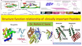
  • 1. Structure-function relationship of clinically important Peptides Dr. Rohini C Sane
  • 2. Protein structure-function relationship • The overall conformation of a protein , the particular position of amino acid side chains in three-dimensional space determines the function of the protein. • The diversity of protein structure and its correlation with function can be explained by the following clinically important proteins. Protein Function Carbonic anhydrase Formation and degradation of carbonic acid Myoglobin(Mb) Stores oxygen in muscle cells Hemoglobin (Hb) Transports oxygen from lungs to the tissue Collagen Providestensile strengthtomanytissueinthebody Elastin(withrubberlikeproperties) Occur in distensible structure
  • 3. Quaternary structure of enzyme regulates its functions Structural conformation of the active site the enzyme precisely oriented for the substrate binding Binding of enzyme to the substrate . enzyme catalysis →product + enzyme set free for substrate binding again
  • 4. Disordered region of enzyme and its Importance Schematic diagram Many Disordered region become ordered region when a specific ligand is bound . Disordered region gives flexibility and performs a vital biological role. Disordered regions of enzyme- catalytic site ordered regions of enzyme Substrate added to enzyme catalyzed reaction mixture Enzyme with Disordered regions set free at the end of reaction Product Enzyme-substrate complex –induced fit +
  • 5. QuaternarystructureofenzymeCarbonicanhydraseregulatesitsfunctions ❖Function of Carbonic anhydrase : catalyzes the reversible hydration of carbon dioxide. H2O + CO2  H2CO3(Carbonic acid) 1.H2O binds to Zinc ion (located in deep cleft of Carbonic anhydrase and is coordinated to Histidine residue present at its active site). Binding of CO2 to histidine residue is close to Zinc ion. 2.H2O→ H+ + OH- (Ionization of water to hydroxyl ion) 3.OH- +CO2(located proximally to OH- ions)→HCO- 3 Carbonic anhydrase facilitates the precise positioning of CO2 molecule and hydroxyl ion OH- for the formation of bicarbonate ion HCO- 3. 4.HCO- 3 + H+ → H2CO3(Carbonic acid) Thus , two substrates CO2 and H2O are brought in close proximity for reaction to proceed and to facilitate the function of Carbonic anhydrase .
  • 8. StepsofenzymecatalysisbyCarbonicanhydrase 1.H2O binds to Zinc ion (located in a deep cleft of Carbonic anhydrase and is coordinated to Histidine residue present at an active site of enzyme ) 2.H2O→ H+ + OH- (Ionization of water) 3. Binding of CO2 toHistidine residue close to Zinc ion of Carbonic anhydrase 4.OH- + CO2( located proximally to OH- ions)→HCO- 3 5.HCO- 3 + H+ →H2CO3(Carbonic acid)
  • 9. Structure of Myoglobin ❖StructureofMyoglobin: ❖ Oxygenbindingcytosolicmetalloprotein functionalwithinskeletal&cardiacmuscle. • hasasinglepolypeptidechainwith153aminoacidsanditcontainsonehemegroup(iron containingporphyrinringsooneFe2+ ionwhichimpartsredcolortomyoglobin– chromoprotein).Itisaglobularproteinwithlowmolecularweight(16700Dalton). • 80%ofitspolypeptidechain isfoldedinto8-helices,whicharelabeledas AtoH. -helicalregionsareterminatedbypresenceofProline(5-memberedringofProcannot beaccommodatedinalpha-helix→Proishelixdestabilizingaminoacid).Restofthe polypeptidechainformsturnsandloopsbetweenhelices. • Thehelicescreatehydrophobicoxygen(O2)bindingpocketcontainingtightlybound hemewithanironatom(Fe2+)initscenter.WithinthepocketofMyoglobin,O2 binds directlytotheFe2+ ionoftheporphyrinring. • hasnobetasheets.Thisstructureisunusual fortheglobularproteins.
  • 10. Myoglobincontentofmuscles Type of muscle Myoglobincontentofmusclesin gm/100gmof muscles Skeletal 2.5 Cardiac 1.4 Smooth 0.3
  • 11. Structure of Myoglobin Structure of Myoglobin(Mb) :hasasinglepolypeptidechainwith153aminoacidsand containsonehemegroup(ironcontainingporphyrinringsooneFe2+ ion-impartingred colortoMyoglobin).Itis a globularproteinwithmolecularweight(16700Dalton) andhas cytosoliclocation.ThisOxygenbindingmetalloproteinproteinfunctionalwithinskeletal& cardiacmuscles.Onemoleculeof(Mb)containsoneoxygenmolecule. JohnKendrewandMaxPerutz(Noble1962):determinedthestructureofMyoglobinby highresolutionX-crystallography.
  • 12. Tertiary Structure of Myoglobin(Mb) Myoglobin:PrimarystructuresimilartosinglemonomericunitofHemoglobin withasinglepolypeptidechainhaving 153aminoacids(molecularweight16700Dalton).It haseightalpha–helices(AtoH)andonehemegroup(iron containing porphyrin)tofacilitateitsfunctionofoxygenstorageincardiacandskeletalmusclesinhumanbody,Whales andSeals.
  • 13. Tertiary Structure of Hemoglobin (Hb) Hemoglobin:Tetramericwith4hemegroups.Eachpolypeptidechainhassimilarstructureto single polypeptidechainofMyoglobin.Ithasaloweraffinity foroxygenthanMyoglobin.FoursubunitsofHb functioncooperatively.TetramericstructureofhemoglobinfacilitatessaturationwithO2inthelungand releaseofoxygenasittravelsthroughthecapillarybed.
  • 14. Functions of Myoglobin ❖Functions of Myoglobin(Mb): acts as storage and transport protein for oxygen in skeletal&cardiacmuscles. In resting muscle cells ,myoglobin binds oxygen (O2)that has been released by Hemoglobin(Hb) under the conditions of oxygen deprivation (in severe exercise for use O2 by muscle). ❖HbO2 (oxyhemoglobin) → → → Mb(Myoglobin) →MbO2(oxymyoglobin)→ tissue → Respiration Oxygen (O2) transport
  • 15. Oxygen dissociation curve of Myoglobin and hemoglobin When the amount oxygen (O2)bound to the Hemoglobin and Myoglobin is plotted against the partial pressure of oxygen (pO2), a hyperbolic curve is obtained for Myoglobin whereas that for Hemoglobin is sigmoidal. When the (pO2) is high both Myoglobin and Hemoglobin are saturated with oxygen. At lower levels of (pO2), however , Myoglobin contains more oxygen O2 than Hemoglobin . Bohr’s effect ,co-operative effect and 2,3 BPG effect are absent for Myoglobin . Mb has higher affinity for Oxygen (O2) than Hb. At PO2 in tissue (30mmHg): Mb is 90% saturated, Hb is 50% saturated . Physical exercise: PO2 in tissue is (5mmHg), when Mb releases all bound Oxygen (O2).
  • 16. Clinical aspects associated with Myoglobin ❖Myoglobin : under physiological conditions , Myoglobin( being a small molecular weight protein)is filtered and excreted in urine. • is a sensitive maker for muscleinjury,makingitpotential cardiacmarkerfor myocardialinfarctioninpatientswithchestpain. • rises rapidly after the chest pain at about the same rate as CK-MB. • is non-specific ,since it is raised following any form of muscle damage. • Estimation ofMyoglobin–usingmonoclonalantibodyforbyRIA/ELIZA/ chemiluminescence • Cost of the analysis has prevented its widespread use. • TemporalpatternofserumMyoglobin&Creatinekinase-2inpatientswithmyocardial infarctionisdepictedinadiagram.
  • 17. SerumMyoglobin&Creatinekinase-2(CK-MB)inpatientswithmyocardialinfarction • The cellular release Myoglobin is often associated with an increase in Creatine kinase (CPK), Aldolase , Lactate dehydrogenase(LDH) , Serum Glutamate Pyruvate Transaminase(SGPT) in patients with myocardial infarction.
  • 18. Myoglobinuria ❖Myoglobinuria : under physiological conditions , Myoglobin being low molecular weight protein, it is filtered and excreted in urine. Presence of excessive Myoglobin in the urine usually associated with muscle destruction or rhabdomyolysis . Urine color becomes dark brown. ❖Damage to muscle( crush injury), releases Myoglobin to the circulation and filtered by the kidney. If too much of Myoglobin is released into circulation , Myoglobin precipitate and obstruct the renal filtration system results in acute renal failure. ❖Myoglobin is released from myocardium during Myocardial infarction (MI) and is elevated in serum(cardiac marker)followed by its excretion in urine.
  • 22. Quaternary structure of Hemoglobin Hemoglobin (HbA1)has 4 Polypeptides chains ( tetramer) associated by non-covalent bonds : 2 Alpha() chains + 2 Beta( )chains It possesses Quaternary structure(oligomeric). Inthis, Rgroupcontactsare presentbetweensimilarside chainsandthereisverylittle contactbetweendissimilar side chains. HbA:Tetramer22 Each chain/subunit has one heme group and so one Fe2+ ion similar to that of myoglobin. Hb is found exclusively in RBC
  • 23. Structure of normal Hemoglobin Hemoglobin variant Structure of normal Hemoglobin Abbreviation Hemoglobin (HbA 1) 2 Alpha() chains and 2 Beta( )chains 22 Hemoglobin (HbA 2) 2 Alpha() chains and 2 delta( )chains 22 Fetal Hemoglobin (HbF) 2 Alpha() chains and 2 gamma ( )chains 22
  • 24. Functions of Hemoglobin ❖Functions of Hemoglobin: • Hemoglobin is found exclusively in RBC .It transports four molecules oxygen(O2) from lungs(high pO2) to the tissues (low pO2) . Myoglobin is still very saturated with oxygen at the (pO2) of tissue. It can transport H + and carbon dioxide (CO2) from peripheral tissues to lungs for its excretion in air. oxygenation Deoxyhemoglobin + 4O2 oxyhemoglobin (in lungs) Deoxygenation is the reverse process by which O2 is liberated. deoxygenation Oxyhemoglobin 4O2+ Deoxyhemoglobin • Hemoglobin is rich in amino acid Histidine ,helps to maintain pH of blood (acts as buffer). • Transports oxygen(O2)by Hb is regulated by 2-3-bisphosphate glycerate(2,3BPG).
  • 25. Functions of Hemoglobin as a transporter of oxygen(O2) from lungs(high pO2) to the tissues (low pO2)
  • 26. Quaternary structure of Hemoglobin favors its functions ❖ Hemoglobin found exclusively in red cells and functions as transporter of oxygen(O2) from lungs to tissue . Each molecule carry four oxygen(O2)molecules from lungs to the cells .This function is favored by presence of one heme unit one each in monomer (4 heme→4 oxygen(O2)/ hemoglobin). ➢Binding of oxygen(O2) to one heme unit of tetramer facilitates oxygen binding by other subunits. Each subunit has a heme binding pocket similar to that of myoglobin . ➢It can transport H+ and CO2 from the tissue to the lung. ➢Binding of H+ and CO2 promotes release of oxygen(O2) from hemoglobin . ➢This allosteric interaction is physiologically important and as Bohr’s effect . ➢Even a single amino acid substitution alters the structure and thereby functions.
  • 27. Co-operative binding of oxygen to hemoglobin • Binding of oxygen to Heme will increase binding of oxygen to other heme • Affinity of oxygen for hemoglobin • Last oxygen binds with affinity 100 time greater than first oxygen • Heme –Heme interaction ( co-operative binding of oxygen to Heme ) • Release oxygen from one Heme will release oxygen from other • As there is communication between Heme groups of hemoglobin • Myoglobin is reservoir (transient )& supplier of oxygen Lung Tissue Oxygen concentration high Oxygen concentration low Oxygen binds to hemoglobin Oxygen is released to tissue
  • 28. Structural changes in hemoglobin on oxygen binding • Studied using x ray crystallographic • Homotropic effect→ binding of oxygen to hemoglobin • Heterotrophic effect → binding of 2,3BPG to hemoglobin • Distance between two Beta-chain decreases oxygenation from 4nm to 2nm • Increasing affinity for oxygen with addition every molecule of oxygen • On oxygenation iron moves in plane of Heme • Decrease in diameter of iron (movement of iron accompanied by pulling of proximal Histidine • affinity of hemoglobin for last oxygen > first oxygen ( 100 times greater ) • Cooperative binding of oxygen to hemoglobin or Heme →Heme interaction • Release of oxygen from one Heme → Release of oxygen from other Heme ❖Therefore communication between Heme groups of hemoglobin.
  • 29. On oxygenation iron moves in plane of Heme → decrease in diameter of Iron →movement of Fe Is accompanied by pulling of proximal site →primary event of Heme –Heme interaction of Hemoglobin Structural changes in Hemoglobin on its Oxygenation and deoxygenation
  • 30. Deoxy –Hb Hb O₂ Hb O₄ Hb O₆ Hb O₈ T form ↓ ↓ ↓ ↓ ↓ ↑ R form Oxygenation of hemoglobin Quaternary structure of Hemoglobin favors its functions :Binding of oxygen(O2) to one heme unit facilitates oxygen binding by other subunits.
  • 31. Structural changes in Hb on oxygen binding • Structural change in one subunit of hemoglobin on oxygenation is communicated to other subunits • Binding of oxygen to one Heme distorts globin chain to which it is attached → distortion in neighboring chain →oxygen binds more easily.
  • 32.
  • 33. Effect of 2,3 BPG on oxygen affinity of Hb • 2,3 BPG :Most abundant phosphate in RBC • Molar concentration of 2,3 BPG = Molar concentration of Hb • Synthesis ( synthesis through Rapport Leu Bering cycle) 2, 3 BPG mutase ( Glycolysis ) 1,3 BPG 2,3 BPG • Reinhold's & Ruth Benesch’s (1967 )→ 2,3 BPG decreases affinity of Oxygen to Hemoglobin • 2,3 BPG regulates the binding of oxygen • 1mole of 2,3 BPG binds to 1mole of Deoxy Hb not to oxy –Hb • Molecular concentration 2,3 BPG = Molecular concentration of hemoglobin • At partial pressure of oxygen(O₂) in tissue: HbO₂+ 2,3 BPG→Hb 2,3 BPG +O₂ (release of O₂) (Oxy–Hb) (De-oxy Hb) • In tissue → 2,3 BPG shift curve towards right
  • 34. 1mole of 2,3 BPG binds to 1mole of Deoxy Hb not to oxy –Hb Regulation of oxygen binding by 2,3 BPG
  • 35. Effect of 2,3 BPG on oxygen affinity of Hb 2,3 BPG decreases affinity of Oxygen to Hemoglobin. In tissue → 2,3 BPG shift curve towards right At partial pressure of O₂ in tissue: HbO₂+ 2,3 BPG→ Hb 2,3 BPG +O₂ (release of O₂) (Oxy –Hb) (De-oxy Hb)
  • 36. 2,3 BPG shift oxygen dissociation curve of hemoglobin towards right to release of oxygen (O₂) in hypoxic conditions ,anemia ,hyperthermia. Effect of 2,3 BPG shift oxygen dissociation curve of hemoglobin
  • 37. Clinical significance of 2,3 BPG:1 ❖Function of 2,3BPG : Release of oxygen to tissue ( supply of oxygen to tissue ) to cope with oxygen demand → varied concentration of 2,3 BPG 1. Hypoxia : concentration of 2,3BPG in RBC increases during chronic hypoxic conditions e.g. a. Adaptation to high altitude b. Obstruction to pulmonary odema ( air flow in bronchial blocked ) 2. Anemia : concentration of 2,3 BPG in RBC increases in chronic anemic conditions → to cope with oxygen (O₂) demand of body even at low Hb concentration.
  • 38. Clinical significance of 2,3 BPG:2 3. Blood Transfusion : storage of blood in acid citrate dextrose → decrease in concentration of 2,3, BPG ( O₂ remains bound to Hb ) • Blood stored in ACD fails to supply O₂ to tissue→ with 24-48 hr. until 2,3 BPG restored • O₂ supply /tissue O₂ demand met adequately after 24-48 hrs. • Blood with (ACD )+ Inosine ( Hypoxanthine Ribose )→ prevent decrease in 2,3, BPG • Inosine→ phosphorylation of tissue→ entry into HMP shunt → get converted to 2,3BPG →increase in Conc in 2,3 BPG → release of oxygen 4.Fetal hemoglobin(HbF) : the binding of 2,3BPG to fetal hemoglobin is very weak. Therefore, HbF has higher affinity for oxygen compared to adult hemoglobin (HbA). This is needed for transfer of oxygen from the maternal blood to fetus.
  • 39. Protein structure-function relationship in Adult Hemoglobin(HbA1) and Fetal Hemoglobin (HbF) • Function of Fetal Hemoglobin (HbF) : The transfer of oxygen from the mother to the fetus. • This function of Fetal Hemoglobin (HbF) is facilitated by structural differences in between the hemoglobin molecule of the mother and that of fetus (HbF). • Adult Hemoglobin(HbA1) : 2 Alpha() chains and 2 Beta()chains (22) • Fetal Hemoglobin (HbF) : consist of 2 Alpha() chains and 2gamma () chains- 22 • The difference in amino acid composition between the beta chain of HbA1 and gamma ( )chains of (HbF) results in structural changes that causes HbF to have lower affinity for 2,3- biphosphoglycerate (2,3 BPG) than HbA1 and thus greater affinity for oxygen. ➢Therefore , the oxygen released from the mother’s HbA1 is readily bound by HbF in the fetus.
  • 40. Protein structure-function relationship in Myoglobin and adult Hemoglobin(HbA1):1 Criteria Myoglobin Hemoglobin(HbA1) Primarystructure SimilartosinglemonomericunitofHemoglobin withasinglepolypeptidechainhaving 153 aminoacids(molecularweight16700)andone hemegroup(ironcontaining porphyrin). Tetramericwith4hemegroups.Each polypeptidechainhassimilarstructure to singlepolypeptidechainof Myoglobin. Affinityforoxygenof oxygenbinding protein Hasahigheraffinity foroxygenthan hemoglobin.ThebindingofO2 tohemegroup ofeachmoleculeisindependentofanother moleculebecauseitcontains onlyoneheme group(onepolypeptidechain). Hasaloweraffinity foroxygenthan myoglobin.Foursubunits of Hb functioncooperatively. Function of metalloprotein Astoragereserve foroxygen,releasesoxygen theboundoxygenforcellularusewhenoxygen supplyisreduced.Storeofoxygenindeep- divingmammals. Allowsefficienttransferofoxygenfrom lungtotissue.Tetramericstructureof hemoglobinfacilitatessaturationwith O2inthelungandreleaseofoxygenas ittravelsthroughthecapillarybed.
  • 41. Structure of Myoglobin and adult Hemoglobin(HbA1) PrimarystructureofMyoglobin:SimilartosinglemonomericunitofHemoglobin withasinglepolypeptide chainhaving 153aminoacids(molecularweight16700)andonehemegroup(ironcontaining porphyrin). PrimarystructureofadultHemoglobin(HbA1):Tetramericwith4hemegroups.Eachpolypeptidechainhas similarstructureto singlepolypeptidechainofMyoglobin.
  • 42. Functions ofMyoglobin and adult Hemoglobin(HbA1) Functionofmyoglobin:Astoragereserve foroxygen,releasesoxygentheboundoxygenforcellularuse whenoxygensupplyisreduced.Storeofoxygenindeep-divingmammals. Functionofadulthemoglobin(hba1):allowsefficienttransferofoxygenfromlungtotissue.Tetrameric structureofhemoglobinfacilitatessaturationwith O2inthelungandreleaseofoxygenasittravelsthrough
  • 43. Protein structure-function relationship of Myoglobin and adult Hemoglobin(HbA1):2 Condition Myoglobin Hemoglobin(HbA1) Highpartialpressureof oxygen(pO2) Saturated saturated Lowpartialpressureof oxygen(pO2 ) ContainsmoreO2thanhemoglobin Containslesser O2thanmyoglobin Agraphplottedwith the amountofO2 boundtothe proteinagainstthepartial pressureofoxygen(pO2 ) Hyperboliccurve Sigmoidcurve Application Stillverysaturatedwith oxygenatthe lowpartialpressureofoxygen(pO2) intissue facilitatingstoragefunction intissue. Effectivetransporterof oxygen,binding withoxygeninthe lungswhere(pO2)is highandreleasingitintissuewhere(pO2) islow .
  • 44. OxygenbindingaffinityofMyoglobinandHemoglobin Hyperboliccurve of oxygenbinding forMyoglobin Sigmoidcurveof oxygenbinding forHemoglobin At Highpartialpressureofoxygen(pO2)both myoglobin and hemoglobinaresaturatedwithoxygen. Application : Myoglobin stillverysaturatedwith oxygenatthelowpartialpressureofoxygen(pO2)in tissue facilitatingstoragefunctionintissue.Hemoglobinis aeffectivetransporterof oxygen,bindingwith oxygeninthe lungswhere(pO2)ishighandreleasingitintissuewhere(pO2)islow .
  • 45. Transport of oxygen by hemoglobin PO₂ (mm) of Hg % saturation Inspired air 158 Alveolar air 100 lung 90 97% Capillary bed 40 60% 37% - 40% O ₂ release of oxygen at tissue level
  • 46. Structural modification of hemoglobin by glycosylation and its application • Glycosylated hemoglobin (Hb A1C): is formed by non –enzymatic reaction of the aldehyde group of Glucose with amino terminal (N –terminal residue) Valine of beta chains of HbA when blood Glucose enters the erythrocytes. • Normal concentration of Glycosylated hemoglobin (Hb A1C): low ( to the extent of 5% of total hemoglobin • Formation of (Hb A1C) is proportional to blood glucose concentration. • Glycosylated hemoglobin (Hb A1C) increases in Diabetes Mellitus : 12% or more of total hemoglobin as serum glucose is high • Since RBC have life span of 120 days , the content of Hb A1C is an indicator of how effectively blood glucose levels have been regulated over the previous 2 or 3 months. • Application of Glycosylated hemoglobin (Hb A1C) estimation : to follow the effectiveness of treatment (management)for Diabetes Mellitus .
  • 47. Glycosylated hemoglobin (Hb A1C) Glycosylated hemoglobin (Hb A1C): is formed by non- enzymatic reaction of HbA with Glucose when blood Glucose enters the erythrocytes. Application of Glycosylated hemoglobin (Hb A1c) estimation : to follow the effectiveness of treatment for Diabetes Mellitus
  • 48. Primary structure of Normal protein determines biological functions Unique amino acid sequence specified by genes in a Normal protein Specific amino acid sequence→ confers specific 3 dimensional structure (conformation ) Specific Function arises from conformation Normal physiologically active protein e.g. Normal hemoglobin
  • 49. Consequences of Altered primary Structure in Abnormal protein Mutation→ altered genetic constitution ( base sequence of DNA) Altered amino acid sequence→ Altered 3 dimensional structure(conformation) Altered or loss of functions arises from Altered conformation of protein Abnormalorphysiologicallyinactiveprotein→diseaseconditione.g.hemoglobinopathy
  • 50. Structure function relationship of proteins Normal protein • Unique amino acid sequence specified by genes • Specific amino acid sequence→ confers specific 3 dimensional structure ( conformation ) • Specific Function arises from conformation • Normal physiologically active protein e.g. Normal hemoglobin Abnormal protein • Mutation→ altered genetic constitution / base sequence of DNA • Altered amino acid sequence →Altered 3 dimensional structure ( conformation ) • Altered or loss of Function arises from Altered conformation • e.g. abnormal or physiologically inactive protein→ disease condition e.g. hemoglobinopathy
  • 51. Altered primary Structure of Abnormal hemoglobin Abnormalhemoglobin→ hemoglobinopathy Altered primary Structure leading anemia in early life Sickle cell hemoglobin ( Hb S) no change in amino acid sequence of  chain 6 th amino acid in beta chain of HbA1 (Glutamic acid replaced by Valine in HbS ) HbM (Methemoglobinemia ) Substitution of Tyrosine with Histidine Hb Chesapeake Arginine is replaced by Leucine at 92th amino acid in alpha chain of Hb A Alpha(  ) thalassemia ( normally the rate of synthesis  and  chains identical ) Deficiency or absence of alpha chains → HbA of fetus has tetramer  4 or 4 chains ( enlargement of live and spleen) Beta ( )thalassemia ( more common inherited disease than Alpha thalassemia ) Deficiency or absence of beta chains →hemoglobin found in RBC are HbA2 (  2 2) and HbF (  2 2 )
  • 52. Deoxygenated HbS • This substitution generates ‘A stick patch’ on the surface of the beta chain of both oxygenated and deoxygenated HbS . • On the surface of deoxygenated normal HbA and deoxygenated HbS , there exists a complement to the sticky patch. • When HbS is deoxygenated ,its sticky patches can bind to the complementary patches on another deoxygenated HbS . • Binding of large number of deoxygenated HbS causes polymerization of deoxygenated HbS forming long fibrous precipitate that mechanically disturbs the red cell→ sickle shaped→ casing lysis and anemia. • This polymerization will not take place when HbS is in oxygenated form as in arterial blood .
  • 53. Molecular basis of Sickle cell anemia Linus Pauling ( 1954 Noble prize ) reported abnormal electrophoretic mobility & peptide mapping Glutamic acid ( sixth position on beta globin chain ) replaced by Valine (Recessive Mutation ) Hb A & Hb F prevent sickling.
  • 54. sixth amino acid in beta() chain of HbA1 (Glutamic acid )replaced by Valine in HbS Altered primary Structure leading anemia in early life
  • 55. Sickle cell disease Binding of large number of deoxygenated HbS causes polymerization of deoxygenated HbS forming long fibrous precipitate that mechanically disturbs the red cell→ sickle shaped→ casing lysis and anemia.
  • 56. HbM (Methemoglobinemia ) ❖HbM (Methemoglobinemia) : Substitution of Tyrosine with Histidine on hemoglobin result in oxidation of ferrous is to ferric in heme . Hemoglobin in blood is oxidized to methemoglobin. This condition leads to Methemoglobinemia .
  • 57. HbM (Methemoglobinemia) :Substitution of Tyrosine with Histidine  Substitution of Tyrosine with Histidine oxidation of ferrous is to ferric in heme→
  • 58. Hb Chesapeake ❖Hb Chesapeake ➢Substitution of Arginine by Leucine at 92th amino acid in alpha chain of Hb A results in increased affinity of hemoglobin for oxygen and does not release as much oxygen to the peripheral tissue as does normal hemoglobin HbA . ➢This leads to tissue hypoxia and polycythemia ( increased number of RBC per unit volume) in ode to meet the oxygen needs.
  • 59. Quaternary structure of Aspartate trans carbamylase Aspartate trans carbamylase : An allosteric enzyme with 2 subunits : catalytic subunit (C) and the regulatory subunit (R)
  • 60. Quaternary structure of Lactate dehydrogenase (LDH ) ❖Quaternary structure of Lactate dehydrogenase (LDH ): • Tetramer • two types of polypeptide chains :H and M type • five isoenzymes : 1. LDH 1 →H4 2. LDH2 → H3M 3. LDH3→ H2M2 4. LDH 4→ HM3 5. LDH 5 →M4
  • 61. Isoenzymes of Lactate dehydrogenase Subunit composition of LDH1 isoenzymes in heart cells favors conversion of Lactate to Pyruvate Subunit composition of LDH5 in muscle cells favors conversion of Pyruvate to Lactate Quaternary structure of Lactate dehydrogenase (LDH ): Tetramer with 2 types of polypeptide chains →H and M type
  • 62. Structure of Human Insulin In 1953 , Frederick Sanger determined primary structure of Insulin ( a pancreatic protein hormone ) and showed for the first time that a protein has a precisely defined amino acid sequence ( primary structure.)
  • 63. Insulin and Glucagon(Polypeptide Hormones) Insulin Hormone secreted by  pancreatic cells Polypeptide and a Dimer with 51 amino acids regulates glucose metabolism and Induces hypoglycemia Amino acid sequence varies in different mammalian species Glucagon Hormone secreted by  pancreatic cells Polypeptide and monomer with 29 amino acids regulates glucose metabolism and Induces hyperglycemia Amino acid sequence is same in all mammalian species
  • 64. Structure of Human Insulin ❖Structure of Human Insulin : described by Sanger(Noble-1955 ) ❖Dipeptide (2 polypeptide chains)of insulin has 51 amino acids A polypeptide chain : 21 amino acids B polypeptide chain : 30 amino acids ❖ Dipeptideofinsulinarerequiredforbiologicalactivityandheldby ➢inter chain disulphide bonds: a. between cysteine residues ( 7th amino acid of A chain and 7th amino acid of B chain ) b. between cysteine residues (20 th amino acid of A chain and 19 th amino acid of B chain) ➢intra chain disulphide bond : between cysteine residues (6 th amino acid of A chain with 11 th amino acid of B chain)
  • 65. Structure of Human Insulin Carboxy terminal end A chain : Asparagine B chain : Threonine Amino terminal end A chain : Glycine B chain : Phenylalanine A polypeptide chain : 21 amino acids B polypeptide chain : 30 amino acids intra chain disulphide bond : between cysteine residues( 6 th amino acid of A chain with 11 th amino acid of B chain) inter chain disulphide bonds   
  • 66. Primary Structure of human Insulin S S A chain H2N Gly Cys Cys Cys Cys Asn 1 6 7 11 20 21 S S S S B chain H2N Phe Cys Cys Thr 1 7 19 30 Pig insulin differs from human insulin in only one position ,30th amino acid is alanine instead of Threonine. Insulin from other animals like cattle ,sheep, horse etc. differ from human insulin in having a different sequence of amino acids in the positions 8-9-10 in A chain . This minor altered sequence does not result inappreciable change biological activity.
  • 67. Amino acid substitution in Primary Structure of human Insulin 8 9 10 Achainof insulin ofspecies Thr Ser Ile Human Ala Ser Val Bovine Thr Ser Ile Pig Ala Gly Val Sheep Thr Gly Ile Horse Aminoacidcompositionat30th amino acidinBchainof insulinofspecies Thr Human Ala Bovine Ala Pig Porcineandhumaninsulinare similar(homologous)exceptC-terminal aminoacidinBchain(Thr→Ala).Itmay produceantibodiesinhumanafter repeated injections.Dealaninatedporcineinsulinwill notproduceanyantibodiesindiabetic patientsevenafterlongtermuse. Aminoacidsequencehasbeenconservedto thegreatextentduringevolution.Human insulinrequiredforreplacementtherapy,is nowsynthesizedbyrecombinantDNA technology.
  • 68. Site of Insulin Biosynthesis ❖Amino acids form primary structure → definite function ❖Peptide < 10 amino acids ❖Polypeptide > 10 amino acids Preproinsulin (singlepolypeptidechainwith108aminoacids,molwt.11500) ↓ Proinsulin(single polypeptide chain with 86 amino acids, molwt.9000) ↓ Human Insulin ( dipeptide 51 amino acids : polypeptide chain A →21 amino acid and polypeptide chain B →30 amino acids held together b interchain disulfide bridges molwt.5734 ) ➢The gene for insulin synthesis : located on chromosome 11 in beta cells of pancreatic cells. Removal of signal sequence in endoplasmic reticulum Removal of C-peptide in Golgi apparatus
  • 69. Pre proinsulin ( single polypeptide chain ): 108 amino acids ,mol wt. 11500 Proinsulin ( single polypeptide chain):86 amino acids ,mol wt. 9000 Insulin ( dipeptide chain ):51 amino acids ,mol wt. 5734 Biosynthesis of Insulin from Preproinsulin:1 Removal of signal sequence in endoplasmic reticulum Removal of C- peptide in Golgi apparatus The gene for insulin synthesis : located on chromosome 11 in beta cells of pancreatic cells.
  • 70. Biosynthesis of Insulin from Preproinsulin:2 In the beta pancreatic cells , insulin and proinsulin combines with Zinc to form complexes. In this form it is stored in the granules of the cytosol which is released in response to various stimuli by exocytosis. Pre proinsulin ( single polypeptide chain ) :108 amino acids ,mol wt. 11500 Proinsulin ( single polypeptide chain):86 amino acids ,mol wt. 9000 Insulin (dipeptide chain):51 amino acids, mol wt. 5734
  • 71. Structure function relationship of C–peptide and insulin C – peptide has no biological activity . Single polypeptide chain with 86 amino acids  Beta cells of panaceas synthesize insulin as a prohormone – Proinsulin . Biologically active insulin (a dipeptide) is formed by removal of C – peptide ( the central potion ) of proinsulin . C–peptide and insulin are synthesized in equimolar concentration. It useful index for the endogenous production of insulin.
  • 73. Biochemical aspects of Collagen ❖Biochemical aspects of Collagen: 1. Most abundant fibrous protein(major macromolecules) in human body :70 kg body weight→ 12-14 kg of total protein→5kg of Collagen (1/3 of total protein) 2. Main Component of : connective tissue, skin(70%) ,bone (90%) tendon(85%), cartilage ,teeth and liver(4%) 3. Synthesized by fibroblast in connective tissue and osteoblasts in bone 4. Made up of small fibrils → tropocollagen( fundamental units ) containing 3 polypeptide chains each of them in left-handed helix with 3 amino acid per turn. 5. rich in Glycine and rare amino acids like hydroxyproline, hydroxylysine 6. Cysteine and Tryptophan absent 7. have a triple helical secondary structure and rich in helix destabilizing amino acids (Glycine ,Proline and Hydroxyproline). These amino acids prevent the formation of the usual - helical and - pleated structure. Instead it forms a triple helical secondary structure.
  • 74. Triple stranded helix Structure of Collagen Made up of small fibrils → Tropocollagen( fundamental units ) containing 3 polypeptide chains each of them in left-handed helix with 3 amino acid per turn.
  • 75. Triple stranded helix Structure of Collagen • Collagenhas3 polypeptidechainswoundarounditself. Eachpolypeptidechainsubunitis calledalpha-chains. EachofAlpha-chain is twistedintoleft-handedhelix ofthreeresidues perturncomparedwith 3.6forright-handedalpha-helix.Threeoftheseleft–handedhelices arethenwoundtoright-handedsuperhelixtoformastiffrodlike molecule(Triplehelical secondarystructure). • Itisrich in helixdestabilizingaminoacids(Glycine ,ProlineandHydroxyproline).Theseamino acids preventtheformationoftheusual- helicaland-pleatedstructure.Instead,it formsa triplehelicalsecondarystructure. • Every3rdresidueis Glycineandtheonlyaminoacidthatcanfitintothetriplestrandedhelix. • QuarterstaggeredtriplestrandedhelixofCollagenis stabilizedbythestericrepulsionof ringshydroxyproline andhydrogenbondsbetweenthem. • Triplehelicalsecondarystructureimpartsthetensilestrengthofsteeltocollagen(has unusualstrength) ❖Types arrangementofcollagenfibril: a. Parallelbundles:in tendons,cartilage b. Sheets:layered atmanyanglesin skin
  • 76. Arrangements of collagen fibers in cartilage of bone
  • 77. Types of Collagen ❖19 different Types of Collagen , composed of 30 distinct polypeptide chains encoded by separate genes. ❖ Numbering for Types of Collagen: Roman numerals I, II, III….XIX ❖Structure of collagen types : in principle , all types of collagen are triple helical structures . The triple helix may occur throughout the molecule or only a part of the molecule. ❖Each one suited to performed specialized function in tissue ❖e.g. Collagen Type I →skin, Collagen Type II → bone
  • 78. Most abundant types of collagen found in human tissue and their distribution Type of collagen Distribution Composition of triple helix I Skin , bone, tendon , cornea 2 -1(I), -2(I) II Articular cartilage, intervertebral disc, vitreous body 3-1(II) III Fetal skin ,cardiovascular system, reticular fibers 3-1(III) IV Basement membrane 2 -1(IV), -2(IV) V Placenta, Skin 2 -1(V), -2(V)
  • 79. Structure of collagen Type 1 ❖ Structure of collagen Type 1: 1. Triple stranded helical structure present throughout the collagen molecule 2. Shape : rod-like molecule → 1.4 nm diameter and 300 nm length 3. Number of Amino acid residues : 1000 per for each polypeptide chain (3000 /molecule) 4. Amino acid contribution : 1/3 rd of amino acids are Glycine (every third amino acid in collagen is Glycine. 5. Repetitive amino acid sequence : (Gly – X –Y )n ,where X and Y represent other amino acids 6. Proline and hydroxyproline : 100 per for each polypeptide chain 7. Function of Proline and hydroxyproline : confer rigidity to the collagen molecule 8. Collagen Fibril formation : Triple helical molecule of collagen assemble to form elongated fibrils . It occurs by a quarter staggered alignment i.e. each triple helix is displaced longitudinally from its neighbor collagen molecule by about one-quarter of its length 9. Collagen Fiber formation : Collagen Fibrils assemble to form rod like fibers . 10. Strength of Collagen Fiber : contributed by covalent cross linking of formed between Lysine and hydroxylysine and also between Proline and hydroxyproline.
  • 80. Collagen molecules in Collagen fibers Triple helical molecule of collagen assemble to form elongated fibrils . Triple stranded helical structure present throughout the collagen molecule Collagen Fibrils assemble to form rod like fibers . Repetitive amino acid sequence (Gly – X –Y )n Proline and hydroxyproline confer rigidity to the collagen molecule
  • 81. Arrangement of Tropocollagen molecules in collagen fibril Heads of Tropocollagen molecules 64 nm Cross striations Sections of Tropocollagen moleculeCollagen Fibril formation : Triple helical molecule of collagen assemble to form elongated fibrils . It occurs by a quarter staggered alignment i.e. each triple helix is displaced longitudinally from its neighbor collagen molecule by about one-quarter of its length.
  • 82. Tropocollagen molecule ❖Tropocollagen : Subunits of Collagen • Shape : rod shaped • Length : 300nm • Thickness : 1.4 nm • Molecular weight : 300,000 • Constituent polypeptides: three helically interwind polypeptides of equal length (each with 1000 amino acid residues) • Primary structure of collagen : all 3 or two out of three chains have identical in amino acid sequence. Rich in Glycine (35%)and Alanine(11%) , Gly-Pro-X or Gly-Hpr-X or • Repetitive amino acid sequence : (Gly – X –Y )n ,where X and Y represent other amino acids • Secondary structure of collagen : Each of three polypeptide chains of tropocollagen is itself -helix. Proline and hydroxyproline form bends in polypeptide chains that they are not compatible with -helix structure.
  • 83. Collagen fibrils ❖Collagen fibril : • Triple helical molecules are associated into Collagen fibrils. • It consists of recurring polypeptide subunits called tropocollagen, arranged head to tail in parallel bundles . The heads of the tropocollagen molecules are staggered along the length of fibers ,accounting for the characteristic 64 nm spacing of the cross striations in most collagens . • A section of tropocollagen molecule shows the backbone of triple helix . Each of three polypeptide chains of tropocollagen is itself -helix whose pitch and spacing is determined by the rigid R group of the numerous Proline and hydroxyproline residue . • The gap between the end of one triple helix and the beginning of the next where there is the deposition of hydroxyapatite crystals in bone formation.
  • 84. Constituent amino acids of triple stranded helix Structure of Collagen • ScvConstituent amino acids of Collagen % of total amino acids Glycine 33 Proline and hydroxy proline 21 Lysine and hydroxy Lysine 3 Alanine 11 Arginine 5 Cysteine and Tryptophan absent Scurvy:vitaminCdeficiency→failure ofhydroxylationofProlineandLysineleadstoreducedhydrogen bonding→weaknessofcollagen→Brittlebonedisease:mutation→replacementofcentralGlycine
  • 85. Triple helical secondary structure of Collagen
  • 86. Forces stabilizing Triple helical secondary structure of Collagen ❖Forces stabilizing Triple helical secondary structure of Collagen: 1. Hydrogen bonds : three left-handed helices are bound together by interchain hydrogen bonds. 2. Lysinonorleucine bond: covalent cross links both within and between triple helical units further stabilize Collagen fibers. 3. Electrostatic interactions 4. Hydrophobic interactions
  • 87. Covalent cross-links in Collagen fibers • Strength of Collagen Fiber : contributed by covalent cross linking formed between Lysine and hydroxylysine and also between Proline and hydroxyproline. • Covalent cross links are formed both within and between triple helical units further stabilize Collagen fibers. • The degree of covalent cross-linking in Collagen molecule increases with age . • In Elder individuals : skin, blood vessels (Collagen containing tissue) become less elastic and more stiff → health complications
  • 88. Skin :Collagen containing tissue In Elder individuals , skin, blood vessels (Collagen containing tissue) become less elastic and more stiff → health complications
  • 89. Collagen and calcific aortic valve stenosis(CAVS)
  • 90. Biosynthesis of collagen ❖Biosynthesis of collagen: collagen is an extracellular protein but synthesized as an intracellular precursor molecule before becoming a mature collagen fibril. • Site : fibroblast ,osteoblasts in bones , chondroblasts in cartilage, odontoblasts in teeth • Cellular location : ribosomes in endoplasmic reticulum (ER) • Precursor : preprocollagen (a single polypeptide chain) with leader peptide at amino terminal 20000 MW and carboxy terminal 35000MW.Both are not present in mature collagen. • Function of preprocollagen: contains a signal peptide which directs the protein to each endoplasmic reticulum (ER) • Synthesis of procollagen : from preprocollagen in (ER) after cleavage of a signal peptide • Post transcriptional modification of procollagen : hydroxylation, glycosylation and disulfide formation . Followed by its secretion in extracellular medium by the way of Golgi complex . • Synthesis of collagen in extracellular medium : from preprocollagen after action of aminopeptidase and carboxypeptidase to remove terminal amino acids. This followed by a spontaneous assembly of polypeptide chains to form triple helical structure (with 1000 amino acids each) of collagen .
  • 91. Types of cross links in collagen Lysine Lysil oxidase Allolysine Allysine Lysine H2O H2O Aldol condensation Schiff base Reduction Lysinonoleucine
  • 93. Structural modification of Collagen during its Synthesis Procollagen Tropocollagen Collagen Glycosylationloss of peptide potion from N-terminal and C-terminal Each of the 3 chains is in a left handed helix with 3 amino acids per turn. 3 Chains are further twisted in right handed way to give cable like structure. Hydroxylation of Proline and Lysine by Lysyl hydroxylase and Proline hydroxylase in presence of vitamin C→ Cross linking of hydroxy proline and hydroxy lysine Since vitamin C is required for collagen synthesis ,a connective tissue , there is a delay in wound healing process in vitamin C deficiency.
  • 94. Intracellular and extracellular alterations of Collagen during post-translational processing Intracellular alterations of Collagen Extracellular alterations of Collagen Hydroxylation of Proline and some Lysine residues Formation of intra and interchain crosslinks Glycosylation of some of the hydroxylysine residue Oxidative deamination of epsilon amino groups of Lysine and hydroxylysine residues Formation of intrachain and interchain disulphide bonds ,mainly in the carboxy and amino terminal ends Cleavage of 25-35 kD portions at both carboxy and amino terminal ends Formation of triple helix Formation of quarter staggered alignment
  • 95. Functions of Collagen ❖Functions of Collagen : triple helical molecules are associated into fibrils. There is gap between the end of one triple helix and the beginning of the next where there is deposition of hydroxyapatite crystals in bone formation. 1. Gives tensile strength, support and shape to tissue . To break a collagen fiber of 1 mm in diameter, a load of 10-40 kg is needed. In disease status tensile strength is reduced. 2. Contributes to proper alignment of cells ,which in turn help in cell proliferation and their differentiation to different tissue and organs . 3. Collagen which is exposed in blood vessels contributes to thrombus formation. ❖Collagen can be converted to a. gelatin by boiling by splitting off some amino acids .Gelatin is highly soluble and easily digestible. It forms gel on cooling and is provided as diet for convalescents and invalids. But it lacks essential amino acid Tryptophan. b. a tough hard substance on treatment with tannic acid (tannic process)
  • 96. Genetic aspects of Collagen Synthesis ❖Genetic aspects of Collagen Synthesis : 1. Complex process 2. Involves at least 30 genes in human 3. about 8 post –transcriptional modifications 4. Inherited diseases due to gene mutations linked with collagen synthesis: a. Ehlers-Danlos syndrome b. Alport syndrome c. Osteogenesis imperfecta d. Epidermolysis bullosa
  • 97. Abnormalities associated with collagen synthesis Disease Abnormalities associated with collagen synthesis Ehlers-Danlos syndrome Inherited disorders characterized by hyperextensibility of skin and abnormal tissue fragility , hypermobile and lax joints Alport syndrome Defect in formation of type IV collagen fibers found in the basement membrane of renal glomeruli→ hematuria and renal disease Osteogenesis imperfecta Characterized by abnormal bone fragility due to deceased synthesis of collagen Epidermolysis bullosa due to alteration in in the structure of type VII collagen fibers→ skin breaks and blister formation even with minor trauma Scurvy Deficiency of vitamin C→ defective post translational modification of collagen→ bleeding gums ,poor wound healing, subcutaneous hemorrhage Lathyrism (disease of bone deformities ) CausedbyconsumptionofLathyrussativa(kesaridal)containingtoxiccompound BetaOxalylAminoAlanine(BOAA).BOAAinhibitsenzymeLysyloxidaseand interfereswiththecrosslinkingoflysineaminoacidresiduesincollagen.
  • 98. Types of Ehlers-Danlos syndrome ❖Types of Ehlers Danlos syndrome : • Ehlers-Danlos syndrome type V: inherited deficiency of Lysyl oxidase(copper requiring enzymes)→prevents cross-linking of collagen→ arterio-vascular and skeletal changes. • Ehlers-Danlos syndrome type VI: inherited deficiency of Lysyl hydroxylase →abnormalities of the eye ,severe scoliosis (abnormal vertebral curvature) and hyperextensibility of skin and joints. • Ehlers-Danlos syndrome type VII: non-serving of procollagen as a substrate for the procollagen amino protease →hip dislocation , increased skin elasticity and short stature.
  • 99. Ehlers-Danlos syndrome : Clinical manifestations Hyperextensibility of skin and joints severescoliosis (abnormalvertebralcurvature) Ehlers-Danlos syndrome
  • 100. Alport syndrome Alport syndrome :Defectinformationof typeIVcollagenfibersfoundinthebasementmembraneof renalglomeruli→ hematuriaandrenaldisease
  • 101. Alport syndrome :Clinical manifestations Visual abnormality Deafness Glomerular Nephritis
  • 103. Osteogenesis imperfecta : Clinical manifestations Osteogenesisimperfecta:Characterizedby abnormalbonefragilityduetodeceasedsynthesisofcollagen
  • 104. Marfan's syndrome ❖An autosomal dominant trait. ❖Molecular basis : defect in the gene coding for fibrillin -1 located on chromosome 15 → deficient of deposition of fibrillin -1 and elastin which are components of microfibrils or defect in the gene coding for fibrillin -2 located on chromosome 5 → deficient of deposition of fibrillin -2→congenital contractual Arachnodactyly. ❖Clinical manifestations of Marfan's syndrome : a. Arachnodactyly (long digits) b. Ectopia lentis(dislocation of lenses) c. Hyperextension of joints d. Aortic aneurism
  • 105. Molecular basis of Marfan's syndrome Molecular basis : defect in the gene coding for fibrillin -1 located on chromosome 15 → deficient of deposition of fibrillin -1 and elastin which are components of microfibrils or defect in the gene coding for fibrillin -2 located on chromosome 5 → deficient of deposition of fibrillin -2→congenital contractual Arachnodactyly. An autosomal dominant trait
  • 106. Clinical manifestations of Marfan's syndrome
  • 108. Hyperhomocysteinemia Accumulation of Homocysteine Reaction of Homocysteine with Lysyl aldehyde formed by Lysyl oxidase Prevention of cross- linking of Lysine residues in connective tissue Skeletal deformities ,vascular and ocular defects
  • 109. Hyperhomocysteinemia ❖Normal Homocysteine levels (blood): 5-15 micromoles/L ❖Hyperhomocysteinemia : homocysteine levels (blood) increased 50-100 times→ increased risk of coronary artery diseases ,urinary excretion of homocysteine increases( >300 mg/24 hr.). ❖Causes of Hyperhomocysteinemia: a. Vitamin B6 and/or B12 deficiency b. Hypothyroidism c. Tobacco smokers d. Alcoholics →chronic pancreatitis e. Congenital diseases f. Pre –eclampsia of Pregnancy g. Elderly persons
  • 110. Congenital Hyperhomocysteinemia ❖Congenital Hyperhomocysteinemia : due Cystathionine beta-synthase deficiency ❖Clinical Signs and symptoms of Hyperhomocysteinemia : a. Mental retardation b. Charley Chaplin gait c. Skeletal deformities d. ocular defects: glaucoma myopia , Ectopia lentis(dislocation/ subluxation of lenses) e. vascular defects: intravascular thrombosis ❖Molecular basis /changes : increased Homocysteine→ activation of Hageman’s factor→ increased platelet adhesiveness → intravascular thrombosis→ life threatening ❖Biochemical changes : increased serum Homocysteine and Methionine levels, increased urinary excretion of Homocysteine( > 300mg/24 hr.), reduced plasma cysteine levels
  • 111. Clinical manifestations of Congenital Hyperhomocysteinemia
  • 112. Management of Hyperhomocysteinemia ❖Management of Hyperhomocysteinemia : a. Dietary supplementation of Vitamin B6( 500mg /per day) and/or B12 b. Diet Low in Methionine and rich in Cysteine supplemented ❖Cyanide – nitroprusside Biochemical test for diagnosis (in urine): positive ➢Other diseases associated with Hyperhomocysteinemia: neurological disorders
  • 113. Protein structure-function relationship of Menke’s kinky hair syndrome ❖Menke’s kinky hair syndrome : • An x-linked defect (affects only male child). • Molecular basis :absence of an intracellular copper binding ATPase protein(mutation in ATP7 A gene)→dietary copper absorbed from GI tract; but cannot be transported to the blood . • Copper that has entered into intestinal cells is not able to get out of the cell and so it gets accumulated there . Therefore , Copper(a constituent of Lysyl oxidase)is not available for metabolism ,resulting in defective cross linking in collagen molecule of connective tissue. • Defective Vascular( weakening of walls of major blood vessels including aorta →aneurysm→ fatal rupture of aorta→ cardiac failure) and connective tissues. • Child dies in infancy. ➢Copper binding ATPase protein present in intestinal cells are different from that present in liver and extrahepatic tissues . Therefore , Clinical manifestations of Wilson’s disease and Menke’s disease are different .
  • 114. Menke’s disease Absence of an intracellular copper binding ATP ase protein( mutation in ATP7 A gene) Accumulation of Copper in intestinal cells (Copper that has entered into is not able to get out of the cell and so it accumulated gets there) Unavailability of Copper for metabolism and function of Lysyl oxidase Defective cross-linking in collagen molecule of connective tissue Defective formation of Vascular and connective tissues Death of the Child in infancy
  • 115. Menke’s kinky hair syndrome
  • 116. Scurvy Scurvy: Deficiency of vitamin C→ defective post translational modification( hydroxylation )of collagen → fragility of blood vessels → bleeding gums ,poor wound healing, subcutaneous hemorrhage Scurvy
  • 119. Degradation of collagen by Collagenase Collagen peptides Amino acids Collagenase ❖ Clinical applications of degradation of Collagen by collagenase: • Reabsorption of bone and cartilage • Osteoporosis • Postpartum involution of uterus • Rickets • Paget’s disease • Osteoarthritis • Rheumatoid arthritis • Scurvy • Gas gangrene • Tumor metastasis ➢ Adult human tissue do not have significant amount of collagenase activity. ➢ Tissue collagenase is active in animals whose tissue undergo a degree of remolding e.g. tadpoles Peptidase ❖ Gas gangrene: • Collagenase produced by Clostridium histolyticum splits each polypeptide chain at the site indicated ( X-Gly-Pro-Y) • Connective tissue barriers destroyed by bacterium →invasiveness ✓ Collagen: a protein resistant to action of by ordinary proteolytic enzymes.
  • 120. Clinical applications of degradation of Collagen structure by Collagenase Gas gangrene : degradation of collagen structure by collagenase by bacterium Clostridium Histolyticum Osteoarthritis and Rheumatoid Arthritis
  • 121. Connective tissue proteins Connective tissue proteins Foundinlarge quantities Functionsof proteins Abnormities associatedwithprotein Elastin Lungs,elasticligaments, arterialbloodvessels Extensibilityand elasticityoftissue Williams'ssyndrome:impairmentofelastinsynthesis duetogenemutation→defectiveconnectivetissue andcentralnervoussystem,pulmonaryemphysema Fibrillin Myofibrilsfoundinvarious tissue extensibilityofmuscles Marfansyndrome:impairmentoffibrillin synthesisduetogenemutation→hyperextensibilityof jointsandskeletalsystem→longdigitsandtallness, Cardiovascularcomplications(e.g.AbrahamLincoln) Fibronectin Connectivetissue InvolvedinInteraction ofcellswithextracellular matrix,celladhesion ,cellmigration Tumorcellmetastasis: impairmentoffibronectin synthesisduetogenemutation→lackofcelladhesion amongtumorcells→cellmigration→metastasis Laminin Basallaminaofglomerular membraneofrenalcells, Extracellularprotein Involvedinneuronal growthandnerve degeneration Alzheimer'sdisease→excessivefibronectinsynthesis duetogenemutation→highconcentrationofLaminin
  • 122. Elastin ❖Elastin: 1. A Connective tissue protein imparting high tensile strength 2. Occurrence : the major component in yellow elastic fibers of connective tissue →lungs, elastic ligament , arterial blood vessels( especially large vessels like aorta ,tendons 3. Formed in large amount in uterus during pregnancy 4. Are hydrolyzed by pancreatic elastase enzyme
  • 123. Amino acid composition of Elastin ❖Tropoelastin(the basis subunit of elastin fibrils): • contains about 800 amino acid residues • Rich in non-polar amino acids such as Alanine, Leucine, Valine , Isoleucine and Proline . • Contain high amounts of Glycine, Proline(like collagen) • One-third the residues are Glycine but No repeat sequence of (Gly-X-Y)n (unlike collagen) • Less hydroxyproline • Do not contain Cysteine, Methionine , Histidine , 5-hydroxylysine, glycosylated hydroxylysine. • No triple helix
  • 124. Biosynthesis of Elastin Biosynthesis : is synthesized as Tropoelastin Tropoelastin Post-translational modifications formation of hydroxyproline Elastin ➢Collagen has aldol cross links, while elastin has Desmosine cross links. ➢When elastin matures , Desmosine cross links are formed from lysine residues. ➢Once elastin matures , elastin is very stable ,turn overrate is very low due to different crosslinks .
  • 125. Desmosine: Cross links of Elastin ❖Cross links of elastin: • More complex than those in collagen. • the major cross links in elastin are Desmosine. • are formed from 4 Lysine residues. Some Lysine residue of Tropoelastin get oxidized by lysine oxidase( copper containing enzyme) to aldehyde derivative of lysine called Allysine. 3Allysine + unmodified Lysine →lysinonoleucine cross links of Desmosine (by condensation) • permit the elastin to stretch in two dimensions and subsequently recoil during the performance of its physiologic functions. • are destroyed by elastase . ✓Deficiency of alpha-trypsin (an inhibitor elastase)can result in Emphysema.
  • 126. Cross links of elastin
  • 127. Emphysema: a clinical condition related to loss of Elastin function Deficiency of alpha-trypsin (an inhibitor elastase)can result in emphysema.
  • 128. Comparison of primary structure of Collagen and Elastin Collagen Elastin Many different genetic type One genetic type It has no capacity to stretch It has capacity to stretch and subsequently to recoil Primarystructurehasrepeating(Gly-X-Y)sequences Primarystructurehasnorepeating(Gly-X-Y)sequences Formation of triple helix secondary structure No triple helix secondary structure Presence of Hydroxylysine absence of Hydroxylysine Presence of Glycosylated hydroxylysine absence of Glycosylated hydroxylysine Formation of intramolecular aldol cross links Formation of intramolecular Desmosine cross links
  • 129. Abnormalities associated with elastin biosynthesis Disease Molecularbasis ClinicalManifestation William-Beurensyndrome Deletionofgeneforelastinonchromosome7 Severedevelopmentalabnormalitiesin connectivetissuealloverthebody. Defectiveconnectivetissueandcentral nervoussystem,pulmonary emphysema Pseudoxanthomaelasticum Inheriteddefectinformationofelastin Inheriteddisorderscharacterizedby hyperextensibilityofskinandabnormal tissuefragility,hypermobileandlax joints(similartoEhlers-Danlos syndrome) Copperdeficiency Blockstheformationofaldehydeswhichare essentialforcross-linking.Somelysineresidues areoxidizedbycoppercontainingLysyloxidase andresultingaldehydederivativewhichcondense withunmodifiedlysinetoformLysinonorleucine. Reducedcrosslinkageofcollagen
  • 130. Human fibrinogen ❖Human fibrinogen ( factor I): 1. Soluble glycoprotein 2. 2-3 % plasma protein(plasma fibrinogen concentration: 0.3g/dl) 3. Consist of 6 polypeptide chains → two A  ,two B , two  4. Structural formula= (A )2 (B )2  2
  • 131. Structure –function relationship of fibrinogen Fibrinogen Fibrin monomer formation Fibrin monomers stick together to form hard clot formation Stabilization of clot formation by cross linking between Glutamine and Lysine Red cells get entangled in fibrin clot → red color of clot Proteolytic cleavage catalyzed Thrombin (IIa)→ Release of fibrinopeptides A and B Prothrombin→ Thrombin(IIa)
  • 132. Fibrinogen Schematic diagram of fibrin clot formation from fibrinogen Fibrin monomer Proteolytic cleavagecatalyzed Thrombin(IIa) → Releaseof fibrinopeptidesAandB Fibrin clot Prothrombin→ Thrombin(IIa) Stabilizationofclotformation bycrosslinkingbetween GlutamineandLysine Redcellsgetentangledinfibrinclot→redcolorofclot
  • 133. Biochemistry of Albumin ❖Biochemistry of Albumin: • Plasma concentration : 3.5 - 5.5 gm/dl(60% of plasma proteins ) • Molecular weight : 69000 • Structure : a single polypeptide with 585 amino acids with 17 disulfide bridges • Site of synthesis : liver ( 12gm/day→25% of total hepatic protein) • Half life : 20 days • Application of Measurement of Plasma concentration→ Liver function test
  • 134. Functions of Albumin (Globular proteins) ❖ Functions of Albumin : 1.Nutritive(serum albumin ,ovalbumin , Lactalbumin): serves as source of amino acids for protein synthesis particularly in nutritional deprivation of amino acids. 2. Transport :binds and transports plasma free fatty acids ,bilirubin, steroid hormones ,Calcium and Copper in circulation 3. Buffering function : among the plasma proteins, albumin has maximum buffering capacity(lower than bicarbonate buffer system). 4.Osmotic function : due to high concentration and low molecular weight .It plays predominant role in maintaining blood/plasma volume and body fluid distribution. It contributes to 75-80% of the total plasma osmotic pressure(25 mm Hg) . Hypoalbuminemia:Lowplasmaalbumin<2g/dl(e.g.kwashiorkor, nephroticsyndrome,cirrhosis )→edema TherapeuticuseAlbumin:treatmentof burns,hemorrhageandkwashiorkor
  • 136. Four orders of protein structure
  • 137. Protein misfolding • The process of Protein folding is complex. ❖Causes of Protein misfolding : 1. Spontaneous 2. Gene mutations ❖Consequences of Protein misfolding : misfolded protein usually get degraded . However ,as the individual age progresses , misfolded protein get accumulated and cause number of diseases. ❖Group of diseases due to Protein misfolding : a. Prion diseases b. Amyloidosis
  • 138. Prion diseases due to Protein misfolding ❖Prion: represents proteinous infectious agents. ❖Prions protein(PrP): 1. the altered forms of normal proteins. 2. No difference in the primary structure (amino acid sequence) and post- translational modifications observed. 3. Certain changes in three –dimensional structure. 4. Major alterations is the replacement of alpha-helices by beta-sheets in PrP which confers resistance to proteolytic digestion of Prions protein. 5. highly infectious agents and can act as template to convert non-infectious proteins with alpha-helices to infectious form . 6. The process continues in exponential manner to accumulate a large number of prion proteins in tissue.
  • 139. Prion: represents proteinous infectious agents Apolipoprotein E 2 (APO-E2) : responsible for production of chaperons of Tau protein→ the risk for Alzheimer's disease.
  • 140. A model for the formation of infectious prions Alpha-helix of a protein(non-infectious) Infectious prion (with beta –sheets} Two molecules of Infectious prions (with beta-sheets) These two molecules of Infectious prions separate and convent another two non- infectious proteins to Infectious prions with beta –sheets. Exponential increase in Infectious prions interaction
  • 141. A model for the formation of infectious prions
  • 142. OXPHOS(Oxidative phosphorylation)Diseases • Defects in mitochondrial genome will lead to myopathies. Leber’s Hereditary Neuropathy (LHON)is caused by a single base mutation which alters one Arginine to Histidine in NADH Coenzyme Q reductase. OXPHOS(Oxidative phosphorylation)Disease Clinical features Leber’s Hereditary Neuropathy (LHON) Complex I defect, blindness ,cardiac conduction defects Leigh’s syndrome Complex I defect , NDUFS gene defect, movement disorders Myoclonic epilepsy ragged red fiber disease (MERRF) Myoclonic epilepsy, myopathy , dementia Mitochondrial encephalopathy lactic acidosis stroke like episodes ( MELAS) Complex I defect , lactic acidosis, stroke , myopathy, seizures, dementia
  • 143. Amyloidosis ❖Amyloidosis: refers to the altered proteins with beta sheets that accumulate in the body particularly in the nervous system. ❖Amyloids : 1. Extracellular Proteins found in tissue and body fluids resembling starch. 2. pathological deposit formed by protein misfolding or due to gene mutations associated with a group of disorders collectively called amyloidosis. 3. pathological deposit exert pressure on the vital organs and eventually cause their death. 4. at least 15 different proteins found in a amyloidosis 5. are not infectious agents as prion proteins. 6. accumulate as the age advances (aging). 7. implicated in degenerative diseases ( e.g. Alzheimer'sdisease) and multiple myeloma. 8. Secondary amyloidosis associated with inflammatory or infectious diseases. 9. Familial amyloidosis: inherited genetic mutations 10. Diagnosis / Detection of amyloidosis: Amyloids + Congo red + polarized light→ Apple green fluorescence
  • 145. Protein misfolding in Amyloidosis
  • 146. Diseases associated with Amyloidosis due to Protein misfolding Diseases Abnormal misfolded Protein Alzheimer’s disease Beta amyloid Cystic fibrosis CFTR Parkinson’s disease Alpha synuclein Huntington’s disease Huntingtin Creutzfeldt Jakob disease Prion
  • 147. ❖Chaperones : • Three dimensional conformation of proteins important for biological functions. • some proteins can generate the functionally active conformation spontaneously e.g. ribonuclease. • Majority of attain correct conformation ,through assistance of certain proteins called Chaperones. Role of Chaperones in protein folding
  • 148. Functions of Chaperones ❖Functions of Chaperones : 1. are heat shock proteins. 2. facilitate and favor interactions on the polypeptide surfaces to finally give the specific conformation of a protein. 3. can reversibly bind to hydrophobic regions of unfolded proteins and folding intermediates. 4. can stabilize intermediates and prevent the formation of incorrect intermediates. 5. prevent undesirable interactions with other proteins. 6. these activities of chaperones help the protein to attain compact and biologically active conformation.
  • 149. Functions of Chaperones Chaperones facilitate and favor interactions on the polypeptide surfaces to finally give the specific conformation of a protein. Chaperones can reversibly bind to hydrophobic regions of unfolded proteins and folding intermediates.
  • 150. Types of chaperones ❖Types of chaperones : 1. heat shock protein (Hsp)system : consist of 70 kDa heat shock protein (Hsp 70) and 40 kDa heat shock protein (Hsp 40) . These proteins can bind individually to the substrate protein and help in formation of protein folding. 2. Chaperonin system : a large oligomeric assembly which forms a structure into which the folded proteins are inserted. It mainly consist of Hsp 60 and Hsp 10 i.e. 60 kDa and 10 kDa Hsp. They are required at later part of protein folding process and work in association with Hsp 70 system.
  • 151. Chaperonin system : a Type of Chaperone It is a large oligomeric assembly which forms a structure into which the folded proteins are inserted It is required at later part of protein folding process and work in association with Hsp 70 system.
  • 152. Heat shock protein (Hsp)system: a Type of Chaperone Heat shock protein (Hsp)system : consist of 70 kDa heat shock protein (Hsp 70) and 40 kDa heat shock protein (Hsp 40) . These proteins can bind individually to the substrate protein and help in formation of protein folding.
  • 153. Protein misfolding and diseases ❖Protein misfolding and diseases : The failure of a protein to fold properly and generally leads to its rapid degradation. Prions (proteinous degraded infectious agents) are aggregates of misfolded proteins or their partially degraded products . Prions exhibit the characteristics of viral and microbial pathogens. ❖Protein misfolding and Cystic fibrosis (CF): 1. A common autosomal recessive disease 2. with mutations that result in abnormal protein cystic fibrosis transmembrane conductance regulator (CFTR) 3. Mutated CFTR cannot fold properly ,not being able to get glycosylated and transported . Therefore ,CTFR gets degraded. ❖Protein misfolding and neurological diseases : Prion are implicated in Alzheimer’s disease , mad cow disease , Huntington’s disease, Creutzfeldt – Jacob disease
  • 154. Cystic fibrosis (CF) and Alzheimer'sdiseasearedueProtein misfolding The failure of a protein to fold properly and generally leads to its rapid degradation.
  • 155. Alzheimer’s disease and chaperones PrionsareimplicatedinAlzheimer’sdisease.Alzheimer'sdiseaseis duecellular accumulationofaggregatesofmisfoldedproteins(withbetasheets)ortheir partially degradedproductsinthebodyparticularlyinthenervoussystem.
  • 156. Alzheimer'sdisease ❖Alzheimer'sdisease was reportedbyAloysiusAlzheimerin1906. ❖Alzheimer'sdisease: 1. aneurodegenerativedisease→seriouspsychologicalproblem. 2. affects5-10%ofthepeopleabove60yearsofage. 3. asthediseaseprogresses,thepatientmayentervegetativestate(affectentirefamily). 4. maydieafter10yearsafterthefirstonsetofthediseasesymptoms/manifestation. ❖Molecularbasis:ApolipoproteinEpromotestheconformationchangeofalphaamyloid tobetaamyloid.Followedbyselfaggregationofbetaamyloidsinthenervoussystem.
  • 157. ClinicalmanifestationsofAlzheimer'sdisease ❖ClinicalmanifestationsofAlzheimer'sdisease: a. Memoryloss/dementia b. Confusion c. Hallucinations d. Personalitychangeswithabnormalbehavior→patients entervegetativestatewithno comprehensiveoftheoutsideworld. e. Requireroundtheclockcareandprotection f. Shakespeare’sKingLear(whoislosinghismemoryandbecomingdisoriented)isawell –knownexample.
  • 158. Genes associated with Alzheimer's disease Genes associated with Alzheimer's disease Located onChromosome number Amyloidprecursorprotein(APP) 21 Presenilin-1 14 Presenilin-2 1 AD-3 14 AD-4 1 APO E4(Apolipoprotein E4) 19 Gene S 182 14
  • 159. PathologicalhallmarksofAlzheimer'sdisease ❖PathologicalhallmarksofAlzheimer'sdiseaseinclude: • Cerebralamyloiddeposition-Amyloid precursor protein (APP) • NeurofibriltanglesinCNS(Tauproteins) • Senileneuroticplaques ➢Inflammationwithinthebrainplaysa roleindevelopmentAlzheimer'sdiseaseandlong termuseofanti-inflammatorydrugwasfoundtoreducetheincidenceofdisease. ➢Aluminumtoxicity:Alpha-helicesofAPPundergoconformationchangetobeta-pleated sheetsinpresenceofAluminum.ThisAPPproteinwithabnormalconformationbeing insolublegetsdepositedinCNSleadingtoDementia.
  • 160. Alpha()secretase( proteolytic enzyme) ❖Alpha ()secretase: 1. Expressed and present on cell surfaces(trans-membrane region). 2. A proteolytic enzyme. 3. Are members of ADAM(a Disintegrins and metalloprotease domains). ➢Secretase complex is a prime target for pharmacological interventions of AD. Amyloid precursor protein(APP- a transmembrane protein ) Alpha ()secretase Beta ()secretase Gamma( )secretase Soluble Alpha protein (sAPP)into the extracellular environment insolubleproteins withAlpha()andbeta ()regions–toxic conformation→getdeposited
  • 161. Structure-functionrelationshipofAmyloid precursor protein(APP) in Alzheimer's disease Criteria Normal Amyloid precursor protein( APP) Amyloid precursor protein (APP) in Alzheimer's disease Genetic aspect Itiscodedbyagenelocatedonalongarmof chromosome21.Normalconstituentofserum andalsofoundintransmembraneregion. Mutation of a gene coding for APP→ substitution of Valine by Isoleucine Solubility Soluble Insoluble(cannot be degraded by Cathepsins and are deposited in neurons)→neurotic plaque Substrate for Alpha ()secretase( proteolytic enzyme) Beta ()secretase( proteolytic enzyme) Product of secretase catalysis Soluble protein(sAPP) Insoluble beta- protein with 40-42 amino acids gets deposited → neurotoxic effect →dementia(Alzheimer's disease)
  • 162. Selfaggregationofbeta-amyloidsinthenervoussysteminAlzheimer'sdisease Action of beta and gamma secretase on the Amyloid precursor protein(APP) can result in toxic conformation , called alpha-beta peptide (40-42amino acids )with neurotoxic activity→ dementia of Alzheimer'sdisease.
  • 163. Structure-functionrelationshipofBeta-Amyloid precursor protein(APP) in Down syndrome Trisomyofchromosome21/mutationofagenecodingfor Amyloidprecursor protein(APP) Substitution of Valine by Isoleucine Increased rate of biosynthesis of Beta-APP Deposition of insoluble Amyloid plaques Dementia
  • 164. sAPP and Alzheimer's disease ❖ sAPP : soluble form of Amyloid precursor protein results from its proteolytic cleavage by -Secretase. It is released from neurons in response to Electrical activity. ❖Functions of sAPP in Modulation of : 1. Synaptic plasticity 2. Synaptogenesis 3. Neuronal excitability 4. Neurite outgrowth 5. Neuronal Cell survival sAPP C-Gmp (guanosine 3’,5’ –cyclic monophosphate ) Modulation of activities of K-channels Signaling pathway Beta APP sAPP : a soluble neuroprotective protein APP with C-terminal end of truncated (s-APP ) +amyloid beta- peptide(A –beta) with neurotoxic activity -Secretase - secretase Alzheimer's disease: is associated with mutation in secretase leading to decreased levels of sAPP protein and or elevated levels of A-beta( APP )protein→ Dementia - secretase amyloid beta peptide with neurotoxic activity
  • 165. Regulation of Beta-Amyloid precursor protein(APP) synthesis by 25- hydroxy cholesterol ➢27- hydroxy cholesterol(27OHC)→ regulate a number of key enzymes within the brain→ regulate synthesis of Beta- Amyloid precursor protein(APP)→ prevent dementia/ Alzheimer's disease . ➢27- hydroxy cholesterol→ suppresses expression of gene responsible for synthesis of Arc( a cytoskeletal-associated protein responsible for memory consolidation). ➢Alzheimer's disease is associated with low levels of Arc.
  • 166. Toxic effects of Beta-Amyloid precursor protein(APP) on neurofibrils ❖Toxic effects of Beta -Amyloid precursor protein(APP) on neurofibrils: 1. Beta -Amyloid precursor protein(APP) cause oxidative injury and changes 2. Changes in intracellular calcium homeostasis 3. Cytoskeletal recognition ➢The gene coding beta-APP is located on chromosome 21 .Therefore , in Trisomy 21(Down syndrome) the rate of production of Beta-Amyloid precursor protein(APP) is increased leading to early onset of Alzheimer’s in patients with Down syndrome.
  • 167. Tauproteins ❖Tauproteins: • Microtubuleassociatedproteinsthatareabundantinneuronsinthecentralnervous system.Itenhancespolymerizationoftubulin. • PhosphorylationofTauisregulatedbyahostofkinasese.g.PKN(Serine/Threonine kinases) ActivationofPKN(Serine/Threoninekinases) PhosphorylationofTau Disruptionofmicrotubuleorganization Pathogenesis of Alzheimer's disease and other Tauopathies Self –assembly of tangles of paired helical filaments and straight filaments HyperphosphorylationofTau(Tauinclusions) Enhanced activity of protein kinases and diminished activity of phosphatases
  • 168. Structure-functionrelationshipofTauproteinsinAlzheimer's disease Criteria Normal Tau proteins Tau proteins in Alzheimer's disease Solubility Soluble and catabolized easily Insoluble(cannot be degraded by Cathepsins and are deposited around neurons) Function Required for Stabilizing axonal microtubules and facilitating the communication channels in nerve fibers. Loss stability of microtubules ,hyper polymerization of tubulins in neurons →deceased synthesis of Acetyl CoA→ dementia . Diabetes mellitus ,hypertension are risk factors for Alzheimer's disease.
  • 169. BiochemicalchangesduetoTauproteinsinAlzheimer's disease Tauopathies:Biochemical changes duetoTau proteins in Alzheimer's disease : 1. Threonine /Serine kinases catalyze phosphorylation of Tau proteins → self-assembly of paired helical filaments of Tau proteins → tangles of Tau→ disruption of microtubules in neurons of CNS → Dementia 2.Changes in intracellular Calcium homeostasis .
  • 170. Dementia :due to Mutation of a gene coding for Transmembrane protein Presenilin-1 and Presenilin-2 Mutation of a gene coding for Transmembrane proteins Presenilin-1 and Presenilin-2 Excessive production of Beta- Amyloid precursor protein(APP) Deposition of insoluble Amyloid plaques in neurofibrils Dementia
  • 171. Apolipoprotein E4 associated with Familial Alzheimer's disease ❖Apolipoprotein E : Arginine rich protein. It is present in chylomicrons, LDL, and VLDL . It is a ligand for hepatic uptake. Normal Blood levels Apo E 2 mg/dL. Gene for Apolipoprotein E is polymeric and located on chromosome 19 . It has has 3 alleles and therefore 6 possible combinations . 1. ABC 2. ACB 3. BAC 4. BCA 5. CAB 6. CBA ❖Liver and also Astrocytes make Apo-E. It is involved in cellular transport of lipids in CNS. Apo-E is associated with lipoprotein glomerulopathy. Apo-E has four isoforms viz. ApoE- I, II, III, IV. ❖Two of these isoforms i.e. Apolipoprotein E- II and Apolipoprotein E- IV, increase the risk for Alzheimer's disease by eight folds. ❖Apo E- IV is implicated in the development of senile dementia and Familial Alzheimer's disease (AD). ❖Familial Alzheimer's disease: 30% cases of (AD) with genetic background are associated with Apolipoprotein E- IV . ➢Apolipoprotein E- II (Apo E -II) : responsible for production of chaperons of Tau protein→ the risk for Alzheimer's disease.
  • 173. Zinc deficiency and Alzheimer's disease ❖Zinc binds to beta amyloid to form plaque →dementia → Alzheimer's disease
  • 174. Glutamate transporters in Alzheimer's disease • L –Glutamate : an excitatory neurotransmitter in mammalian CNS. • EAAT : high affinity glutamate transporters (5 types viz EAAT 1-5) • Neural EAATs : play specialized roles at neural synapses. • The transporters EAAT1 and EAAT2 present in glial neural cells and are responsible for majority of Glutamate uptake. • Dysfunction of EAAT is implicated in pathology of neurogenerative conditions including Alzheimer's disease( other conditions : Huntington’s disease ,epilepsy , ischemic stroke, amyotrophic lateral sclerosis). • Trafficking ,splicing, and post-transcriptional modifications of EAAT are exploited in treatments of these conditions.
  • 175. Clinical aspects of N-acetyl –D-aspartate receptors and Transcription factor NFKB ❖Genetic mutations or age-related changes in N-acetyl –D-aspartate receptors and Transcription factor NFKB may promote neuronal degeneration indicated in Alzheimer's disease by increasing production of A-beta and or decreasing levels of neuroprotective s-APP-alpha .
  • 176. Role of Glutathione in prevention of Alzheimer's disease Alzheimer's disease: is due cellular accumulation of aggregates of misfolded proteins or their partially degraded products . The term Prion ( proteinous infectious agents ) is coined to collectively represent them. Reduced Glutathione prevents amyloid formation by activating enzyme Glyoxalase and thus restores memory.
  • 177. Diagnostic tests for Alzheimer's disease ❖Diagnostic tests for Alzheimer's disease 1. Elevated levels of Tau proteins in CSF 2. Elevated levels of soluble -Amyloid precursor protein Beta in CSF (sAPP) 3. Decreased levels of (s-APP) 4. Serum DHEA ➢APP-beta mutations are linked to some inherited forms of Alzheimer's disease. ➢ s-APP is approximately 100-fold more potent than (s-APP) in protecting hippocampal neurons against excitotoxicity.
  • 178. Management of Alzheimer's disease 1. N –methyl aspartate an antagonist → slows the progression of disease 2. Omega-3-fatty acids → prevention disease 3. Long term anti-inflammatory drugs→ reduce incidence of disease 4. Beta() and Gamma ()secretase inhibitors ➢ Limitation of Gamma ()secretase inhibitor therapy : Gamma ()secretase is required by variety of physiological substrates therefore ,there is a need to develop substrate specific compounds.
  • 179. Alpha-synuclein and Parkinson’s disease ❖Parkinson’s disease: • A degenerative disease –affecting muscular coordination. • Two genes are associated with this disease 1. Gene for Alpha-synuclein is mutated in Parkinson’s disease. Alpha- synuclein is found in Lewy bodies(=inclusion bodies) found in many regions of the brain in this disease . Lewy bodies are responsible for neuronal degeneration of Parkinson’s and Alzheimer’s disease. 2. Gene Codes for protein- Parkin which is associated with juvenile form of Parkinson’s disease.
  • 180. Cystic Fibrosis ❖Cystic Fibrosis (CF) is a multisystem disease that presents • in neonates ,with failure to pass the first feces containing bile, intestinal debris and mucus ( meconium ileus ) • in early childhood with respiratory infections • in the adults ❖Inheritance of Cystic Fibrosis (CF) : autosomal recessive disorder
  • 181. Pathogenesis of Cystic Fibrosis ❖Cystic Fibrosis arises due to mutations in gene located on chromosome 7 encoding the Cystic Fibrosis transmembrane regulator (CFTR ) protein that regulates transmembrane chloride transport . ❖ The most common mutation is F 508 mutation which refers to deletion of three base pairs , resulting in the absence of Phenylalanine at position 508 in amino acid sequence of CFTR .
  • 182. Structure-functionrelationshipofCystic Fibrosis transmembrane regulator (CFTR ) ❑Absence of Cystic Fibrosis transmembrane regulator (CFTR ) /chloride channel leads to following Consequences : • Exocrine pancreatic insufficiency with impaired secretion of sodium, bicarbonates and water resulting in increased viscosity (mucoviscoidosis) ,obstruction of pancreatic duct , pancreatic fibrosis and obstruction of pancreatic tissue. • Chronic airways infection that affects mucus secretion in the bronchi with recurrent respiratory infections ,bronchiectasis and chronic lung disease • Malabsorption ,cirrhosis of liver and cholelithiasis due to defective secretion of chloride and water • abnormal sweat gland function due to excessive excretion of sodium and chloride in sweat • Abnormal urogenital functions
  • 183. Diagnosis of Cystic Fibrosis ❖Diagnosis of Cystic Fibrosis is based on: 1. Clinical symptoms 2. Measurement of pilocarpine induced sweat electrolyte concentration: 3. Na ⁺ and Cl⁻ in sweat ( 70 mmols /L or mequ /L ) 4. Neonatal screening test : increased plasma immunoreactive trypsin 5. Prenatal screening test : for F 508 mutation
  • 184. Diagnosis of Cystic Fibrosis
  • 185. Sweat test for Diagnosis of Cystic Fibrosis