2. L.M. Montes-Rincón et al. / Acta Tropica 164 (2016) 100–106 101
Fig. 1. Study area. Map of Mexico, Guanajuato state marked in dark gray. The patients’ municipality of origin is marked in light gray. Positive cases of pregnant women are
marked with an asterisk. Congenital transmission cases are marked with a dot. HRGL marks the hospital’s municipality location.
by parasitological and molecular techniques, without comparing
them, since this type of studies have been reported by others (Mora
et al., 2005; Velázquez et al., 2014).
2. Materials and methods
2.1. Sample size and study design
This study was done in Guanajuato state, located in north-
central Mexico and which represents a 1.6% of the Mexican
territory. Samples were collected over a yearly period, from
February 2014 to March 2015 at the Obstetrics and Gynecology
Department of Hospital General Regional de León (HGRL), which
provided medical care to the population of Guanajuato state (Fig. 1).
This study was performed with a sample size of 520 pregnant
women, which was calculated from a population of 777,217 women
of child-bearing age reported in Guanajuato state according to the
2012 census (INEGI, 2012). Our final sample exceeded the calcu-
lated sample size of 384 according to the prevalence reported in
a Mexican community (0.8%) (Sosa-Estani et al., 2008), with the
equation:
n =
Z2pq
E2
Where: Z = Absolute precision; p = prevalence reported; q = (1-p);
E = Standard error. The absolute precision was 1.96, confidence level
of 97.5% and E = 1.36 (Walpole et al., 2011).
A questionnaire was given to the enrolled participants to
determine the epidemiological profile of the dwellings related to
vectorial transmission and other individual risk factors associated
with potential non-vectorial transmission, such as their age, blood
transfusion (or blood products), gestational age, birthplace and
time of residence, and number of deliveries. The questionnaire
ascertained information on their place of birth, age, sex, and region
and location of home village. Another set of questions (independent
variables) was designed according to the methodology suggested
by the Official Mexican Standards for surveillance, prevention, and
control of vector-transmitted diseases (NOM, 2012) to evaluate
the household characteristics and pet ownership (dogs and cats).
Knowledge of triatomine bugs inside or around dwellings was eval-
uated showing a set of color photos of eggs, each triatomine nymph
stadia and adults (male and female) of representative species of the
region.
2.2. Inclusion criteria
Pregnant women were invited to be included in the congenital
CHD protocol, at arrival t to HRGL, of which 520 accepted to partic-
ipate, whereas 285 refused to be included on this study. Inclusion
criteria for pregnant women included (i) the mother’s acceptance
to participate in this research; (ii) being a resident of the study
area; and (iii) a gestational age among 6th–9th month, accord-
ing to Capurro method and the last menstrual period (Capurro
et al., 1978; Berg, 1991). Informed consent forms were signed after
detailed interviews, and a sample of maternal blood was collected
in addition to a sample of the umbilical cord blood at the time of
delivery and they allowed a follow-up of their newborn children.
Women under 18 years old were included only with the consent
of their parents. Exclusion criteria were (i) no acceptance or return
to the study protocol, and (ii) inconclusive (when only one of two
serological tests used is positive, or when they do not express the
positive or negative standards of the methods) or negative (when
both serological test used express the negative control).
2.3. Serologic test for pregnant women
Blood samples were taken in vacutainers (B & D) from the
peripheral vein of women at the time of delivery. The serum was
separated by centrifugation (1200g for 10 min), and the samples
were stored at −20 ◦C until further analyses by serological tests
(Galavíz-Silva et al., 2009). Enzyme-linked immunosorbent assay
(ELISA; Chagatest ELISA recombinant v. 4.0; Wiener Laboratory,
Rosario, Argentina) was performed according to the manufacturer’s
protocol. The absorbance was measured spectrophotometrically
at 450/620 nm (Multiscan MS; Thermo Labsystem, Waltham, MA).
Each test was performed in duplicate. Positive sera from chronic
chagasic patients from Brazil and Mexico, provided by the Instituto
Nacional de Cardiología Ignacio Chavez, D.F., Mexico, and nega-
tive sera from healthy individuals were used as the controls for
each test. The cut-off (CO) value was calculated according to the
3. 102 L.M. Montes-Rincón et al. / Acta Tropica 164 (2016) 100–106
Table 1
Birthplace of mothers sampled at Hospital General Regional de León, the prevalence of T. cruzi infection and transmission rate of Chagas disease to newborns.
Birthplace of mothers Samples by locality n (%) Seropositive mothers (%) Positive cord PCR (%)
− Foreign mothers*
Chiapas 2 (0.4%) 1 (0.2%) -
Mexico City 5 (1%) 2(0.4%) 1 (0.2%)
Hidalgo 2 (0.4%) - -
Jalisco 4 (0.8%) 1 (0.2%) -
Michoacán 3 (0.6%) - -
Puebla 1 (0.2%) - -
− Mothers born in Guanajuato
Apaseo el Alto 1 (0.2%) - -
Dolores Hidalgo 8 (1.5%) 2 (0.4%) -
Irapuato 12 (2.3%) - -
León 195 (37.5%) 1 (0.2%) -
Pénjamo 4 (0.8%) 1 (0,2%) -
San Diego 51 (9.8%) 3 (0.6%) 1 (0.2%)
San Felipe 57 (10.9%) 5 (1%) 1 (0.2%)
San Luis de la Paz 44 (8.4%) 2 (0.4%) -
Santa Catarina 33 (6.3%) 1 (0.2%) -
Silao 82 (15.8%) - -
Victoria 9 (1.7%) - -
Xichú 5 (1%) 1 (0.2%) 1 (0.2%)
Yuriria 2 (0.4%) - -
Total: 520 (100%) 20 (4.0%) 4 (0.8%)
n = samples by locality; - = Negative test.
*
Foreign mothers: Defined as women that were born in another state, but are currently living in Guanajuato.
manufacturer’s instructions using the equation CO = NC + 0.3 OD,
where NC = the average absorbance of the negative controls and
OD = optical density. The indirect hemagglutination assay (IHA;
SERODIA-Chagas; Fujirebio, Tokyo, Japan) was performed accord-
ing to the manufacturer’s instructions. Reactive samples at dilution
≥1:32 were considered a positive test. All samples were ana-
lyzed in duplicate, including the control positive and negative sera
described above. A positive sample is defined as a sample clearly
positive by two different serological techniques, without inconclu-
sive results (WHO, 2002; CDC, 2014).
2.4. Identification of Trypanosoma cruzi infection by
parasitological and PCR tests in newborns
For microhaematocrit test, six heparinized capillary tubes were
filled with 70 l of cord blood each one at delivery; tubes were cen-
trifuged at 3000g for 1 min and after centrifugation, each tube was
cut with a diamond point marker between the buffy coat and the
erythrocyte pellet. The buffy coat was placed in a slide and covered
with a coverslip, and the complete coverslip area was analyzed at
40× and 100× under the microscope (Velázquez et al., 2014).
The PCR was performed with a blood sample of 0.5 ml from the
umbilical cord, which was immediately mixed with EDTA 0.2 M, pH
8 and stored at 4 ◦C until processing. Total DNA was extracted from
300 l of whole blood sample with DNAzol (Invitrogen, San Diego,
California) and incubated for 24 h. Next, 300 l of chloroform was
added and incubated for 15 min at 25 ◦C. After incubation, samples
were centrifuged at 12,000 rpm for 10 min, and the aqueous phase
was poured into a fresh tube. The DNA was concentrated via ethanol
precipitation, and the pellet was washed once with 70% ethanol,
resuspended in 50 l TE (10 mM Tris-HCl, 1 mM EDTA) and stored
at 4 ◦C until further use. A PCR test targeted to the minicircle region
of kDNA was performed using primers S35 and S36 to amplify a
repetitive sequence of 330 base pair (Virreira et al., 2003). The PCR
reactions were performed using 2 l of extracted DNA as a tem-
plate (10–15 ng), 1 U of Taq DNA polymerase (QIAGEN, MA), 0.2 mM
of each dNTP, 1.5 mM MgCl2, 5 l of 10× reaction buffer provided
by the manufacturer, and RNasa-free deionized water to obtain
a total volume of 50 l. An initial DNA denaturalization step was
performed at 94 ◦C for 5 min, followed by 35 cycles of DNA denat-
uralization at 94 ◦C for 30 s, alignment at 57 ◦C for 10 s and chain
elongation at 72 ◦C for 30 s, ending with a final elongation period at
72 ◦C for 5 min (Ramsey et al., 2012). The reactions were performed
in a DNA multigene thermocycler (Labnet, MA, USA). The nega-
tive control contained DNA extracted from healthy newborns from
T. cruzi-seronegative mothers, and the positive control contained
epimastigotes T. cruzi DNA from a culture in LIT medium. To moni-
tor the quality of the extraction and the presence of PCR inhibitors,
each sample was also tested for the amplification of the housekeep-
ing gene with -glob-forward (5′-CCTTTGTTCCCTAAGTCCAA-3′)
and -glob-reverse (5′-CCTCACCTTCTTTCATGGAG-3′) amplifying a
product of 200 bp (Virreira et al., 2003). The PCR products were elec-
trophoresed in 2% agarose gels in TAE buffer (40 mM Tris, 40 mM
acetate, 1 mM EDTA), stained with 0.5 g/ml ethidium bromide and
imaged under UV light (Sambrook and Russell, 2001).
2.5. Statistical analyses
The association between positive samples and risk factors was
calculated by constructing contingency tables; in addition, the 2
(Chi-squared) with a significance level of 95% was calculated. The
odd ratios (OR) were calculated using SPSS version 17 (Chicago, IL).
3. Results
The overall seroprevalence of T. cruzi infection among the 520
mothers was 4% with 20 positives detected by both techniques
without inconclusive results. Nearly all of the women (503, 96.7%)
were autochthonous of Guanajuato state, from 13 different munic-
ipalities, and 16 women demonstrated anti-T. cruzi antibodies. The
rest of the sampled population (17/3.3%) was from another state of
the country, but their current residence place was Guanajuato state,
four of whom were seropositive. Most of the seropositive cases of
mothers who attended the HGRL came from San Felipe (5/57) and
San Diego (3/51), followed by Dolores Hidalgo and San Luis de la Paz
(Table 1). Although most pregnant women lived in Leon and Silao
(195 and 82, respectively), only one positive mother was reported
from Leon. In this study, four positive umbilical cords were found
in three autochthonous mothers from Guanajuato (San Felipe, San
4. L.M. Montes-Rincón et al. / Acta Tropica 164 (2016) 100–106 103
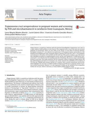
Fig. 2. PCR from the umbilical cords of newborns from T. cruzi-seropositive moth-
ers. M = DNA Marker (100-bp); P1 and P2 = Positive controls; N = Negative control;
1–4 = PCR samples of newborns’ umbilical cords.
Diego and Xichú); and the last positive umbilical cord was obtained
from a mother born in Mexico City (Table 1).
With regard to the main risk factor associated with CHD
transmission in positive mothers, the presence of pets (OR = 1.9;
P = 0.017) was found as the highest significant risk factor, followed
by the residence place located at rural areas (OR = 1.1; P = 0.040),
walls constructed of brick and adobe (OR = 2.1; P = 0.044), and
ceiling constructed of concrete (OR = 2.2; P = 0.049) but all them
unplastered. The type of floor construction material was not sig-
nificant (OR = 1.0; P = 0.141, Table 2). Moreover, 16.9% of pregnant
women (88/520) from the rural area of Guanajuato were able to
identify the vectors of CHD, and 11 of the women (12.5%, 11/88)
reported having been bitten by triatomines.
Approximately 6.3% of dwellings (31–33/520) showed
neglected public services (potable water or drainage and sewer-
age), and although this factor may not have any direct effect on
T. cruzi infection, three seropositive pregnant women were found
without any of these services (Table 2).
The youngest women aged 14–24 years showed the highest risk
factor associated with congenital transmission (OR = 2.4; P = 0.000),
particularly if they were between their first or second delivery
(OR = 1.7; P = 0.003). Four women were seropositive (16%, 4/25)
with only a newborn positive for T. cruzi by PCR from the group
of twenty five mothers (25/520) who reported their participa-
tion as a blood donor; but no significance was found as a risk
factor for vertical transmission (P = 0.880). Two women (11.76%)
were seropositive for anti-T. cruzi antibodies from the group of
17 mothers who had participated as blood receptors, however, no
PCR-positive results were found in samples from umbilical cords
(Table 3).
The PCR results from the umbilical cord blood of T. cruzi-
seropositive mothers showed four positive cases (20%, Fig. 2).
Amplification of the housekeeping gene was obtained in all of the
analyzed samples; however, microhaematocrit tests were nega-
tives.
4. Discussion
This is the first systematic study of seroprevalence of anti-T. cruzi
antibodies in pregnant women and congenital transmission of CHD
performed in Guanajuato state. This study demonstrated a total
seroprevalence of T. cruzi antibodies of 4% in pregnant women with
an index of 20% positive umbilical cords by PCR from 20 seropos-
itive mothers. In addition, the main risk factors involved in the
CHD transmission, included building materials of houses, dwellings
located in rural places and the presence of pets. The scarcity of
information on the prevalence of T. cruzi infection in pregnant
women and newborns indicated that this disease is underestimated
because a routine analysis was not performed in the obstetrics or
gynecology hospitals to diagnose infected mothers or newborns.
Thus, most children who are congenitally infected will remain
without treatment (Mu˜noz et al., 2007). The control and prevention
of CHD transmission, particularly via the congenital route, is one
of the main goals of the World Health Organization (WHO, 2013),
which indicates that opportune diagnostics of mother and baby are
important issues to prevent this pathology and avoid the spread of
disease.
According to our epidemiological surveys, 88 mothers reported
the presence of intra domiciliary triatomines (16.9%), but only
three seropositives were confirmed to have been bitten by bugs.
Although this finding was not significant, we thought that it could
have an effect on the prevalence of infection associated with the
rural setting of domiciles, where triatomines have domestic and
peridomestic habitats (Ramsey et al., 2000). Furthermore, a higher
seroprevalence of anti-T. cruzi antibodies was found in pregnant
women from rural areas of Guanajuato state (15 mothers), where
the inhabitants are easily infected by the presence of infected vec-
tors (Cucunubá et al., 2014). Other significant risk factors identified
in this study included the building materials of dwellings, such
as unplastered walls and ceiling, because these surfaces are easily
used as appropriate hiding by triatomines (Bustamante et al., 2009;
Sandoval-Ruiz et al., 2014). In Guanajuato state, the presence of the
triatomines species has been reported and is considered among the
most epidemiologically important areas due to the higher vecto-
rial capacity. Such species include Triatoma barberi, T. dimidiata,
T. longipennis and T. pallidipennis (López-Cárdenas et al., 2005;
Ramsey et al., 2015). The presence of pets associated with the trans-
mission of T. cruzi was significant, and domestic dogs and cats have
been previously identified as major sources of parasites for domes-
Table 2
Risk factors associated with building materials of dwellings, public services, and pets.
Variable Total Positives OR 95% CI X2 P-value
Ceiling construction material
Concrete vs. roofing/lamine 420/100 14/6 2.2 2–8.4 7.2 0.049
Wall construction material
Plastering vs. brick and adobe 147/373 9/11 2.1 0.8–5.1 5.1 0.044
Flooring construction material
Cement floor vs. Tile floor 426/94 18/2 1 0.9–1 2 0.141
Basic services
Potable water supply Yes/No 487/33 19/1 0.8 0.1–0.9 0.1 0.633
Drainage and sewerage vs. Latrine 489/31 17/3 0.5 0.1–0.6 0.6 0.338
Pets
Yes/No 377/143 15/5 1.9 0.3–2.5 5 0.017
Livestock
Yes/No 46/474 3/17 0.5 0.2–1.9 10.2 0.710
Domiciliary triatomines
Yes/No 88/432 3/17 0.9 0.1−1.1 6.4 0.815
Place of residence
Rural or urban 455/65 15/5 1.1 0.9–1.8 6.8 0.040
5. 104 L.M. Montes-Rincón et al. / Acta Tropica 164 (2016) 100–106
Table 3
Risk factors directly associated with pregnant women and their seropositivity of Chagas disease in Guanajuato state, Mexico.
Variable Total Positives OR CI P
Women’s Age: 14–24 vs. 25–48 327/193 16/4 2.4 0.8–7.40 0.000
N◦
of deliveries: 1–2 vs. 3–6 467/53 17/3 1.7 0.5–5.8 0.003
Blood donor: Yes vs. No 25/495 4/16 0.2 0.1–0.7 0.880
Recipient of Blood transfusion: Yes or No 17/503 2/18 0.3 0.2–0.8 0.713
Bitten by kissing-bugs: Yes or No 11/509 3/17 0.6 0.5–1.1 0.691
tic transmission in Mexico, Texas and Argentina (Kjos et al., 2013;
Enriquez et al., 2014; Odagiu and Mayer, 2015).
Only two T. cruzi-infected mothers (3.8%, 2/520) received blood
transfusions at least once in their lifetime, while four T. cruzi-
infected mothers participated as blood donors (0.8%, 4/520). The
overall prevalence reported by blood banks in Latin America was
1.3%, with 0.6% specifically for Mexico (PAHO, 2006). The estimated
participation as blood donors in this study reached a rate of 4.8%
(25/520). Although this result could suggest the low probability of
acquiring the infection by this route, it highlights the necessity to
install a universal screening for blood donors, particularly for preg-
nant women, and within this scenario, our health authorities could
improve their efforts to reduce the prevalence of CHD in the coun-
try. This is recommended by the Initiative of Central America and
Mexico (PAHO, 2014), particularly in non-endemic states, where
it remains poorly recognized and most physicians consider it to
be an “exotic” disease. Thus, they cannot provide a diagnosis and
treatment to prevent and remediate this pathology (Bonney, 2014).
This study demonstrated a direct association among the sero-
prevalence of T. cruzi antibodies and younger-age mothers with low
number of deliveries; both factors were highly significant, which
may be because parasitemia declines with age, as the frequency of
transmission would be greater among young mothers (Bittencourt,
1992). This highlights the importance of performing an earlier
detection of CHD as previously described (PAHO, 2014). Congen-
ital transmission of T. cruzi cannot be prevented during pregnancy;
however, diagnosis and early treatment in newborns is a bet-
ter recommended strategy (Mu˜noz et al., 2007). Pregnant women
between 22 and 26 years of age from Colombia and Paraguay
showed similar seroprevalence of CHD (2–4%) compared to our
results (4%). Nevertheless, the congenital transmission reported
was higher (20–30%) (Russomando et al., 2005; Manrique-Abril
et al., 2013; Cucunubá et al., 2014). The rate of congenital trans-
mission reported for other endemic and non-endemic regions of
South America ranges from 1% to 12% (Carlier and Torrico, 2003;
Torrico et al., 2004). Although, it appears that in Mexico, this epi-
demiologic threat is characterized by the highest rates reported
to Oaxaca, Jalisco and Mexico states where the prevalence among
mothers was 4.4, 4.12, and 12.02%, respectively, and transmissions
to their newborns were between 11.9 and 20% (Jiménez-Cardoso
et al., 2012). Mothers and children from Mayan communities in
Yucatan ranged between 4.4% and 0.7% (Gamboa-León et al., 2014),
which supports the importance of CHD in pregnant women and
newborns. Taking these rates as a whole, it is obvious that T. cruzi
transmission is active in several regions of Mexico.
Although there are no guaranteed mechanisms to avoid con-
genital transmission in endemic countries, a control over the major
routes of transmission is suggested, such as the vectorial and trans-
fusional, in addition to the recommended anti-parasitic treatment
(nifurtimox and benznidazol) to reduce the prevalence in infected
women. In addition, a warranty over a control strategy based on
laboratory diagnosis of infected neonates is suggested to prevent
mortality and side effects by congenital CHD and to decrease pre-
and post-neonatal morbidity (Carlier and Torrico, 2003).
Several studies have indicated that over 50% of congenitally
infected infants developed clinical manifestations with a fatality
rate of up to 14% (Carlier and Torrico, 2003). Although the totality
of congenital cases reported here does not have strong clinical evi-
dence, some 60–90% of congenital cases can present non-specific or
heterogeneous symptoms (Freilij and Altcheh, 1995; Altcheh et al.,
2005; Brutus et al., 2008), or oligosymptomatic cases, i.e., babies
who present with severe cases of meningoencephalitis, myocardi-
tis or respiratory distress syndrome (RDS) (White et al., 2000;
Torrico et al., 2004). In addition, other congenital infections may be
commonly identified with the acronym TORCH (Toxoplasma gondii,
rubella virus, cytomegalovirus, and herpes simplex virus), and in
untreated cases, the infection could progress to chronicity (Carlier
et al., 2012).
Currently there is an official consensus to consider parasitolog-
ical test close to birth as the gold standard test for the diagnosis of
congenital Chagas disease, and although PCR technique is under
evaluation and has not been validated for the diagnosis of con-
genital infection (Carlier et al., 2011) it might improve the early
detection (Velázquez et al., 2014; Rendell et al., 2015). Over more,
the PCR could be considered also as an early diagnostic method
besides the microhaematocrit and hemoculture, and with this
group of tests might establish evidence for congenital T. cruzi infec-
tion before the mother and child leave the maternity ward (Mora
et al., 2005). Although is considered that parasitological techniques
offer a rapid and definitive diagnosis allowing a faster initiation
of treatment, recent reports had shown several disadvantages for
them (1) it requires skilled personnel and assured quality control,
which may not be available in primary health care facilities (Carlier
et al., 2011) (2) parasitological methods are highly specific, but lack
sensitivity if parasitemias are too low (<40) for direct detection (3)
microhaematocrit likely misses around of 50% of infected children
and hemoculture may easily increase this proportion (Mora et al.,
2005; Velázquez et al., 2014).
We obtained negative results for microhaematocrit test, per-
haps due to the low parasitic loads obtained in the four positive
cases. Since the current standardized technique is based in micro-
haematocrit it had demonstrated lower level of detection at low
parasite load (Feilij et al., 1983), and over more, few blood par-
asites in some children are below of the limit of sensibility or
specificity (40 parasites/ml) (Velázquez et al., 2014), which should
be considered in diagnostic procedures since it has been reported
a congenital transmission in chronic phase occurs in pregnant
women with parasitemias among 10–20 parasites/ml (Rendell
et al., 2015). Also, the uneven distribution of few parasites in small
samples can show negative results in parasitological diagnostic
(Mora et al., 2005). A new consideration might be taken account for
PCR, because this technique demonstrate much higher sensitivity
for early diagnosis (Mora et al., 2005; Bern et al., 2011; Velázquez
et al., 2014). Thus in this study, using PCR technique, we determined
four positive samples for T. cruzi infection from newborns, currently
they are follow up in HGRL for medical care and clinical observation,
which will continue until their second year of life using serologic
techniques that can be assure the absence of antibodies, (Jackson
et al., 2010), because has been proved the effectiveness of treat-
ment of CHD in infected children during the first year of life yielded
therapeutic success in nearly 100% of cases (Moya et al., 2005),
6. L.M. Montes-Rincón et al. / Acta Tropica 164 (2016) 100–106 105
the treatment for T. cruzi-infected pregnancy in women remains
controversial (Sosa-Estani et al., 2009).
5. Conclusion
This study determined for the first time the seroprevalence of
T. cruzi in pregnant women in Guanajuato as well as the rate of
congenital transmission by PCR, as it has been underestimated as
a public health problem in Mexico. The calculated index of con-
genital transmission was 20% (4/20), from 20 seropositive mothers
and four PCR-positive umbilical cords. These findings were similar
to those reported from Oaxaca, an endemic state, although Gua-
najuato state is not considered endemic. In addition, the calculated
incidence in this study of the congenital transmission of Chagas dis-
ease was 770 cases per 100,000 births during a period of 12 months.
This finding highlights the importance of performing serological
tests in all populations of pregnant women to prevent the trans-
mission from one generation to another (vertical transmission).
Conflict of interest
The authors declare that there is no conflict of interest regarding
the publication of this study.
Ethics statement
This study was performed in compliance with the Declara-
tion of Helsinki in its V amendment and with respect for human
rights, as stipulated in the Constitution of the United Mexican
States. This protocol was approved by both the Ethics Commit-
tee and Research School of Biological Sciences at the Universidad
Autonoma de Nuevo Leon (UANL) and by the HGRL ethics commit-
tee. Analyses were performed according to the Biosafety laboratory
Experimental and Molecular Pathology protocol.
Acknowledgments
We are very grateful to the gynecology and obstetrician team
of the HRGL for their help and support with all of our activities.
We also thank all of the pregnant women who participated in this
study. We specifically thank the “Red de Cuerpos Academicos SEP-
PROMEP” for the project “Zoonosis Parasitarias” 2016, grant N◦
103.5/15/11043.
References
Altcheh, J., Biancardi, M., Lape˜na, A., Ballering, G., Freilij, H., 2005. Congenital
Chagas disease: experience in the hospital de Ni˜nos, Ricardo Gutierrez, Buenos
Aires, Argentina. Rev. Soc. Bras. Med. Trop. 38, 41–45.
Berg, A.T., 1991. Menstrual cycle length and the calculation of gestational age. Am.
J. Epidemiol. 133, 585–589.
Bern, C., Martin, D.L., Gilman, R.H., 2011. Acute and congenital Chagas disease. Adv.
Parasitol. 75, 19–47, http://dx.doi.org/10.1016/B978-0-12-385863-4.00002-2.
Bittencourt, A.L., 1992. Possible risk factors for vertical transmission of Chagas’
disease. Rev. Inst. Med. Trop. de São Paulo 34, 403–408.
Bonney, K.M., 2014. Chagas disease in the 21st century: a public health success or
an emerging threat? Parasite 21, 11, http://dx.doi.org/10.1051/parasite/
2014012.
Brutus, L., Schneider, D., Postigo, J., Romero, M., Santalla, J., Chippaux, J.P., 2008.
Congenital Chagas disease: diagnostic and clinical aspects in an area without
vectorial transmission Bermejo, Bolivia. Acta Trop. 106, 195–199.
Bustamante, D.M., Monroy, C., Pineda, S., Rodas, A., Castro, X., Ayala, V., Qui˜nónes,
J., Moguel, B., Trampe, R., 2009. Risk factors for intradomiciliary infestation by
the Chagas disease vector Triatoma dimidiata in Jutiapa, Guatemala. Cad. Saude
Publica 25, 83–92.
CDC (Centers for Disease Control and Prevention, 2014. Parasites – American
Trypanosomiasis (also known as Chagas Disease): Diagnosis, http://www.cdc.
gov/parasites/chagas/health professionals/dx.html (accessed 14.03.03.).
Capurro, H., Konichezky, S., Fonseca, D., Caldeyro-Barcia, R., 1978. A simplified
method for diagnosis of gestational age in the newborn infant. J. Pediatr. 93,
120–122.
Carlier, Y., Torrico, F., 2003. Congenital infection with Trypanosoma cruzi: from
mechanisms of transmission to strategies for diagnosis and control. Rev. Soc.
Bras. Med. Trop. 36, 767–771.
Carlier, Y., Truyens, C., 2010. Maternal–fetal transmission of Trypanosoma cruzi. In:
Telleria, J., Tibayrenc, M. (Eds.), American Trypanosomiasis – Chagas Disease.
One Hundred Years of Research. Elsevier, London, UK, pp. 539–581.
Carlier, Y., Truyens, C., 2015. Congenital Chagas disease as an ecological model of
interactions between Trypanosoma cruzi parasites, pregnant women, placenta
and fetuses. Acta Trop. 151, 103–115.
Carlier, Y., Torrico, F., Sosa-Estani, S., Russomando, G., Luquetti, L., Freilij, H.,
Albajar-Vinas, P., 2011. Congenital Chagas disease: recommendations for
diagnosis, treatment and control of newborns, siblings and pregnant women.
PLoS Negl. Trop. Dis. 5, e1250, http://dx.doi.org/10.1371/journal.pntd.0001250.
Carlier, Y., Truyens, C., Deloron, P., Peyron, F., 2012. Congenital parasitic infections:
a review. Acta Trop. 121, 55–70.
Cucunubá, Z.M., Valencia-Hernández, C.A., Puerta, C.J., Sosa-Estani, S., Torrico, F.,
Cortés, J.A., Ramirez, J.D., Veraf, M.J., Acostag, B.X., Álvarez, C.A., Muller, E.Á.,
Beltránj, M., Bermúdez, M.I., Berríoj, M., Camacho-Morenok, G., Castellanos,
Y.Z., Criollo, I., Flórez, A.C., Guerra-Morales, P., Herazoa, R.A., Hernández, D.C.,
Leóna, C.M., Medina-Camargo, M., Medina-Alfonso, M., Pachón, E.,
Paez-Fonseca, B., Parra, M.L., Pavia, P.X., Quiróz, F.R., Ríos, L.C., Roar, N.L.,
Torres, F., Uribe-Rivero, L.M., 2014. Primer consenso colombiano sobre Chagas
congénito y orientación clínica a mujeres en edad fértil con diagnóstico de
Chagas. Infectio 18, 50–65.
Enriquez, G.F., Bua, J., Orozco, M.M., Wirth, S., Schijman, A.G., Gürtler, R.E., Cardinal,
M.V., 2014. High levels of Trypanosoma cruzi DNA determined by qPCR and
infectiousness to Triatoma infestans support dogs and cats are major sources of
parasites for domestic transmission. Infec. Genet. Evol. 25, 36–43.
Feilij, H., Muller, L., Gonzalez-Cappa, S.M., 1983. Direct micromethod for diagnosis
of acute and congenital Chagas’ disease. J. Clin. Microbiol. 18, 327–330.
Freilij, H., Altcheh, J., 1995. Congenital Chagas’ disease: diagnostic and clinical
aspects. Clin. Infect. Dis. 21, 551–555.
Galavíz-Silva, L., Molina-Garza, D.P., González-Santos, M.A., Mercado-Hernández,
R., González-Galavíz, J.R., Rosales-Encina, J.L., Molina-Garza, Z.J., 2009. Update
on seroprevalence of anti-Trypanosoma cruzi antibodies among blood donors
in northeast Mexico. Am. J. Trop. Med. Hyg. 81, 404–406.
Gamboa-León, R., Gonzalez-Ramirez, C., Padilla-Raygoza, N., Sosa-Estani, S.,
Caamal-Kantun, A., Buekens, P., Dumonteil, E., 2011. Do commercial serologic
tests for Trypanosoma cruzi infection detect Mexican strains in women and
newborns? J. Parasitol. 97, 338–343.
Gamboa-León, R., Ramirez-Gonzalez, C., Pacheco-Tucuch, F.S., O’Shea, M.,
Rosecrans, K., Pippitt, J., Buekens, P., 2014. Seroprevalence of Trypanosoma
cruzi among mothers and children in rural Mayan communities and associated
reproductive outcomes. Am. J. Trop. Med. Hyg. 91, 348–353.
INEGI (Instituto Nacional de Estadística y Geografía), 2012. http://www3. inegi.org.
mx/sistemas/mexicocifras/default.aspx?e=11 (accessed 15.10.10.).
Jackson, Y., Alirol, E., Getaz, L., Wolff, H., Combescure, C., Chappuis, F., 2010.
Tolerance and safety of nifurtimox in patients with chronic Chagas disease.
Clin. Infect. Dis. 51, e69–e75, http://dx.doi.org/10.1086/656917.
Jiménez-Cardoso, E., Campos-Valdéz, G., Cortes-Campos, A., de la Luz-Sanchez, R.,
Rivera-Mendoza, C., Plascencia-Hernández, A., Hernández-Ramírez, M.,
Ruiz-Habana, J., Bonilla-González, E., Damian-Matzumura, P., Carlier, Y., 2012.
Maternal fetal transmission of Trypanosoma cruzi: a problem of public health
little studied in Mexico. Exp. Parasitol. 131, 425–432.
Kjos, S.A., Marcet, P.L., Yabsley, M.J., Kitron, U., Snowden, K.F., Logan, K.S., Dotson,
E.M., 2013. Identification of bloodmeal sources and Trypanosoma cruzi
infection in triatomine bugs (Hemiptera: reduviidae) from residential settings
in Texas, the United States. J. Med. Entomol. 50, 1126–1139.
López-Cárdenas, J., Gonzalez-Bravo, F.E., Salazar-Schettino, P.M.,
Gallaga-Solorzano, J.C., Ramírez-Barba, E., Martinez-Mendez, J.,
Sánchez-Cordero, V., Peterson, A.T., Ramsey, J.M., 2005. Fine-scale predictions
of distributions of Chagas disease vectors in the state of Guanajuato, Mexico. J.
Med. Entomol. 42, 1068–1081.
Manrique-Abril, F., Ospina, J.M., Herrera, G., Florez, A.C., Pavia, P.X., Montilla, M.,
Nicholls, R.S., Puerta-Bulac, C., 2013. Diagnóstico de enfermedad de Chagas en
mujeres embarazadas y recién nacidos de Moniquirá y Miraflores Boyacá,
Colombia. Infectio 17, 28–34.
Mora, M.C., Sanchez-Negrette, O., Marco, D., Barrio, A., Ciaccio, M., Segura, M.A.,
Basombrío, M.A., 2005. Early diagnosis of congenital Trypanosoma cruzi
infection using PCR, hemoculture, and capillary concentration, as compared
with delayed serology. J. Parasitol. 91, 1468–1473.
Moya, P., Basso, B., Moretti, E., 2005. Congenital Chagas disease in Córdoba,
Argentina: epidemiological, clinical, diagnostic, and therapeutic aspects.
Experience of 30 years of follow up. Rev. Soc. Bras. Med. Trop. 38, 33–40.
Mu˜noz, J., Portús, M., Corachan, M., Fumado, V., Gascon, J., 2007. Congenital
Trypanosoma cruzi infection in a non-endemic area. Trans. R. Soc. Trop. Med.
Hyg. 101, 1161–1162, http://dx.doi.org/10.1016/j.trstmh.2007.06.011.
NOM, 2012. Norma Oficial Mexicana-017-SSA2-2012, Para la vigilancia
epidemiológica. http://dof.gob.mx/nota detalle.
php?codigo=5288225&fecha=19/02/2013 (accessed 02.01.14.).
Odagiu, S., Mayer, J.D., 2015. Chagas’ disease in Mexico: factors, surveillance, and
recommendations. Proceedings of the National Conference On Undergraduate
Research (NCUR), https://ncurdb.cur.org/ncur2015/search/display ncur.
aspx?id=93432 (accessed 15.12.09.).
7. 106 L.M. Montes-Rincón et al. / Acta Tropica 164 (2016) 100–106
Oliveira, I., Torrico, F., Mu˜noz, J., Gascon, J., 2010. Congenital transmission of
Chagas disease: a clinical approach. Expert. Rev. Anti. Infect. Ther. 8, 945–956,
http://dx.doi.org/10.1586/eri.10.74.
PAHO (Pan American Health Organization), 2006. Estimación cuantitativa de la
enfermedad de Chagas en las Américas, http://www.paho.org/hq/index.
php?option=com content&view=article&id=5856&Itemid=40687&lang=es
(accessed 15.11.04.).
Pan American Health Organization (PAHO), 2014. Regional Consultation on
Organization and Structure of Health Care for the sick or infected with. In:
Chagas’ Disease: Scientific and Technical Materials, http://www.paho.org/hq/
index.php%3Foption%3Dcom docman%26task%3Ddoc
view%26gid%3D23129%26Itemid%3D%26lang%3Dfr+&cd=9&hl=es-
419&ct=clnk (accessed 15.09.27.).
Ramsey, J.M., Ordo˜nez, R., Cruz-Celis, A., Alvear, A.L., Chavez, V., Lopez, R., Pintor,
J.R., Gama, F., Carrillo, S., 2000. Distribution of domestic Triatominae and
stratification of Chagas disease transmission in Oaxaca, Mexico. J. Med. Vet.
Entomol. 14, 1–12, http://dx.doi.org/10.1046/j.1365-2915.2000.00214.x.
Ramsey, J.M., Gutiérrez-Cabrera, A.E., Salgado-Ramírez, L., Townsend, P.A.,
Sánchez-Cordero, V., Ibarra-Cerde˜na, C.N., 2012. Ecological connectivity of
Trypanosoma cruzi reservoirs and Triatoma pallidipennis hosts in an
anthropogenic landscape with endemic Chagas disease. PLoS ONE 7, e46013,
http://dx.doi.org/10.1371/journal.pone.0046013.
Ramsey, J.M., Peterson, A.T., Carmona-Castro, O., Moo-Llanes, D.A., Nakazawa, Y.,
Butrick, M., Tun-Ku, E., de la Cruz-Félix, K., Ibarra-Cerde˜na, C.N., 2015. Atlas of
mexican triatominae (Reduviidae: hemiptera) and vector transmission of
chagas disease. Mem. Inst. Oswaldo Cruz. 110, 339–352.
Rassi, A., Rassi Jr., A., Marin-Neto, J.A., 2010. Chagas disease. Lancet 375,
1388–1402.
Rendell, V.R., Gilman, R.H., Valencia, E., Galdos-Cardenas, G., Verastegui, M.,
Sanchez, L., Acosta, J., Sanchez, G., Ferrufino, L., La Fuente, C., Abastoflor, M.C.,
Colanzi, R., Bern, C., 2015. Trypanosoma cruzi-infected pregnant women
without vector exposure have higher parasitemia levels: implications for
congenital transmission risk. PLoS One 10 (3), e0119527, http://dx.doi.org/10.
1371/journal.pone.0119527 (eCollection).
Russomando, G., Almirón, M., Candia, N., Franco, L., Sánchez, Z., de Guillen, I., 2005.
Implementación y evaluación de un sistema localmente sustentable de
diagnóstico prenatal que permite detectar casos de transmisión congénita de
la enfermedad de Chagas en zonas endémicas del Paraguay. Rev. Soc. Bras.
Med. Trop. 38, 49–54.
Sambrook, J., Russell, D.W., 2001. Molecular Cloning. In: A Laboratory Manual,
third ed. Cold Spring Harbor Laboratory Press, New York.
Sandoval-Ruiz, C.A., Guevara, R., Ibá˜nez-Bernal, S., 2014. Household risk factors
associated to infestation of Triatoma dimidiata the Chagas disease vector in
Central Region of Veracruz, Mexico. Salud Publica Mex. 56, 213–220.
Sosa-Estani, S., Gamboa-León, M.R., del Cid-Lemus, J., Althabe, F., Alger, J.,
Almendares, O., Cafferata, M.L., Chippaux, J.P., Dumonteil, E., Gibbons, L.,
Padilla-Raygoza, N., Schneider, D., Belizán, J.M., Buekens, P., Working Group,
2008. Use of a rapid test on umbilical cord blood to screen for Trypanosoma
cruzi infection in pregnant women in Argentina, Bolivia, Honduras and Mexico.
Am. J. Trop. Med. Hyg. 79, 755–759.
Sosa-Estani, S., Cura, E., Velazquez, E., Yampotis, C., Segura, E.L., 2009. Etiological
treatment of young women infected with Trypanosoma cruzi, and prevention of
congenital transmission. Rev. Soc. Bras. Med. Trop. 42, 484–487.
Torrico, F., Alonso-Vega, C., Suarez, E., Rodriguez, P., Torrico, M.C., Dramaix, M.,
Truyens, C., Carlier, Y., 2004. Maternal Trypanosoma cruzi infection, pregnancy
outcome, morbidity, and mortality of congenitally infected and non-infected
newborns in Bolivia. Am. J. Trop. Med. Hyg. 70, 201–209.
Velázquez, E.B., Rivero, R., De Rissio, A.M., Malagrino, N., Esteva, M.I., Riarte, A.R.,
Ruiz, A.M., 2014. Predictive role of polymerase chain reaction in the early
diagnosis of congenital Trypanosoma cruzi infection. Acta Trop. 137, 195–200.
Virreira, M., Torrico, F., Truyens, C., Alonso-Vega, C., Solano, M., Carlier, Y., Svoboda,
M., 2003. Comparison of polymerase chain reaction methods for reliable and
easy detection of congenital Trypanosoma cruzi infection. Am. J. Trop. Med.
Hyg. 68, 574–582.
WHO (World Health Organization), 2002. Control of Chagas Diasease, http://apps.
who.int/iris/bitstream/10665/42443/1/WHO TRS 905.pdf (accesed 13.01.08.).
WHO (World Health Organization), 2013. Chagas Disease (American
Trypanosomiasis), http://www.who.int/mediacentre/factsheets/fs340/en/
(accesed 15.12.11.).
WHO (World Health Organization), 2015. Chagas Disease in Latin America: An
Epidemiological Update Based on 2010 Estimates, http://www.who.int/wer/
2015/wer9006/en/ (accessed 15.03.12.).
Walpole, R.E., Myers, R.H., Myers, S.L., Keying, Y., 2011. Probability & Statistics for
Engineers & Scientists, 9th ed. Prentice Hall Boston.
White, I.J., Souabni, A., Hooper, N.M., 2000. Comparison of the
glycosyl-phosphatidylinositol cleavage/attachment site between mammalian
cells and parasitic protozoa. J. Cell Sci. 113, 721–727.