Kingdom Protista
•
17 likes•13,374 views

- Protists are a diverse group of mostly single-celled eukaryotes that are classified into several different kingdoms. - They exhibit an enormous diversity of structures and metabolic capabilities, including photosynthesis, heterotrophy, and mixotrophy. - Common protist groups discussed in the document include protozoa, alveolates, stramenopiles, and chlorophytes. Many important parasites are protists, including those that cause malaria, African sleeping sickness, and Chagas disease.

Recommended
Recommended
More Related Content
What's hot
What's hot (20)
Viewers also liked
Viewers also liked (19)
Similar to Kingdom Protista
Similar to Kingdom Protista (20)
More from Nattapong Boonpong
More from Nattapong Boonpong (20)
Recently uploaded
Recently uploaded (20)
Kingdom Protista
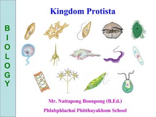
- 1. Kingdom Protista B I O L O G Y Mr. Nattapong Boonpong (B.Ed.) Phlabphlachai Phitthayakhom School
- 2. Overview: A World in a Drop of Water • Even a low-power microscope – Can reveal an astonishing menagerie of organisms in a drop of pond water 50 m
- 3. The Protozoa
- 4. Characteristic Protist • These amazing organisms – Belong to the diverse kingdoms of mostly single- celled eukaryotes informally known as protists • Advances in eukaryotic systematics – Have caused the classification of protists to change significantly. • Protists are more diverse than all other eukaryotes – And are no longer classified in a single kingdom • Most protists are unicellular – And some are colonial or multicellular
- 5. Characteristic Protist (Continuous) • Protists, the most nutritionally diverse of all eukaryotes, include – Photoautotrophs, which contain chloroplasts – Heterotrophs, which absorb organic molecules or ingest larger food particles – Mixotrophs, which combine photosynthesis and heterotrophic nutrition • Protist habitats are also diverse in habitat • And including freshwater and marine species
- 7. Reproduction of Protist • Reproduction and life cycles – Are also highly varied among protists, with both sexual and asexual species
- 8. A Sample of Protist Diversity
- 9. Protozoa • Eukaryotic • Unicellular • Chemoheterotrophs • Vegetative form is a trophozoite • Asexual reproduction by fission, budding, or schizogony • Sexual reproduction by conjugation • Some produce cysts
- 10. I. Diplomonadida & Parabasala • No mitochondria, ER, golgi complex, & centriole • Multiple flagella • Are adapted to anaerobic environments • Lack plastids
- 11. Diplomonads & Parabasalids • Diplomonads – Have two nuclei and multiple flagella 5 µm Giardia intestinalis, a diplomonad (colorized SEM)
- 13. Diplomonads & Parabasalids • Parabasalids include trichomonads – Which move by means of flagella and an undulating part of the plasma membrane – Have parabasal body Flagella Undulating membrane 5 µm Trichomonas vaginalis, a parabasalid (colorized SEM)
- 15. Diplomonads & Parabasalids • Have one nucleus and multiple flagella • Have parabasal body Trichonympha sp., a parabasalid
- 16. II. Euglenozoa • Move by flagella • Photoautotrophs – Euglenoids • Chemoheterotrophs – Trypanosoma • Undulating membrane, transmitted by vectors – Leishmania • Flagellated form in sand fly vector, ovoid form in vertebrate host
- 19. Kinetoplastids • Kinetoplastids – Have a single, large mitochondrion that contains an organized mass of DNA called a kinetoplast – Include free-living consumers of bacteria in freshwater, marine, and moist terrestrial ecosystems
- 20. Kinetoplastids
- 21. Kinetoplastids • The parasitic kinetoplastid Trypanosoma – Causes sleeping sickness in humans 9 m
- 22. Kinetoplastids Trypanosoma brucei gambiense Trypanosoma brucei rhodesiense Trypanosoma cruzi Trypanosomiasis, African (African sleeping sickness) Trypanosomiasis, American (Chagas disease)
- 23. Trypanosoma
- 24. Trypanosomiasis (African Sleeping Sickness); T. brucei gambiense & T. brucei rhodesiense
- 25. Trypanosomiasis (American Chagas Disease)
- 26. III. Alveolata • Alveolates have sacs beneath the plasma membrane • Members of the clade Alveolata – Have membrane-bounded sacs (alveoli) just under the plasma membrane 0.2 µm Alveoli Flagellum
- 27. Alveolata: Dinoflagellate • Dinoflagellates • Cellulose in plasma membrane • Unicellular • Chlorophyll a and c, carotene, xanthins • Store starch • Some are symbionts in marine animals • Neurotoxins cause paralytic shellfish poisoning
- 29. Dinoflagellate: Red tide Bloom Dinoflagellate หลายชนิด เช่น Gymnodinium & Trichodesmium จะทาให้เกิดปรากฏการณ์“red tide”ในทะเล และมหาสมุทรบางแห่ง และสร้างสารพิษออกมา ทาให้สัตว์นา ตายครังละมาก ๆ โดยจะออกฤทธิ์ที่ระบบประสาท นอกจากนี Gymnodinium & Gonyaulax ยังสร้างพิษทาให้ปลาตายและ มนุษย์ที่ได้รับสารพิษเข้าไปจะเสียชีวิตได้
- 30. Dinoflagellate Peridinium Trichodesmium Gymnodinium Gonyaulax
- 31. Alveolata: Apicomplexa • Nonmotile • Intracellular parasites • Complex life cycles • Plasmodium * Plasmodium vivax * Plasmodium malariae * Plasmodium ovale * Plasmodium falciparum
- 32. Alveolata: Apicomplexa; Plasmodium sp. Sporozoites 1 Infected mosquito bites 2 Sporozoites in salivary human; sporozoites undergo gland migrate through schizogony in bloodstream to liver cell; liver of human merozoites are produced 9 Resulting sporozoites migrate to salivary glands of mosquito 3 Merozoites Sexual released into reproduction bloodsteam from liver may infect Asexual new red blood 8 In mosquito’s cells Zygote digestive tract, reproduction gametocytes unite to form Intermediate host Female gametocyte zygote 4 Merozoite develops Male into ring stage in red gametocyte blood cell Ring 5 Ring stage stage grows and Definitive host divides, 7 Another mosquito bites producing 6 Merozoites are released merozoites infected humnan and when red blood cell ingests gametocytes ruptures; some merozoites infect new red blood cells, and some develop into male and female gametocytes Merozoites
- 35. Alveolata: Ciliates • Ciliates, a large varied group of protists – Are named for their use of cilia to move and feed – Have large macronuclei and small micronuclei • The micronuclei – Function during conjugation, a sexual process that produces genetic variation • Conjugation is separate from reproduction – Which generally occurs by binary fission
- 36. Alveolata: Ciliates • Structure and Function in the Ciliate Paramecium caudatum FEEDING, WASTE REMOVAL, AND WATER BALANCE Paramecium, like other freshwater Contractile Vacuole Paramecium feeds mainly on bacteria. protists, constantly takes in water Rows of cilia along a funnel-shaped oral by osmosis from the hypotonic environment. groove move food into the cell mouth, Bladderlike contractile vacuoles accumulate where the food is engulfed into food excess water from radial canals and periodically vacuoles by phagocytosis. expel it through the plasma membrane. Oral groove Cell mouth Thousands of cilia cover 50 µm Food vacuoles combine with the surface of Paramecium. lysosomes. As the food is digested, the vacuoles follow a looping path Micronucleus through the cell. Macronucleus The undigested contents of food vacuoles are released when the vacuoles fuse with a specialized region of the plasma membrane that functions as an anal pore.
- 37. Alveolata: Ciliates CONJUGATION AND REPRODUCTION 1 Two cells of compatible 2 Meiosis of micronuclei mating strains align side produces four haploid 3 Three micronuclei in each cell by side and partially fuse. micronuclei in each cell. disintegrate. The remaining micro- nucleus in each cell divides by mitosis. MEIOSIS 4 The cells swap one micronucleus. Macronucleus Haploid micronucleus Compatible Diploid mates micronucleus Diploid micronucleus MICRONUCLEAR FUSION 5 The cells separate. 9 Two rounds of cytokinesis 8 The original macro- 7 Three rounds of 6 Micronuclei fuse, partition one macronucleus nucleus disintegrates. mitosis without forming a diploid Key and one micronucleus Four micronuclei cytokinesis micronucleus. into each of four daughter cells. become macronuclei, produce eight Conjugation while the other four micronuclei. remain micronuclei. Reproduction
- 38. Ciliates Stentor Paramecium
- 39. Ciliates Stentor polymorphus Stentor roeseli Epistylis rotans Vorticella Paramecium bursaria Suctoria Strombidium Trichodina pediculus Euplotes
- 40. IV. Stramenopila (Algae) • Stramenopiles have “hairy” and smooth flagella • The clade Stramenopila - Includes several groups of heterotrophs as well as certain groups of algae • Most stramenopiles - Have a “hairy” flagellum paired with a “smooth” flagellum Hairy flagellum Smooth flagellum 5 µm
- 41. Algae
- 42. Stramenopila: Brown algae (Phaeophyta) • Are the largest and most complex algae • Are all multicellular, and most are marine • Brown algae (kelp) • Cellulose + alginic acid cell walls • Multicellular • Chlorophyll a and c, xanthophylls; fucoxanthrin • Store carbohydrates • Harvested for algin
- 43. Stramenopila: Brown algae • Kelps, or giant seaweeds – Live in deep parts of the ocean
- 44. Brown algae Laminaria saccharia - สกัดเอา algin มาทายา อาหาร เส้นใย ยาง สบู่ ฯลฯ - ใช้ทาปุ๋ย K ได้ Padina & Fucus -ใช้ทาปุ๋ย K ได้ Sargassum (สาหร่ายทุ่น) - มีไอโอดีนสูง และยังให้ algin Laminaria & Sargassum - สามารถนามาตากแห้ง ต้มนาดื่มแก้ร้อนใน คอพอก และฟอกเลือด Giant kelp - สร้างสาร algin มาทายา อาหาร เส้นใย กระดาษ ยาง สบู่ และเป็นสาหร่ายที่มีขนาดใหญ่ที่สุด เป็นที่อยู่อาศัย หลบภัยและอาหารของสัตว์นานานาชนิดใต้ท้องทะเล Laminaria Padina Fucus
- 45. Brown algae Sargassum Kelp
- 46. Brown algae (a) The seaweed is grown on nets in shallow coastal waters. (b) A worker spreads the harvested sea- weed on bamboo screens to dry. (c) Paper-thin, glossy sheets of nori make a mineral-rich wrap for rice, seafood, and vegetables in sushi.
- 47. Stramenopila: Diatoms (Bacillariophyta) • Diatoms • Pectin and silica cell walls • Unicellular • Chlorophyll a and c, carotene, xanthophylls • Store oil • Fossilized diatoms formed oil • Produce domoic acid
- 48. Diatomaceous earth • Accumulations of fossilized diatom walls – Compose much of the sediments known as diatomaceous earth
- 49. Diatoms Pinnidaria Navicular Asterionella Melorista
- 50. V. Rhodophyta (Red algae) • Red algae • Cellulose cell walls • Most multicellular • No flagella stage • Chlorophyll a and d, phycobiliproteins (phycocyanin & phycoerythrin) • Store glucose polymer • Harvested for agar and carrageenan
- 52. Red algae Porphyra (จี่ฉ่าย) - ใส่แกงจืดเป็นอาหาร Gracilaria (สาหร่ายผมนาง หรือ สาหร่ายวุ้น) - นามาสกัดวุ้นผสมในอาหารเลียงเชือจุลินทรีย์ (agar) - ใช้เพาะเลียงเนือเยื่อ - ทาแคปซูลยา ทายา ทาเครื่องสาอางค์ ครีมโกนหนวด ฯลฯ Chondrus Plumoria Andrus Corellina Polysiphinia Gelidium Grinnellia Porphyra Polysiphinia
- 53. Red algae Gelidium Chodrus crispus
- 54. VI. Chlorophyta (Green algae) • Green algae • Cellulose cell walls • Unicellular, multicellular or colonial form • Chlorophyll a and b • Store glucose polymer • Gave rise to plants • Are named for their grass-green chloroplasts • Are divided into two main groups: chlorophytes and charophyceans • Are closely related to land plants
- 58. Chlorophyta (Green algae) Spirogyra
- 59. Chlorophyta (Green algae) Micrasterias rotata Cell division Micrasterias fimbriata Staurastrum Hyalotheka dissilens Closterium lunula Staurodesmus convergens Euastrum verrucosum Volvox aureus Draparnadia platyzonata Mougeotia Pediastrum
- 61. Chlorophyta (Green algae) Chara sp., Overlap between Protista with Plant
- 62. VII. Mycetozoa (Slim Molds) • Cellular slime molds • Plasmodial slime molds – Resemble amoebas, – Multinucleated large ingest bacteria by cells phagocytosis – Cytoplasm separates – Cells aggregate into into stalked sporangia stalked fruiting body. – Nuclei undergo – Some cells become meiosis and form spores uninucleate haploid spores
- 65. Slim Molds
- 66. Slim Molds
- 67. Slim Molds
- 68. Slim Molds
- 69. Slim Molds
- 70. Slim Molds
- 72. Amoebiasis
- 73. Acanthamoeba spp. & Balamuthia mandrillaris
- 74. Entamoeba • Entamoeba coli - in Large intestine; colon • Entamoeba gingivalis - in Mouth; teeth & gill • Entamoeba histolytica - in Small intestine
- 75. Entamoeba histolytica • Entamoeba histolytica; Amoebic Dysentery • Amoeba feeds on RBCs and GI tract tissues • Diagnosis by observing trophozoites in feces • Treated with metronidazole
- 76. The End