
Recommended
More Related Content
What's hot
What's hot (20)
Similar to Chap 5 Cleavage.pptx
Similar to Chap 5 Cleavage.pptx (20)
Recently uploaded
Recently uploaded (20)
Chap 5 Cleavage.pptx
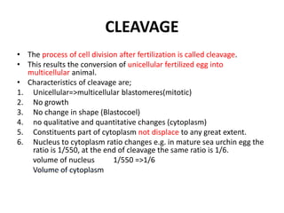
- 1. CLEAVAGE • The process of cell division after fertilization is called cleavage. • This results the conversion of unicellular fertilized egg into multicellular animal. • Characteristics of cleavage are; 1. Unicellular=>multicellular blastomeres(mitotic) 2. No growth 3. No change in shape (Blastocoel) 4. no qualitative and quantitative changes (cytoplasm) 5. Constituents part of cytoplasm not displace to any great extent. 6. Nucleus to cytoplasm ratio changes e.g. in mature sea urchin egg the ratio is 1/550, at the end of cleavage the same ratio is 1/6. volume of nucleus 1/550 =>1/6 Volume of cytoplasm
- 2. • Although the chemical changes during cleavage are limited but following changes takes place. • 1. SYNTHESIS OF DNA • 2. SYNTHESIS OF RNA • 3. SYNTHESIS OF PROTEINS
- 3. Chemical changes during cleavage • SYNTHESIS OF DNA • The number of nuclei doubled with every new division. • It occurs at the expense of synthesis of DNA. • G1 period becomes limited. • DNA in synthesized at the expense of cytoplasmic RNA in the presence of an enzyme Ribonucleotide reductase. • If the embryo is treated with H3 uridine after some time, radioactivity will appear in DNA, it means that RNA is involved in the synthesis of DNA. • Second source of DNA are the low density precursors. e.g: Cytidine is the precursor for purines.
- 4. • SYNTHESIS OF RNA • Synthesis of RNA takes place but it is very limited, especially synthesis of mRNA takes place. • If the egg is treated with Actinomycin D, it will stop the synthesis of RNA, but the cleavage will not stop.
- 5. • SYNTHESIS OF PROTEINS • Much of the proteins, produced during cleavage are that which is directly involved in the process of cell multiplication. e.g. nuclear histones. • Other protein which is responsible for the formation of spindle fibre is also formed which is tubulin. • The third important protein formed during cleavage is Ribonucleotide Reductase. • To prove that protein synthesis is important or not treat the embryo with puromycine which is responsible to stop the protein synthesis, the cleavage will also stop. • This proves that protein synthesis is very important.
- 6. PATTERNS OF CLEAVAGE • The division of egg into the daughter blastomeres is usually very regular. • Generally the plane of first division is vertical i.e. It passes through the animal vegetal axis of the egg. • The plane of the second division is also vertical, but it is at right angles to the first plane of cleavage. • As a result the first 4 blastomeres all lie side by side. • The plane of the third division is at right angles of the first two planes i.e. it is horizontal to the equator of the egg. • Now there are eight cells. • Four cells upward and for cells downwards. • The upper four cells comprising the animal hemisphere of embryo, and the lower four cells comprising the vegetal hemisphere. • Pattern of cleavage is different in different animals. • Some of the main types of cleavage are
- 7. • RADIAL CLEAVAGE • If each of the blastomeres of the upper tier lie exactly over the corresponding lower blastomeres, the pattern of the blastomeres is radially symmetrical. • Radially type of cleavage is found in sea cucumber (Synapta digitata).
- 8. Radial cleavage with almost equal size of blastomeres in sea cucumber Synapta digitata.
- 9. • SPIRAL TYPE CLEAVAGE • In the annelids, molluscs and some of the planarians the blastomeres are not exactly on one another. • The upper cells move spirally. • If the movement is in clockwise direction, the cleavage is called dextral type of spiral cleavage. • If the upper tiers move in anti-clockwise direction the cleavage is called sinistral type of spiral cleavage.
- 10. Spiral cleavage In the mollusc Trochus.
- 11. • BILATERAL TYPE • Bilateral type of cleavage is found tunicates and nematodes. • In these animals two of first four blastomeres become larger than the other two, thus forming a plane of bilateral symmetry giving a shape of T.
- 12. Holoblastic cleavage mammals - rotational cleavage Figure 5.9 - first cleavage is meridional - second cleavage is meridional in one blastomere, but equatorial in the other one = rotation of cleavage plane - divisions are NOT synchronized
- 13. • MEROBLASTIC OR INCOMPLETE CLEAVAGE • In the eggs of birds & reptiles the cleavage is always incomplete. • Only the active cytoplasm divides.
- 14. Meroblastic cleavage birds - telolecithal egg - discoidal cleavage chicken embryo - cleavage furrows appear at animal pole of the oocyte to form the blastodisc - inner cells are continuous with the yolk - egg is laid during blastoderm stage Figure 5.15
- 15. • HOLOBLASTIC OR COMPLETE CLEAVAGE • In amphibian holoblastic type of cleavage is found. • whole of the egg becomes subdivided into blastomeres • SUPERFICIAL CLEAVAGE • In case of arthropods the division of nucleus takes place, but the cytoplasm remains undivided. • In this way a number of nuclei are formed. • The nuclei later on separate from each other and arrange themselves at the periphery along with a little cytoplasm. As a result blastoderm is formed.
- 16. Meroblastic cleavage insects Drosophila - centrolecithal eggs - superficial cleavage - repeated mitosis without cytokinesis - multiple nuclei in endoplasm nuclei move towards the yolkfree periplasm - pole cells form at posterior pole - become primordial germ cells nuclei & cytoplasm form syncytial blastoderm - cellularization - formation of cellular blastoderm Figure 5.17
- 18. Patterns of embryonic cleavage: holoblastic and meroblastic cleavage (evenly distributed yolk) 8-7 8-25, 26 8-36, 38 Mesolecithal - amphibians
- 20. Holoblastic (complete cleavage) Isolecithal Mesolecithal C Meroblastic (incomplete cleavage) Radial (echinoderms, amphioxis) Spiral (annelids, molluscs, flatworms) Bilateral (tunicates) Rotational (mammals, nematodes) Radial (amphibians) Bilateral (cephalopod molluscs) Telolecithal Discoidal (fish, reptiles, birds) Centrolethical Superficial (most insects)
- 21. CHARACTERISTICS OF NUCLEI DURING CLEAVAGE • Weisman gave the theory of germplasm according to which there are present certain particles in an egg which are responsible to determine the cleavage. • These particles are called determinants. • He said that these determinants are present on chromosomes and these may be hereditary material. • During cleavage these determinants are disturbed in the blastomeres. • All these determinant are necessary to develop a normal embryo. • It means that all the blastomeres are necessary to develop into a normal embryo.
- 22. • Horstadius & Wolsky took the sea urchin egg at 4 blastomeres stage and separate the 4 blastomeres. • In this way he divides one of the four blastomeres, may develop into a whole embryo but reduce in size. • According to germplasm theory the complete set of determinants is necessary to develop into normal embryo. • But they observed that all the four blastomeres are able to develop into complete individual. • This is against germplasm theory.
- 23. • SPEMANN • He took the fertilized egg of Triturus (newt) and divide it into two halves with a fine hair, just as they were about to begin cleave. • The constriction was not carried out completely in such a way that the two halves were still connected to each other by a narrow bridge of cytoplasm. • Cleavage started in the one half containing nucleus, but the one half without nucleus remained uncleave. • At 16 –cells stage he allowed nucleus to go into the other half which had no nucleus as a result this half also begin to cleave. • In a number of cases as a result of this experiment two complete normal embryos were developed. • This experiment proves that at 16-cells stage & even 32- cells stage every nucleus has a complete set of hereditary factors which are responsible for the normal development.
- 24. • 2) EXPERIMENT ON DRAGON FLY • In dragon fly cleavage is incomplete. • The nucleus divides first, the cytoplasm remains uncleaved. • After first division of the nucleus one moves toward the anterior end where as the other move towards the posterior end. • After first cleavage if any of two nuclei is destroyed with a narrow beam of ultraviolet rays, we find that the other nucleus continue to divide to form a complete embryo. • This thing is once again against the germplasm theory. • This thing can also be further proved by using the technique of transplantation.
- 25. • 3). TECHNIQUE OF TRANSPLANTATION • Take a ripe egg from oviduct of a frog. • Prick it with a fine glass needle to activate it, and then exclude nucleus. • Now take a nucleus from the cell of an advanced embryo with the help of a pipette. • Now inject this nucleus in enucleated egg. • 80% of such egg starts cleaving. • This experiment proves that nuclei of the later stages have all the factors responsible for development. • In further experiments the potential of the nuclei of cells which were in more advance stage were tested by transplanting then into enucleated eggs. e.g. Nuclei from the neural plate of well developed tadpoles of frog were transplanted into enucleated egg. • Normal embryos were developed. • In further experiments the potentialities of nuclei of cells, which were even more advanced in the process of differentiation were tested.
- 26. • 4). NUCLEI FROM ADULT TISSUE • When nuclei from the cells of adult advance tissue were taken and transplanted into enucleated eggs they did not show further development. • If adult cells cultured in vitro, they lose the properties of different cells and start reproducing mitotically and if their nuclei are then transplanted into enucleated eggs, the embryo develop abnormally. • If nuclei of cells of healthy parts of the abnormal embryos are transplanted into eggs, the development of second generation embryos takes place and many normally developing embryos are produced.
- 27. • Gurdon in 1968 took the nerve cell (which is highly differentiated cell and is unable to undergo mitosis) and transplanted this nucleus in the enucleated eggs. • He observed that the highly specialized nucleus is capable to develop into a complete embryo. • He obtained same results from skin, kidney and intestine nuclei. • When the nucleus enters the enucleated egg the size of nucleus is increased to 30 folds in initial few minutes and during this interval the nucleus is changed into the undifferentiated nucleus, and actually this is the cytoplasm of egg which is capable to change the fate of nucleus.
- 28. DISTRIBUTION OF CYTOPLASMIC SUSTANCES IN THE EGG DURING CLEAVAGE • For the normal development of the embryo, in the cytoplasm there are determinants which are important. • First approach was from Spemann in 1928. • He was awarded noble prize in the developmental biology. • Spemann observed that the distribution of pigments is not equal in the amphibians egg. • The animal pole is heavily pigmented where as the vegetal pole is poor in pigment or complete white. • On one side of the egg the marginal zone broader then other, and this region is known as the gray crescent. • This area forms the dorsal lip of blastopore he noted that after first nuclear division, if we divide the egg into two parts each part having half grey crescent area and one nucleus. • Both parts will develop into normal embryos.
- 29. • On the other hand if we divide the egg into two parts in such a way that one half has grey crescent area and other is devoid of it, the egg having grey crescent area will develop into embryo and other will not develop into it. • It means that the grey crescent has some importance for the normal development of the embryo.
- 30. • The importance of cytoplasmic substances in the egg for normal development is further proved by a number of experiments carried on the eggs of various animals. • Wilson (1904): The cytoplasm of egg of a mollusc Dentallium has three zones. • A clear cytoplasm at the animal pole of the egg • A second clear cytoplasm at the vegetal pole end • In between there is a pigmented granular cytoplasm
- 31. • After first cleavage the egg divides into two cells. The division is such that one cell has only two parts i.e. • Animal clear cytoplasm +pigmented area • Other cell has all three parts i-e animal clear cytoplasm +pigmented area+ vegetal clear cytoplasm. • The reason is that during cleavage the vegetal clear cytoplasm is pushed off as a polar lobe and remains attach to the cell CD. • Cell AB is without this polar lobe. • If we separate these two cells, the cell containing all three parts will be able to develop into normal embryo. • It means all parts of cytoplasm are necessary for normal development.
- 32. • At four cell stage only one cell has polar lobe i-e when AB divides into A and B there is no polar lobe. • When CD divides into C and D, the D contains polar lobe . • if we separate these four cells only the cell D having polar lobe will develop into normal embryo, although nucleus is present in all four cells.
- 33. • This is very clear proof that it is the cytoplasm and not the nucleus that makes blastomere D, different from other three blastomeres and able to produce normal embryo. • This is proved further if we separate the polar lobe of blastomer ‘D’ then it will also give abnormal embryo. • Conklin (1905): In the egg of ascidian Styela partita the eggs has four different zones. • The animal pole consists of clear transparent zone. • The vegetal pole consists of slaty grey cytoplasm. • At the center there are other two zones one consists of light grey and other is yellow zone.
- 34. • During cleavage the four kinds of cytoplasm are distributed to different cells which for different tissues of the embryo. • Clear zone forms ectoderm • Yellow zone forms mesoderm • Slaty grey forms endoderm • Light grey forms nerve cord +notochord • He took the egg of Styela and centrifuges it in such a way that the position of various zones is changed. • Normally the animal pole is responsible for the formation of ectoderm. • But when as a result of centrifuge the animal pole is dislocated to the lower part (vegetal pole) here it starts to form ectoderm. • This clearly determined that fate of embryo is determined by cytoplasm.
- 35. BIOCHEMICAL SIGNIFICANCE OF THESE AREAS • Muscles are formed from mesoderm. • The blastomeres which form muscles contain marker enzyme acetylcholine esterase. • Dopa oxidase is marker enzyme of the eye and statocysts. • Both these enzymes appear in the embryo nine hours after fertilization. • Treat the dividing embryo with cytochalasin B or with colchicine. • This will stop cell division but we will see that enzymes will appear in the correct blastomeres at the fixed time as in normal embryo. • Now treat the embryo with puromycin, this will stop protein synthesis. • Now there is no formation of these enzymes. • This clearly proves that enzymes must be synthesized during the development and is not already present in the cytoplasm.
- 36. • If we treat the embryo with Actinomycin D this will stop transcription i-e the formation of mRNA stopped. • The embryo does not develop the enzymes. • This proves that transcription process occurring after 7th hour is very necessary for the formation of mRNA, which after 8 hours is used for the formation of enzymes. • We can further prove that the cytoplasm plays its role in the egg in determining the differentiation of cells during development by the following experiments.
- 37. • Germ Cells: In insects at vegetal pole there are certain cells called germ plasms or polar plasm. • They are called germ plasms because at later stages of development these cells enter into gonads and form germ cells for reproduction. • That is why they are also called “primordial germ cells”. • To prove this the location of cells in cytoplasm is important? • Mahowald (1974) transplant these cells to the animal pole. • He saw that they were unable to enter into gonads although they are differentiated into polar plasms. • In the second experiment he transplanted the germ plasms from animal pole to vegetal pole in another embryo. • The cells moved & enter to the gonads, and formed germ cells. • The flies developing from second experiment were mated and they produced off spring. • This shows that the location of cells in the cytoplasm is important
- 38. Meroblastic cleavage insects Drosophila - centrolecithal eggs - superficial cleavage - repeated mitosis without cytokinesis - multiple nuclei in endoplasm nuclei move towards the yolkfree periplasm - pole cells form at posterior pole - become primordial germ cells nuclei & cytoplasm form syncytial blastoderm - cellularization - formation of cellular blastoderm Figure 5.17
- 39. ROLE OF THE EGG CORTEX • In sea urchin, Arbacia the egg contain red pigment. • We can disturb or change the position of pigment by the process of centrifugation. • Morgon (1927) took the egg of Arbacia and centrifuged it. • Now the position of the pigment was changed. • He saw that the process of invagination started at normal position i-e at the vegetal pole it has no influence on the site of invagination so the distribution of pigment is not important for normal development.
- 40. • Morgan done another experiment. • Egg of mollusc Dentallium has three zones • Two clear zones at upper and lower side and a pigmented zone in the middle. • At the vegetal pole, formation of vegetal lobe takes place. • At vegetal pole yolk is maximum. • He changed the position of yolk with the help of centrifugation in such a way that the amount of yolk is now maximum at the animal pole. • But the formation of vegetal lobe takes place at the normal position i-e at the vegetal pole. • Although the gradients of polar lobe will be different from normal vegetal lobe (polar lobe). • So it is concluded that polar lobe is not dependent on the yolk. • It is clear from the above experiment that the polar factor responsible for the organization of the cytoplasm is not linked with • 1) position of the yolk. • 2) distribution of the pigment
- 41. • It is also clear that the factor is such which is not disturbed with centrifugation. • It is considered that there may be some factors which are not disturbed with centrifugation • It may be “skeletal system” but a system of “skeletal fibre” is not formed in many kinds of eggs. • The alternative is the cortex of the eggs which is not displaced by centrifugation • Many embryologist suggest that the cortex is actually responsible for the organization of cytoplasm
- 42. • If we centrifuge the egg of ascidian the whole cytoplasm is disturbed but the cortex is not disturbed. • But later on we see that all the cytoplasm is reorganized i-e cortex has capability reorganize the cytoplasm.
- 43. • In the egg of mollusc Aplysia there are granules which contains ascorbic acid (vitamin c) probably associated with Golgi bodies. • In immature oocytes the vitamin c granules are evenly disturbed in the cytoplasm. • During maturation the granules are located on the equator just under the cortex forming a ring. • We can easily disturb the layer of vitamin C by centrifugation. • After few hours this layer will be rearranged to its original position. • We know that normal arrangement of cytoplasm is necessary for development of normal embryo and the cortex has capability to rearrange the cytoplasm.
- 44. Redistribution of vitamin C granules in eggs of Aplysia limacina during maturation and following centrifugation. a, Immature egg; b, mature egg; c, egg immediately after centrifugation. d-h, Consecutive stages of recovery of the centrifuged egg, with the vitamin c granules returning to their normal position.
- 45. • If we disturb the cortex then normal development does not occur. • So the most important part of the cytoplasm is cortex that has capability to rearrange the egg of cytoplasmic inclusions and is very necessary for the normal development of the embryo.
- 47. THE MORPHOGENETIC GRADIENTS IN THE EGG CYTOPLASM • The cytoplasm and cortex, starts the chain of reactions which eventually leads to the differentiation of parts of the embryo. • The structure of the future embryo is not rigidly determined by local differences in the cytoplasm of the egg. • The local peculiarities of the egg cytoplasm are only some of the factors necessary for the formation of organ rudiments. • e.g. examining the development of the sea urchin.
- 48. • At 16-cell stage of the sea urchin, the blastomeres are of three different sizes. 1. The animal hemisphere consists of 8 blastomeres of medium size, the mesomeres, form ectoderm. 2. The vegetal hemisphere consists of 4 very large blastomeres, the macromeres, form endoderm & some ectoderm 3. and of 4 very small blastomeres, the micromeres, which lie at the vegetal pole of the egg. • In a subsequent cleavage the future ectoderm and endoderm are segregated from each other into an upper tier of macromeres and a lower tier of macromeres.
- 49. • The upper tier, lying immediately under the equator of the embryo, contains the ectodermal material; • the lower tier, lying nearer to the vegetal pole, contains the endodermal material. • The micromeres develop into mesenchyme, which later produces the larval skeleton consisting of calcareous spicules. • In the sea urchin Paracentrotus lividus, the cytoplasm of the macromeres possesses red pigment granules. • The red pigment is already present in the egg at the beginning of cleavage as a broad subequatorial zone.
- 51. • BLASTULA STAGE • In the blastula stage, the cytoplasmic substances are found in the same arrangement as at the beginning of cleavage. • Subsequently, the descendants of the micromeres migrate into the blastocoele where they develop the skeletal spicules; • the descendants of the lower tier of macromeres invaginate, forming a pocket-like cavity-the gut; • and the ectoderm produces a ciliary band, serving for locomotion, and a tuft of rigid cilia at the former animal pole. • The ectoderm also sinks inward to produce a stomodeum, coming into communication with the endodermal gut at its anterior end, while the original opening of the pocket-like invagination (the blastopore) becomes the anal opening.
- 52. • The embryo of the sea urchin develops into a larva called a pluteus. • If the blastomeres of the sea urchin are separated in the two-cell stage or in the four- cell stage, each of them develops into a complete pluteus of diminished size. • This may be related to the fact that the first two cleavage planes are meridional, passing through the main axis of the egg.
- 53. • All of the first four blastomeres therefore get equal portions of the three cytoplasmic regions of the egg (ectodermal, endodermal, and mesenchymal). • A different result is observed if the egg is separated into the animal and vegetal halves after the third cleavage, so third cleavage is equatorial or horizontal. • So the both halves do not have equal properties of all three kinds of cytoplasmic substances i-e ectoderm, endoderm and mesoderm.
- 54. • MORULA STAGE • If we divide the morula into two halves transversely, the vegetal pole develops into large endodermal gut and a small ectodermal vesicle, because it contained the material of endoderm and a small material of ectoderm. • The gut is not present inside the ectodermal vesicles because it is evaginated to the exterior(turned inside out). This phenomenon is known as exogastrulation. • The mesenchymal cells produce no skeletal spicules. • Once again this is due to the fact that both halves do not have same parts . • The parts are not going to develop some things because they do not have material for them.
- 57. • If the fertilized sea urchin egg (morula) is treated with lithium (Li) salt in solution, the embryo once again develops abnormally as in the previous case. • There is exogastrulation. • This is due to the fact that lithium salt combines with animal pole and makes it weakened. • Similarly sodium thiocynate weakened the vegetal pole and as a result there is no normal embryo.
- 58. • From above discussion we concluded that in the egg of sea urchin there are present two factors which are antagonistic and yet interact with each other at the same time and also that development of normal embryo is dependent on a equilibrium between these two factors. • The activities of these factors are maximum at the poles and decreases as we move away from the poles, i.e. each has its center of activity at one of the poles of the egg.
- 59. • One factor is present at animal pole where its ability is maximum and decreases onwards vegetal pole. • The other factor is present at vegetal pole where its ability is maximum and decreases towards the animal pole. • As far as such kinds of activities are concerned, the factors are called gradients. • So there are two gradients animal gradient with maximum activity at animal pole and vegetal gradient with a center of activity at vegetal pole.
- 60. • Normal development occurs when these both are present in an equilibrium condition. • If the animal gradient is weakened (lithium salt e.g.) the vegetal gradient becomes predominant and the embryo is vegetalized, i.e the vegetal pole develops in excess such as gut. • If the vegetal gradient is weakened the animal pole is predominant and the embryo is animalized, i.e It develops in excess e.g. ectoderm. • Ciliary tuft and other structures such as ciliary band and the skeleton can develop if both gradients are active and in equilibrium.
- 61. PHYSIOCHEMICAL NATURE OF ANIMAL-VEGETAL GRADIENTS SYSTEM IN SEA URCHIN EGGS • The existence of animal-vegetal gradients can be proved by using different dyes. • Slightly stain the embryos of sea urchin with Janus green (diethyl safranin azo dimethyl aniline) and place them in a small chamber and sealed them with petroleum jelly. • Embryos will consume all free oxygen present in the chamber in respiration. • Then the embryos will respire using Janus green as an acceptor of hydrogen. • The dye will first reduce to a red dye and further to a colorless substances. • The light grayish blue (colour of Janus green) first changes to red i-e (diethyl safranin) and then to a colorless substance (leucosafranin).
- 62. • The reduction of the dye takes place in a specific pattern in the late blastula and early gastrula, the change in colour first appears at the vegetal pole especially at micromeres which form mesenchyme. • In the long run color changes at whole vegetal pole and reaches the equator. • At this stage a spot of changed color appears at animal pole and also gradually increases. • The red color then begins to disappear in the same sequence, starting from vegetal pole and then from animal pole. • If we treat the embryos with lithium salts, this will suppress the animal pole. • As a result there will be no reduction in the anima pole. • Similarly if we treat the vegetal pole by thiocynate sodium, it will weakened the vegetal pole and there will be no reduction in the vegetal pole. • This proves that there are present animal-vegetal gradients in the sea urchin egg.
- 63. • Micromeres are center of vegetal gradient • Micromeres are carrier of the highest point of the vegetal gradient and they retain this property after transplantation. • Accordingly the micromeres can serve as the center of a reduction gradient. • If a group of four micromeres is implanted laterally and the embryo is tested after reduction of Janus green. • It can be seen in addition to two normal gradients, a third gradient appears having the implanted micromere as its center, but spreading out from these to the adjacent cells. • It means that micromeres are able to establish a vegetal gradient in the adjoining parts of blastoderm, we should expect that they do so by producing some substances which diffuses into adjacent cells.
- 64. • Micromere produce RNA while other blastomeres produce DNA • After the 4th division of the egg the cleavage is slow down in the micromeres. • During this time the macromere and mesomeres continue rapid division and synthesize DNA. • The micromeres in the next division start synthesizing RNA. • This can be proved if we use radioactively tagged uridine, we see only the micromere take up the labeled uridine.
- 65. • If the micromere are then transplanted into an isolated animal half of 16 or 32-cell embryo, radioactivity can be detected as being widely spread in the cytoplasm of the host half embryo. • This clearly shows that the RNA synthesize by micromeres has diffuse into the cytoplasm of the adjoining cells. • This RNA is necessary for gut formation and it can be proved by the following experiment.
- 66. • If a 16-cell embryo, instead of being provided uridine is supplied with 8-azaguanine, an analogue of uridine, this substance is incorporated into the RNA by the micromeres. An abnormal RNA is produced. • After the treatment the progeny of micromeres, (the primary mesenchyme) develops normally but the gut completely fails to develop. • This proves that the RNA diffusing from the micromere is essential for gut development and that if the RNA is abnormal, it cannot perform its function.
- 67. • It is suggested that the spreading of substances from the micromere into the other parts of the blastoderm occurs directly from cell to cell and not through the fluid surrounding the cells. • There are present tiny channels called gap junctions between the cells. • Each gap junction is about 20A˚. • Molecules of mol. wt. over 1000 are passed through these channels from cell to cell.
- 68. • The animal and the vegetal gradient in the sea urchin embryo can be considered to be system of metabolic reactions which involve oxidations and that these reactions are maximum at animal and vegetal pole. • We can also suppress the animal pole (vegetalization) by using sodium azide and dinitrophenol. • Sodium azide inactivates the cytochrome oxidase system and dinitrophenol disturbs the oxidation by preventing the oxidative phosphorylation i-e there is no formation of ATP. • This means the vegetalization is based on disturbance of oxidative reactions in the embryo especially a disturbance of phosphorylation.
- 69. • Lithium salt treatment decreases the oxygen consumption which occurs normally at the beginning of gastrulation. • Furthermore inorganic phosphate accumulates the sea water during the development of lithium ion treated embryos. • This again shows that the embryos are incapable of utilizing the energy of oxidation for phosphorylation and synthesis of ATP.
- 70. • In addition to sodium thiocynate we can animalize the embryo with the following agents. • Some metals e.g. zinc, mercury. • Some proteolytic enzymes: Trypsin, chymotrypsin. • Many acidic dyes e.g. congo red, rose bengal, uranin • Some sulphonated compounds: germanin • Some anionic (acid) detergents.
- 71. • Gradients are protein in nature • The common property of all these agents is that they form compounds with proteins especially basic proteins. • Their acidic group becomes attach to the functional group of protein molecules. • Zinc and mercury also combines to the side groups of proteins. • They block the functional site of proteins i.e. they immobilize the protein molecules. • In this way the protein molecules cannot interact with other cells constituents. • This clearly shows that the gradients are protein in nature.
- 72. • In case of animalizing agents the dyes first stain the primary mesenchyme cells at the vegetal pole. • Later on the adjoining presumptive endodermal cells show the staining too. • It is thus quite clear that the animalizing is due to damage to the center of the vegetal gradient.
- 73. • A more direct approach involve to isolate animalizing and vegetalizing substances from the embryo itself. • By homogenizing sea urchin unfertilized egg or early cleavage stages and by separation of the materials by centrifugation and chromatography, some factors were obtained which can animalize the developing embryos. • Some vegetalizing activity was also recorded. • The exact chemical nature of these substance s is yet not known but the absorption curve of one function in ultraviolet lights resembles that of amino acid tryptophan while the absorption of another suggest nucleotide structure. • These tryptophan and nucleotide structures are considered to be animalizing agents.