Space analysis
•
22 likes•6,287 views

The Indian Dental Academy is the Leader in continuing dental education , training dentists in all aspects of dentistry and offering a wide range of dental certified courses in different formats.for more details please visit www.indiandentalacademy.com

Recommended
More Related Content
What's hot
What's hot (20)
Viewers also liked
Viewers also liked (20)
Similar to Space analysis
Similar to Space analysis (20)
More from Indian dental academy
More from Indian dental academy (20)
Recently uploaded
Recently uploaded (20)
Space analysis
- 2. Space analysisSpace analysis • Space analysis is one of the essential diagnosticSpace analysis is one of the essential diagnostic aidsaids • Helps to visualize patients occlusion from allHelps to visualize patients occlusion from all aspects & also make necessary measurementsaspects & also make necessary measurements of teeth & dental arches & basal bonesof teeth & dental arches & basal bones • Study cast analysis is a three – dimensionStudy cast analysis is a three – dimension assessment of the maxillary and mandibularassessment of the maxillary and mandibular dental arches and the occlusal relationships.dental arches and the occlusal relationships. www.indiandentalacademy.comwww.indiandentalacademy.com
- 3. Advantages of study cast analysisAdvantages of study cast analysis 11. Degree of Malocclusion can be diagnosed in the. Degree of Malocclusion can be diagnosed in the dimensions..dimensions.. a) Midsaggital plan – Transverse planea) Midsaggital plan – Transverse plane b) Tuberosity plan – A-P planeb) Tuberosity plan – A-P plane c) Occlusal plan – Vertical planec) Occlusal plan – Vertical plane 2. Inter arch irregularities. Inter arch relationship2. Inter arch irregularities. Inter arch relationship 3.To view lingual occlusion3.To view lingual occlusion 4. Transverse discrepancies.4. Transverse discrepancies. 5. Motivation of patient.5. Motivation of patient. 6. Prognosis of the case – patient and doctor.6. Prognosis of the case – patient and doctor. 7. Treatment planning – must surgery.7. Treatment planning – must surgery. 8. Dental health education.8. Dental health education. 9. Assessment of the palatal vault.9. Assessment of the palatal vault. www.indiandentalacademy.comwww.indiandentalacademy.com
- 4. Preparation of study modelsPreparation of study models • Study models are reasonably accurateStudy models are reasonably accurate positive replica of teeth & the associatedpositive replica of teeth & the associated structures used primarily for the purposestructures used primarily for the purpose of display &demonstrationof display &demonstration www.indiandentalacademy.comwww.indiandentalacademy.com
- 5. Trimming of study modelsTrimming of study models www.indiandentalacademy.comwww.indiandentalacademy.com
- 6. Gnathostatic model by simon 1922Gnathostatic model by simon 1922 • The gnathostatic method of trimming casts was, in part,The gnathostatic method of trimming casts was, in part, an effort to respond to this problem by relating thean effort to respond to this problem by relating the models to the orientation of the dentition in the head withmodels to the orientation of the dentition in the head with reference to the Frankfort plane, the mid-sagittal planereference to the Frankfort plane, the mid-sagittal plane and the preauricular plane.and the preauricular plane. • The method fell into disuse partly because Simon wasThe method fell into disuse partly because Simon was discredited, partly because the method was morediscredited, partly because the method was more sophisticated than orthodontic treatment at that time, andsophisticated than orthodontic treatment at that time, and partly because it was too difficult and time-consuming.partly because it was too difficult and time-consuming. • Making models from plaster impressions and theMaking models from plaster impressions and the gnathostatic technique probably did more to discouragegnathostatic technique probably did more to discourage careers in orthodontics than any other single factor.careers in orthodontics than any other single factor. www.indiandentalacademy.comwww.indiandentalacademy.com
- 7. Principles of space analysisPrinciples of space analysis Space analysis requires aSpace analysis requires a comparison between the amountcomparison between the amount of space available for theof space available for the alignment of the teeth & thealignment of the teeth & the amount of space required to alignamount of space required to align them properlythem properly www.indiandentalacademy.comwww.indiandentalacademy.com
- 8. Principles of space AnalysisPrinciples of space Analysis Space analysis requires a comparision between the amountSpace analysis requires a comparision between the amount of space available for the alignment of the teeth and theof space available for the alignment of the teeth and the amount of space required to align them properly .amount of space required to align them properly . Space available space requiredSpace available space required CompareCompare Space excess ok space deficiencySpace excess ok space deficiency www.indiandentalacademy.comwww.indiandentalacademy.com
- 9. ANTERIOR DENTAL ARCH LENGTH.ANTERIOR DENTAL ARCH LENGTH. • The anterior arch lengthThe anterior arch length according to Korkhaus (Lu inaccording to Korkhaus (Lu in the maxilla,Ll in the mandible)the maxilla,Ll in the mandible) is definded as theis definded as the perpendicular from the mostperpendicular from the most anterior labial surface of theanterior labial surface of the central incisors to thecentral incisors to the connecting line of theconnecting line of the referance points of the anteriorreferance points of the anterior arch width . The measurementarch width . The measurement should reveal theshould reveal the anteroposterior malpositioninganteroposterior malpositioning of the anterior teeth.of the anterior teeth. www.indiandentalacademy.comwww.indiandentalacademy.com
- 10. CORRELATION BETWEEN MAXILLARY ANDCORRELATION BETWEEN MAXILLARY AND MANDIBULAR ANTERIOR ARCH LENGTHS.MANDIBULAR ANTERIOR ARCH LENGTHS. • The anterior arch length of theThe anterior arch length of the mandible (LL) by themandible (LL) by the labiolingual width of the incisallabiolingual width of the incisal edge of the upper centraledge of the upper central incisor.incisor. • As a rule the followingAs a rule the following relationship applies:relationship applies: • LL = LU – 2mmLL = LU – 2mm www.indiandentalacademy.comwww.indiandentalacademy.com
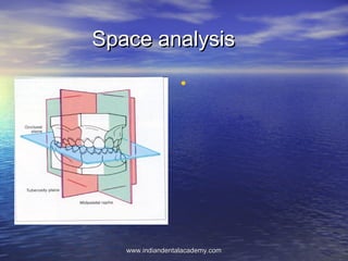
- 11. INTRAMAXILLARYINTRAMAXILLARY SYMMETRY.SYMMETRY. • These symmetryThese symmetry analyses estimate theanalyses estimate the right-left differences inright-left differences in transverse andtransverse and anteroposterior toothanteroposterior tooth positionspositions (Korbitz1909)(Korbitz1909) • Midpalatal raphe &Midpalatal raphe & tuberosity planetuberosity plane www.indiandentalacademy.comwww.indiandentalacademy.com
- 12. Symmetrograph of BernklauSymmetrograph of Bernklau www.indiandentalacademy.comwww.indiandentalacademy.com
- 13. Analysis of Transverse symmetryAnalysis of Transverse symmetry • Symmetric / asymmetric widthSymmetric / asymmetric width development between right & left sides ofdevelopment between right & left sides of the archthe arch • Congruence / incongruence betweenCongruence / incongruence between dental midline & skeletal midlinedental midline & skeletal midline www.indiandentalacademy.comwww.indiandentalacademy.com
- 14. MidlineMidline • Dental midline deviation in theDental midline deviation in the upper arch.upper arch. • The contact point of the upperThe contact point of the upper central incisors is shifted to thecentral incisors is shifted to the right, in relation to the midsagittalright, in relation to the midsagittal plane, i.e. to the side with lack ofplane, i.e. to the side with lack of space for the canine.space for the canine. • Reichenbach and Bruckel, 1967).Reichenbach and Bruckel, 1967). dental midline shift in the mandibulardental midline shift in the mandibular arch.The contact point of the lowerarch.The contact point of the lower central incisors is deviated to thecentral incisors is deviated to the left as the result of tooth drift: in anleft as the result of tooth drift: in an otherwise well aligned arch, theotherwise well aligned arch, the lower right laterallower right lateral www.indiandentalacademy.comwww.indiandentalacademy.com
- 15. Mixed dentition analysisMixed dentition analysis • Huckabas analysisHuckabas analysis • Hixon&Oldfathers analysisHixon&Oldfathers analysis • Nance Caryes analysisNance Caryes analysis • Moyers Mixed dentition analysisMoyers Mixed dentition analysis • Tanaka johnstoneTanaka johnstone • Total space analysisTotal space analysis www.indiandentalacademy.comwww.indiandentalacademy.com
- 16. Mixed dentitionMixed dentition • Three approaches have been employed to estimate theThree approaches have been employed to estimate the mesiodistal crown widths of unerupted canines andmesiodistal crown widths of unerupted canines and premolars:premolars: • (1) use of measurements from erupted teeth(1) use of measurements from erupted teeth • (2) use of measurements from radiographs(2) use of measurements from radiographs • (3) use of a combination of measurements from erupted(3) use of a combination of measurements from erupted teeth and from radiographs of unerupted teethteeth and from radiographs of unerupted teeth • This last approach is considered to be the most accurateThis last approach is considered to be the most accurate since it generally has the lowest standard error ofsince it generally has the lowest standard error of estimate.estimate. www.indiandentalacademy.comwww.indiandentalacademy.com
- 17. • The Moyers' 1967 probability tables for computing theThe Moyers' 1967 probability tables for computing the sizes of unerupted canines and premolars weresizes of unerupted canines and premolars were formulated at the University of Michigan from a sampleformulated at the University of Michigan from a sample consisting of northern European white subjects and areconsisting of northern European white subjects and are currently used worldwide.currently used worldwide. www.indiandentalacademy.comwww.indiandentalacademy.com
- 18. • According to Proffit and Fields, the accuracy withAccording to Proffit and Fields, the accuracy with Moyers' method is fairly good for northern EuropeanMoyers' method is fairly good for northern European white children on which the data is based, despite awhite children on which the data is based, despite a tendency to overestimate the size of unerupted teeth.tendency to overestimate the size of unerupted teeth. • Sexual dimorphism has also been confirmed in severalSexual dimorphism has also been confirmed in several studies, with specific teeth statistically significantly largerstudies, with specific teeth statistically significantly larger in males than females.in males than females. www.indiandentalacademy.comwww.indiandentalacademy.com
- 19. Hixon & OldfatherHixon & Oldfather • 19771977 Kaplan, Smith, and KanarekKaplan, Smith, and Kanarek compared the prediction methodscompared the prediction methods of Hixon and Oldfather, Moyers, and Tanaka and Johnston byof Hixon and Oldfather, Moyers, and Tanaka and Johnston by regression analysis and found the Hixon and Oldfather estimate toregression analysis and found the Hixon and Oldfather estimate to be the most accurate in their sample of 104 white children.be the most accurate in their sample of 104 white children. • 1979 Gardner1979 Gardner found that the methods of Nance, Moyers, andfound that the methods of Nance, Moyers, and Tanaka and Johnston tended to overpredict by 1 to 3 mm, whereasTanaka and Johnston tended to overpredict by 1 to 3 mm, whereas the Hixon and Oldfather technique was more likely to underpredictthe Hixon and Oldfather technique was more likely to underpredict by about 0.5 mmby about 0.5 mm • Hixon and OldfatherHixon and Oldfather prediction did not appear to be seriouslyprediction did not appear to be seriously influenced by sex or the type of dental occlusion.influenced by sex or the type of dental occlusion. www.indiandentalacademy.comwww.indiandentalacademy.com
- 20. • The M-D width of the mandibular 1&2 from castsThe M-D width of the mandibular 1&2 from casts • Determine width of 3,4,5 from radiographDetermine width of 3,4,5 from radiograph • Sum up the width of the central & lateral incisorSum up the width of the central & lateral incisor along with the width of unerupted premolar ofalong with the width of unerupted premolar of that sidethat side • The estimated sum total width of the cuspids &The estimated sum total width of the cuspids & bicuspids of that particular side can be obtainedbicuspids of that particular side can be obtained from the chartfrom the chart • Every measured sum width of incisors &Every measured sum width of incisors & bicuspids has corresponding sum width of thebicuspids has corresponding sum width of the cuspids & bicuspids in the chartcuspids & bicuspids in the chart • www.indiandentalacademy.comwww.indiandentalacademy.com
- 21. Nance Careys analysisNance Careys analysis • Measure M-D width of the erupted permanentMeasure M-D width of the erupted permanent teethteeth • Measure (3,4,5) from radiographsMeasure (3,4,5) from radiographs • The total M-D width of all the teeth in eachThe total M-D width of all the teeth in each quadrant will indicate space required toquadrant will indicate space required to accommodate the permanentaccommodate the permanent • Using brass wire, measure the arch perimeterUsing brass wire, measure the arch perimeter • Compare the space required & space availableCompare the space required & space available to arrive at the arch length discrepancyto arrive at the arch length discrepancy www.indiandentalacademy.comwww.indiandentalacademy.com
- 22. Tanaka and Johnston predictionTanaka and Johnston prediction values 1974values 1974 One half of the M-D width of the four lower incisors + 10.5 = Estimated width of mandibular Canine & premolar in one quadrant + 11.0 = Estimated width of maxillary Canine & premolar in one quadrant www.indiandentalacademy.comwww.indiandentalacademy.com
- 23. Huckaba 1967Huckaba 1967 True width of primary molar = true width of unerupted premolarTrue width of primary molar = true width of unerupted premolar apparent width of primary apparent width of unerupted premolarapparent width of primary apparent width of unerupted premolar molarmolar www.indiandentalacademy.comwww.indiandentalacademy.com
- 24. Total space analysis-MerrifieldTotal space analysis-Merrifield19781978 Anterior areaAnterior area Tooth measurementTooth measurement • Measurement of mandibular incisorsMeasurement of mandibular incisors widths on the cast were added to valueswidths on the cast were added to values obtained from the radio graphicobtained from the radio graphic measurements of the canines.measurements of the canines. www.indiandentalacademy.comwww.indiandentalacademy.com
- 25. • Cephalometric correction wasCephalometric correction was calculated for the Tweedcalculated for the Tweed methodmethod • FMIA was taken intoFMIA was taken into considerationconsideration • The incisors were repositionedThe incisors were repositioned and the difference in the actualand the difference in the actual and proposed FMIA isand proposed FMIA is determined.determined. • The difference in angulation isThe difference in angulation is multiplied by 0.8 to get themultiplied by 0.8 to get the difference in mmdifference in mm www.indiandentalacademy.comwww.indiandentalacademy.com
- 26. • Soft tissue modificationSoft tissue modification • Upper lip thickness from the vermilionUpper lip thickness from the vermilion border of the upper lip to the greatestborder of the upper lip to the greatest curvature of the labial surface of thecurvature of the labial surface of the central incisorcentral incisor • The total chin thickness from the softThe total chin thickness from the soft tissue chin to the N-B linetissue chin to the N-B line www.indiandentalacademy.comwww.indiandentalacademy.com
- 27. • If the lip thickness is greater than chinIf the lip thickness is greater than chin thickness the diff is determined andthickness the diff is determined and multiplied by 2 and added to the spacemultiplied by 2 and added to the space required. If it is less than or equal to chinrequired. If it is less than or equal to chin thickness no soft tissue modification isthickness no soft tissue modification is necessarynecessary www.indiandentalacademy.comwww.indiandentalacademy.com
- 28. • Measure the Z angle of Merrifield and addMeasure the Z angle of Merrifield and add the cephalometric correction to it.the cephalometric correction to it. • If the corrected Z angle is greater than 80If the corrected Z angle is greater than 80 the mandibular incisor angulation wasthe mandibular incisor angulation was modified as necessary upto an IMPA of 92modified as necessary upto an IMPA of 92 • If the corrected angle is less than 75If the corrected angle is less than 75 additional uprighting of the mandibularadditional uprighting of the mandibular incisor is necessaryincisor is necessary www.indiandentalacademy.comwww.indiandentalacademy.com
- 29. Middle areaMiddle area • Measure the M-D with of the 1Measure the M-D with of the 1stst permanentpermanent molar of the castmolar of the cast • Curve of SPEE, the deepest pointCurve of SPEE, the deepest point between the flat surface and the occlusalbetween the flat surface and the occlusal surface is measured on both sidessurface is measured on both sides www.indiandentalacademy.comwww.indiandentalacademy.com
- 31. Posterior AreaPosterior Area • MD width of the 2MD width of the 2ndnd and 3and 3rdrd molar is obtained frommolar is obtained from the radiograph.the radiograph. • Wheelers method is used for calculationWheelers method is used for calculation Y – XY – X11 X=X= YY11 X –estimated value of 3rd molarX –estimated value of 3rd molar X1 –wheelers value of 3rd molarX1 –wheelers value of 3rd molar Y - actual size of 6Y - actual size of 6 Y 1- wheelers value for 6Y 1- wheelers value for 6 www.indiandentalacademy.comwww.indiandentalacademy.com
- 32. Permanent dentition analysisPermanent dentition analysis • Ponts indexPonts index • Korkhaus analysisKorkhaus analysis • Linder Harth indexLinder Harth index • Arch perimeter analysisArch perimeter analysis • Bolton tooth size ratioBolton tooth size ratio • Howes analysisHowes analysis • Peck& peck indexPeck& peck index www.indiandentalacademy.comwww.indiandentalacademy.com
- 33. Ponts index 1909Ponts index 1909 • Pont in 1909 suggested a method for determining thePont in 1909 suggested a method for determining the ideal dental arch width from the combined M-D width ofideal dental arch width from the combined M-D width of the maxillary central incisorsthe maxillary central incisors • Ideal arch width in the premolar regionIdeal arch width in the premolar region X x 100X x 100 8080 Ideal arch width in the molar regionIdeal arch width in the molar region X x 100X x 100 6464 www.indiandentalacademy.comwww.indiandentalacademy.com
- 34. • InferenceInference • If calculated value is greater than theIf calculated value is greater than the measure value ,then arch is narrow formeasure value ,then arch is narrow for sum of incisors-needs expansionsum of incisors-needs expansion • If measured value is greater - arch wide –If measured value is greater - arch wide – no scope for expansionno scope for expansion www.indiandentalacademy.comwww.indiandentalacademy.com
- 35. Linder Harth indexLinder Harth index • Similar to PontsSimilar to Ponts • Ideal arch width in the premolar regionIdeal arch width in the premolar region X x 100X x 100 8080 • Ideal arch width in the molar regionIdeal arch width in the molar region X x 100X x 100 6464 www.indiandentalacademy.comwww.indiandentalacademy.com
- 36. Korkhaus analysis 1939Korkhaus analysis 1939 • Using Linder Harth measurement introduced a thirdUsing Linder Harth measurement introduced a third measurement from the midpoint of the inter-premolar linemeasurement from the midpoint of the inter-premolar line of upper arch to point incison.of upper arch to point incison. • For a particular width of incisors there is a specific valueFor a particular width of incisors there is a specific value of the distance from incison to the inter-premolar lineof the distance from incison to the inter-premolar line • An orthometer was devised which directly measures theAn orthometer was devised which directly measures the ideal arch width in premolar and molar region &alsoideal arch width in premolar and molar region &also perpendicular distance from the inter –premolar line toperpendicular distance from the inter –premolar line to the incison for a given sum mesio-distal width of thethe incison for a given sum mesio-distal width of the maxillary incisors (21/12)maxillary incisors (21/12) www.indiandentalacademy.comwww.indiandentalacademy.com
- 37. Ashleys HowesAshleys Howes • According to Howe, crowding is not onlyAccording to Howe, crowding is not only due to tooth sise ,but as a result whendue to tooth sise ,but as a result when there is inadequate apical basethere is inadequate apical base • PMBAW x 100PMBAW x 100 TMTM www.indiandentalacademy.comwww.indiandentalacademy.com
- 38. • InferenceInference • The patient values should fall within the suggested rangeThe patient values should fall within the suggested range • If PMD>PMBAW, expansion is contraindicatedIf PMD>PMBAW, expansion is contraindicated • If PMBAW>PMD, expansion is indicatedIf PMBAW>PMD, expansion is indicated • If PMBAW x 100If PMBAW x 100 TMTM Less than 37% -basal arch deficiency—extractionLess than 37% -basal arch deficiency—extraction If 44% --ideal case—extraction not requiredIf 44% --ideal case—extraction not required If between 37% - 44% boderline caseIf between 37% - 44% boderline case www.indiandentalacademy.comwww.indiandentalacademy.com
- 39. Arch perimeter analysisArch perimeter analysis • This analysis helps to find theThis analysis helps to find the difference between the basaldifference between the basal bone & the tooth material.bone & the tooth material. • The soft wire is contoured,fromThe soft wire is contoured,from the mesio-buccal line angle ofthe mesio-buccal line angle of molar & pass along themolar & pass along the contacts of the premolar&contacts of the premolar& through the incisive papilla onthrough the incisive papilla on an imaginary repositioned archan imaginary repositioned arch .. • Tooth material - spaceTooth material - space requiredrequired • Arch perimeter - spaceArch perimeter - space availableavailable www.indiandentalacademy.comwww.indiandentalacademy.com
- 40. Careys analysis (1949)Careys analysis (1949) InferenceInference • If the amount of discrepancy is betweenIf the amount of discrepancy is between 0 to 2.5mm – non-extraction case0 to 2.5mm – non-extraction case • If it is between 2.5 to 5mm –extraction ofIf it is between 2.5 to 5mm –extraction of second premolars is recommendedsecond premolars is recommended • If it is more than 5mm – extraction of firstIf it is more than 5mm – extraction of first premolar is recommendedpremolar is recommended www.indiandentalacademy.comwww.indiandentalacademy.com
- 41. Peck and Peck indexPeck and Peck index • According to Peck ideal incisal arrangement had smallerAccording to Peck ideal incisal arrangement had smaller mesiodistal & comparatively larger labio lingual widthmesiodistal & comparatively larger labio lingual width than in persons with incisal crowding .than in persons with incisal crowding . • On the basis of this Peck suggested certain clinicalOn the basis of this Peck suggested certain clinical guidelines.guidelines. MD x 100MD x 100 LLLL Mean value for lower central incisor should be 88% to 92%Mean value for lower central incisor should be 88% to 92% Mean value for lateral incisors – 90% to 95%Mean value for lateral incisors – 90% to 95% www.indiandentalacademy.comwww.indiandentalacademy.com
- 43. Boltons analysisBoltons analysis • In 1958, Bolton published his work on interpretingIn 1958, Bolton published his work on interpreting mesiodistal tooth size dimensions and their effect onmesiodistal tooth size dimensions and their effect on occlusion.occlusion. • Bolton selected 55 cases with excellent occlusions, mostBolton selected 55 cases with excellent occlusions, most of which (44) had been treated orthodonticallyof which (44) had been treated orthodontically (nonextraction).(nonextraction). • The mesiodistal widths of the 12 maxillary teeth (firstThe mesiodistal widths of the 12 maxillary teeth (first molar to first molar) were totaled and compared with themolar to first molar) were totaled and compared with the sum derived by the same procedure carried out on thesum derived by the same procedure carried out on the 12 mandibular teeth.12 mandibular teeth. • The ratio derived between the two is the percentageThe ratio derived between the two is the percentage relationship of mandibular arch length to maxillary archrelationship of mandibular arch length to maxillary arch length.length. www.indiandentalacademy.comwww.indiandentalacademy.com
- 44. • He concluded that anHe concluded that an overall ratio of 91.3 andoverall ratio of 91.3 and an anterior ratio of 77.2an anterior ratio of 77.2 were necessary forwere necessary for proper coordination of theproper coordination of the maxillary and mandibularmaxillary and mandibular teeth.teeth. www.indiandentalacademy.comwww.indiandentalacademy.com
- 45. • If the overall ratio is greater than 91.3%If the overall ratio is greater than 91.3% ,then there is an excess of mandibular,then there is an excess of mandibular tooth materialtooth material • Actual mand 12 – Corrected mand 12 =Actual mand 12 – Corrected mand 12 = • Actual max 12 _ corrected maxActual max 12 _ corrected max www.indiandentalacademy.comwww.indiandentalacademy.com
- 46. • He computed the specific ratios of theHe computed the specific ratios of the mesiodistal widths must exist betweenmesiodistal widths must exist between maxillary and mandibular teeth from bothmaxillary and mandibular teeth from both canine-canine and first molar-first molar tocanine-canine and first molar-first molar to obtain optimum occlusion and to achieveobtain optimum occlusion and to achieve proper occlusal interdigitation in theproper occlusal interdigitation in the finishing stages of orthodontic treatment.finishing stages of orthodontic treatment. www.indiandentalacademy.comwww.indiandentalacademy.com
- 47. • Black was one of the first investigators to measure toothBlack was one of the first investigators to measure tooth sizes and his tables of mean tooth sizes are still usedsizes and his tables of mean tooth sizes are still used today.today. • The tooth size measurements of Wheeler also areThe tooth size measurements of Wheeler also are frequently used.frequently used. • Ballard measured 500 sets of models, evaluatingBallard measured 500 sets of models, evaluating asymmetry in tooth sizes. Ninety percent of his sampleasymmetry in tooth sizes. Ninety percent of his sample showed a right-to-left discrepancy of 0.25 mm or more inshowed a right-to-left discrepancy of 0.25 mm or more in the mesiodistal width of one or more pairs of teeth.the mesiodistal width of one or more pairs of teeth. • His observations led to the conclusion that asymmetry isHis observations led to the conclusion that asymmetry is the rule, not the exception, and that judicious enamelthe rule, not the exception, and that judicious enamel reduction or "stripping" is sometimes necessary,reduction or "stripping" is sometimes necessary, particularly in the anterior segments to gain properparticularly in the anterior segments to gain proper interdigitation of teethinterdigitation of teeth www.indiandentalacademy.comwww.indiandentalacademy.com
- 50. • Lundström 1954 studied 319 13-year-old children andLundström 1954 studied 319 13-year-old children and reported on the variation in intermaxillary tooth widthreported on the variation in intermaxillary tooth width ratio. The mesiodistal widths were recorded and theratio. The mesiodistal widths were recorded and the dispersion for the three tooth size indices weredispersion for the three tooth size indices were calculated:calculated: • His results demonstrated a large biologic dispersion inHis results demonstrated a large biologic dispersion in the tooth width ratio. It was great enough to have anthe tooth width ratio. It was great enough to have an impact on the final tooth position, teeth alignment, andimpact on the final tooth position, teeth alignment, and overbite and overjet relationships in a large number ofoverbite and overjet relationships in a large number of these patients. This same formula originally wasthese patients. This same formula originally was developed by Bolton to observe mesiodistal tooth sizedeveloped by Bolton to observe mesiodistal tooth size discrepancies.discrepancies. www.indiandentalacademy.comwww.indiandentalacademy.com
- 52. TOTAL DENTITION SPACE ANALYSISTOTAL DENTITION SPACE ANALYSIS • MerrifieldMerrifield • lSince the original diagnosis and treatment plan must accept thelSince the original diagnosis and treatment plan must accept the dimensions of the denture presented in the original malocclusiondimensions of the denture presented in the original malocclusion when musculature is normal (i.e., Class I), a total dentition spacewhen musculature is normal (i.e., Class I), a total dentition space analysis allows the clinician to develop a differential diagnosis thatanalysis allows the clinician to develop a differential diagnosis that respects the dimensions of the denture concept during the treatmentrespects the dimensions of the denture concept during the treatment planning process.planning process. • (1) anterior, (2) midarch, and (3) posterior.(1) anterior, (2) midarch, and (3) posterior. • (1) simplicity in identifying the area of space deficit or space(1) simplicity in identifying the area of space deficit or space surplus,surplus, • (2) a more accurate differential diagnosis.(2) a more accurate differential diagnosis. www.indiandentalacademy.comwww.indiandentalacademy.com
- 53. ANTERIOR SPACE ANALYSISANTERIOR SPACE ANALYSIS • space available in the mandibular arch from canine to canine and aspace available in the mandibular arch from canine to canine and a measurement of the six anterior teeth mesiodistally.measurement of the six anterior teeth mesiodistally. • The difference is referred to as a surplus or a deficit.The difference is referred to as a surplus or a deficit. • Tweed's diagnostic facial triangle is also used to further analyze thisTweed's diagnostic facial triangle is also used to further analyze this area.area. • A head film discrepancy, based on the amount of mandibular incisorA head film discrepancy, based on the amount of mandibular incisor uprighting that is needed to restore facial balance, is added to theuprighting that is needed to restore facial balance, is added to the anterior space measurement. The total, if a deficit, is referred to asanterior space measurement. The total, if a deficit, is referred to as anterior discrepancy. Anterior discrepancies are most easilyanterior discrepancy. Anterior discrepancies are most easily resolved, if they are the overriding consideration of theresolved, if they are the overriding consideration of the malocclusion, by removal of the first premolar teeth and by using themalocclusion, by removal of the first premolar teeth and by using the resulting space to move the canines distally to obtain the space toresulting space to move the canines distally to obtain the space to upright and align the incisors.upright and align the incisors. www.indiandentalacademy.comwww.indiandentalacademy.com
- 54. MIDARCH ANALYSISMIDARCH ANALYSIS • Careful analysis of this area can show mesially inclined first molars,Careful analysis of this area can show mesially inclined first molars, rotations, spaces, deep curves of Spee, crossbites, missing teeth, habitrotations, spaces, deep curves of Spee, crossbites, missing teeth, habit abnormality, blocked out teeth, and occlusal disharmonies.abnormality, blocked out teeth, and occlusal disharmonies. • Crowding, deep curves of Spee, end-on, and Class II occlusions notCrowding, deep curves of Spee, end-on, and Class II occlusions not accompanied by anterior discrepancy, all indicate a need for secondaccompanied by anterior discrepancy, all indicate a need for second premolar extraction in the lower arch.premolar extraction in the lower arch. • careful measurement of the space from the distal of the canine to the distalcareful measurement of the space from the distal of the canine to the distal of the first molar should be recorded as available midarch space.of the first molar should be recorded as available midarch space. • To this is added the space required to level the curve of Spee. From theseTo this is added the space required to level the curve of Spee. From these measurements one can determine the space deficit or surplus in this area.measurements one can determine the space deficit or surplus in this area. • Many diagnosticians have suggested that they extract second premolarMany diagnosticians have suggested that they extract second premolar teeth to eliminate facial retrusion. This is faulty reasoning. These casesteeth to eliminate facial retrusion. This is faulty reasoning. These cases have, as a rule, very little anterior discrepancy, and the second premolarshave, as a rule, very little anterior discrepancy, and the second premolars are removed because their space is most advantageously used for theare removed because their space is most advantageously used for the midarch problems that these cases usually demonstrate. The midarchmidarch problems that these cases usually demonstrate. The midarch space analysis is critical in proper differential diagnosis.space analysis is critical in proper differential diagnosis. www.indiandentalacademy.comwww.indiandentalacademy.com
- 55. POSTERIOR SPACE ANALYSISPOSTERIOR SPACE ANALYSIS • The posterior denture area has great importance, and has at times beenThe posterior denture area has great importance, and has at times been ignored or mistreated by our specialty. The required space in the posteriorignored or mistreated by our specialty. The required space in the posterior space analysis is the mesiodistal width of the second molars and the thirdspace analysis is the mesiodistal width of the second molars and the third molars in the mandibular arch. The available space is more difficult tomolars in the mandibular arch. The available space is more difficult to ascertain on the immature patient. It is a measurement in millimeters of theascertain on the immature patient. It is a measurement in millimeters of the space distal to the mandibular first molars along the occlusal plane to thespace distal to the mandibular first molars along the occlusal plane to the anterior border of the ramus, plus an estimate of posterior arch lengthanterior border of the ramus, plus an estimate of posterior arch length increase, based on both age and sex.increase, based on both age and sex. • There are certain variables that must be considered in estimating theThere are certain variables that must be considered in estimating the increase in posterior space available. These variables are as follows:increase in posterior space available. These variables are as follows: • 1. Rate of mesioocclusal migration of the mandibular first molar.1. Rate of mesioocclusal migration of the mandibular first molar. • 2. Rate of resorption of the anterior border of the ramus.2. Rate of resorption of the anterior border of the ramus. • 3. Time of cessation of molar migration.3. Time of cessation of molar migration. • 4. Time of cessation of ramus resorption.4. Time of cessation of ramus resorption. • 5. Sex.5. Sex. • 6. Age.6. Age. www.indiandentalacademy.comwww.indiandentalacademy.com
- 56. • A review and study of the literature10-12 reveals that a consensus ofA review and study of the literature10-12 reveals that a consensus of researchers suggests 3 mm of increase in the posterior denture area occursresearchers suggests 3 mm of increase in the posterior denture area occurs per year until age 14 years for girls and age 16 years for boys. This is a 1.5per year until age 14 years for girls and age 16 years for boys. This is a 1.5 mm increase on each side per year after the full eruption of the first molars.mm increase on each side per year after the full eruption of the first molars. In the mature patient, girls beyond 15 years and boys beyond 16 years, oneIn the mature patient, girls beyond 15 years and boys beyond 16 years, one can measure from the distal of the first molar to the anterior border of thecan measure from the distal of the first molar to the anterior border of the ramus at the occlusal plane and have an accurate determination of theramus at the occlusal plane and have an accurate determination of the space available in the posterior area. It is of extreme importance to knowspace available in the posterior area. It is of extreme importance to know whether there is a surplus or deficit of space in this area during diagnosiswhether there is a surplus or deficit of space in this area during diagnosis and treatment planning. It is imprudent to create a posterior discrepancyand treatment planning. It is imprudent to create a posterior discrepancy while making adjustments in other areas— the midarch, or in the anteriorwhile making adjustments in other areas— the midarch, or in the anterior area. It is equally imprudent not to use a posterior space surplus to helparea. It is equally imprudent not to use a posterior space surplus to help alleviate midarch and anterior deficits. The most easily recognizablealleviate midarch and anterior deficits. The most easily recognizable symptom of a posterior deficit on the young patient is the late eruption of thesymptom of a posterior deficit on the young patient is the late eruption of the second molar. If space is not available for this tooth by the age of its normalsecond molar. If space is not available for this tooth by the age of its normal eruption, then one can pretty well ascertain that there is a posterior spaceeruption, then one can pretty well ascertain that there is a posterior space problem. A good lateral jaw radiograph can immediately confirm the clinicalproblem. A good lateral jaw radiograph can immediately confirm the clinical observation by using the above-mentioned guidelines.observation by using the above-mentioned guidelines. www.indiandentalacademy.comwww.indiandentalacademy.com
- 57. • In summary, a total space analysis that analyzes the anterior, midarch, andIn summary, a total space analysis that analyzes the anterior, midarch, and posterior denture areas is a valuable diagnostic tool. It enables theposterior denture areas is a valuable diagnostic tool. It enables the orthodontic specialist to treat within the dimensions of the denture in theorthodontic specialist to treat within the dimensions of the denture in the case with normal muscular balance. A total dentition space analysis, usedcase with normal muscular balance. A total dentition space analysis, used within the dimensions of the denture framework, enables the orthodontist towithin the dimensions of the denture framework, enables the orthodontist to make correct differential diagnostic decisions.make correct differential diagnostic decisions. • Diagnosis, by definition, is both subjective and objective. Webster definesDiagnosis, by definition, is both subjective and objective. Webster defines diagnosis as a "determination of a disease from symptoms, data, or testsdiagnosis as a "determination of a disease from symptoms, data, or tests and the decisions and judgements made prior to treatment." Thus theand the decisions and judgements made prior to treatment." Thus the determination made in regard to whether, when, and which teeth need to bedetermination made in regard to whether, when, and which teeth need to be eliminated for proper space management is a differential diagnosticeliminated for proper space management is a differential diagnostic process. When diagnostic guidelines or decisions are suggested, they canprocess. When diagnostic guidelines or decisions are suggested, they can appropriately be called "one man's opinion." The following diagnostic spaceappropriately be called "one man's opinion." The following diagnostic space management guidelines are suggested for use and should not bemanagement guidelines are suggested for use and should not be considered as rules. These space management suggestions are based onconsidered as rules. These space management suggestions are based on space analysis only. Any complete diagnostic scheme has to consider thespace analysis only. Any complete diagnostic scheme has to consider the facial pattern and the skeletal pattern.facial pattern and the skeletal pattern. www.indiandentalacademy.comwww.indiandentalacademy.com
- 58. • Lower incisor space analysis - Harris, Vaden, and WilliamsLower incisor space analysis - Harris, Vaden, and Williams • ---------------------------------------------------------------- • The common situation in which the incisors are labially displaced against the corticalThe common situation in which the incisors are labially displaced against the cortical plate is difficult to assess from the casts alone. During uprighting of the incisors, theplate is difficult to assess from the casts alone. During uprighting of the incisors, the radius of the anterior arch decreases and, along with it, the actual space available.radius of the anterior arch decreases and, along with it, the actual space available. This sort of error can be minimized by inspecting the incisor positions on the lateralThis sort of error can be minimized by inspecting the incisor positions on the lateral head film and adjusting the space available accordingly. One such method is tohead film and adjusting the space available accordingly. One such method is to calculate a ''head film discrepancy value." This is the millimetric distance the lowercalculate a ''head film discrepancy value." This is the millimetric distance the lower incisors must be uprighted in order to be placed over basal bone and into a positionincisors must be uprighted in order to be placed over basal bone and into a position of balance with the facial structures.24 Typically this procedure is indicated becauseof balance with the facial structures.24 Typically this procedure is indicated because often all six anterior teeth need to be retracted and uprighted— and the increase inoften all six anterior teeth need to be retracted and uprighted— and the increase in the required space has to be gained from extractions in the midarch. A case in pointthe required space has to be gained from extractions in the midarch. A case in point has been illustrated (Fig. 2, B); the casts exhibit spacing of the lower anterior teeth,has been illustrated (Fig. 2, B); the casts exhibit spacing of the lower anterior teeth, but they are proclined to an IMPA of 97° and are at the anterior limit of the alveolarbut they are proclined to an IMPA of 97° and are at the anterior limit of the alveolar bone. Treatment involved uprighting the lower incisors 9° and retracting them 6 mm (Ibone. Treatment involved uprighting the lower incisors 9° and retracting them 6 mm (I to NP decreased 6 mm).to NP decreased 6 mm). www.indiandentalacademy.comwww.indiandentalacademy.com
- 59. • Source: AJO-DO on CD-ROM (Copyright © 1998 AJO-DO), VolumeSource: AJO-DO on CD-ROM (Copyright © 1998 AJO-DO), Volume 1987 Nov (375 - 380): Lower incisor space analysis - Harris, Vaden,1987 Nov (375 - 380): Lower incisor space analysis - Harris, Vaden, and Williamsand Williams • ---------------------------------------------------------------- • One other diagnostic consideration that warrants attention is theOne other diagnostic consideration that warrants attention is the soft-tissue profile and its relation to tooth position. A compensationsoft-tissue profile and its relation to tooth position. A compensation should be made in any diagnostic scheme for a maldistribution ofshould be made in any diagnostic scheme for a maldistribution of soft-tissue thicknesses. One approach25 is to compare total chinsoft-tissue thicknesses. One approach25 is to compare total chin thickness (Pg' measured normal to the nasion-B-point line) withthickness (Pg' measured normal to the nasion-B-point line) with upper lip thickness. Integumental compensation is needed in theupper lip thickness. Integumental compensation is needed in the direction of more upright lower incisors in those patients possessingdirection of more upright lower incisors in those patients possessing less total chin thickness than upper lip thickness. Compensation isless total chin thickness than upper lip thickness. Compensation is not indicated when the total chin and upper lip thicknesses arenot indicated when the total chin and upper lip thicknesses are equal.25equal.25 www.indiandentalacademy.comwww.indiandentalacademy.com
- 60. • Source: AJO-DO on CD-ROM (Copyright © 1998 AJO-DO), VolumeSource: AJO-DO on CD-ROM (Copyright © 1998 AJO-DO), Volume 1994 Nov (535 - 542): Dimensions of the denture - Merrifield1994 Nov (535 - 542): Dimensions of the denture - Merrifield • ---------------------------------------------------------------- • INTRODUCTION TO DEFICITS AND DECISIONS SpaceINTRODUCTION TO DEFICITS AND DECISIONS Space management guidancemanagement guidance • A. Anterior surplus or deficit:+ to -2mmA. Anterior surplus or deficit:+ to -2mm SpaceSpace Management NonextractionManagement Nonextraction • 3 to 5 mm without crowding.3 to 5 mm without crowding. Extract:Extract: • 3 to 5 mm with crowding.3 to 5 mm with crowding. Extract:Extract: • 5 to 7 mm with less than 3 mm anterior crowding.5 to 7 mm with less than 3 mm anterior crowding. Extract:Extract: • 5 to 7 mm with more than 3 mm anterior crowding.5 to 7 mm with more than 3 mm anterior crowding. Extract:Extract: • 7 to 15 mm anterior deficit.7 to 15 mm anterior deficit. Extract:Extract: • 16 mm and above.16 mm and above. Extract:Extract: www.indiandentalacademy.comwww.indiandentalacademy.com
- 61. • Source: AJO-DO on CD-ROM (Copyright © 1998 AJO-DO), Volume 1994Source: AJO-DO on CD-ROM (Copyright © 1998 AJO-DO), Volume 1994 Nov (535 - 542): Dimensions of the denture - MerrifieldNov (535 - 542): Dimensions of the denture - Merrifield • ---------------------------------------------------------------- • B. Midarch surplus or deficit: An anterior deficit or surplus overrides aB. Midarch surplus or deficit: An anterior deficit or surplus overrides a midarch deficit so the first determination is a decision on the anterior deficit.midarch deficit so the first determination is a decision on the anterior deficit. • + to 3 mm+ to 3 mm NonextractionNonextraction • 3 to 5 mm without crowding.3 to 5 mm without crowding. Extract:Extract: • 3 to 5 mm with Class II molar.3 to 5 mm with Class II molar. Extract:Extract: • 5 to 7 mm with upper anterior protrusion.5 to 7 mm with upper anterior protrusion. Extract:Extract: • 5 to 7 mm5 to 7 mm Extract:Extract: • 8 to 15 mm8 to 15 mm Extract:Extract: • Over 15 mmOver 15 mm Extract:Extract: • *(Use X for all molars: first, second, and third.)*(Use X for all molars: first, second, and third.) www.indiandentalacademy.comwww.indiandentalacademy.com
- 62. • Source: AJO-DO on CD-ROM (Copyright © 1998 AJO-DO), Volume 1994 Nov (535 - 542): Dimensions of theSource: AJO-DO on CD-ROM (Copyright © 1998 AJO-DO), Volume 1994 Nov (535 - 542): Dimensions of the denture - Merrifielddenture - Merrifield • ---------------------------------------------------------------- • C. Posterior surplus or deficit: The space analysis m this area is of great importance, although in correctiveC. Posterior surplus or deficit: The space analysis m this area is of great importance, although in corrective procedures, anterior and midarch deficits are overriding. The posterior space must be carefully measured andprocedures, anterior and midarch deficits are overriding. The posterior space must be carefully measured and protected. No orthodontic treatment is complete until all decisions and treatment procedures are completed in thisprotected. No orthodontic treatment is complete until all decisions and treatment procedures are completed in this area.area. • + to -5 mm with good position of the third molars. Await full development of the third molars.+ to -5 mm with good position of the third molars. Await full development of the third molars. • + to -5 mm with poor position of third molars.+ to -5 mm with poor position of third molars. Extract:Extract: • Note: Wait for maxillary third molars until age 16 years. Have the mandibular third molars removed immediately ifNote: Wait for maxillary third molars until age 16 years. Have the mandibular third molars removed immediately if other treatment is necessary.other treatment is necessary. • 5 to 15 mm.5 to 15 mm. Extract:Extract: • (Determine the timing of these third molar extractions in relationship to symptoms and other treatment that is(Determine the timing of these third molar extractions in relationship to symptoms and other treatment that is necessary.)necessary.) • Consistent, quality orthodontic treatment results are based on fundamental concepts. The concept of dimensionsConsistent, quality orthodontic treatment results are based on fundamental concepts. The concept of dimensions of the denture is predicated on the conviction that the teeth and their supporting structures should be in a state ofof the denture is predicated on the conviction that the teeth and their supporting structures should be in a state of maximum environmental harmony (dynamic equilibrium). Total dentition space analysis, based on the dimensionmaximum environmental harmony (dynamic equilibrium). Total dentition space analysis, based on the dimension of the denture concept, is a valuable tool that can help the orthodontic specialist produce a consistently highof the denture concept, is a valuable tool that can help the orthodontic specialist produce a consistently high quality result that meets the needs and expectations of the patient.quality result that meets the needs and expectations of the patient. www.indiandentalacademy.comwww.indiandentalacademy.com
- 63. • Anterior space analysis: Anterior space analysis includes the measurment inAnterior space analysis: Anterior space analysis includes the measurment in millimeters of the space available in the mandibular arch from canine tomillimeters of the space available in the mandibular arch from canine to canine and a measurment of the mesiodistal dimension of each of the sixcanine and a measurment of the mesiodistal dimension of each of the six anterior teeth. The difference is referred to as a surplus or deficit. Theanterior teeth. The difference is referred to as a surplus or deficit. The Tweed diagnostic facial triangle is also used to further analyze this area.Tweed diagnostic facial triangle is also used to further analyze this area. Lateral headfilm discrepancy is the amount of space required to position theLateral headfilm discrepancy is the amount of space required to position the mandibular incisors for facial balance. This value is added to the anteriormandibular incisors for facial balance. This value is added to the anterior space measurement.space measurement. • The thickness of the soft tissue (upper lip versus total chin) must also beThe thickness of the soft tissue (upper lip versus total chin) must also be considered as part of the anterior space analysis. Total chin thicknessconsidered as part of the anterior space analysis. Total chin thickness should equal upper lip thickness. If it is less than upper lip thickness, theshould equal upper lip thickness. If it is less than upper lip thickness, the anterior teeth must be uprighted further to create a more balanced profileanterior teeth must be uprighted further to create a more balanced profile because lip retraction follows tooth uprighting.because lip retraction follows tooth uprighting. • The sum of the anterior tooth arch surplus or deficit, the cephalometricThe sum of the anterior tooth arch surplus or deficit, the cephalometric discrepancy, and the soft tissue thickness imbalance is referred to as thediscrepancy, and the soft tissue thickness imbalance is referred to as the anterior discrepancy. Each of the three values in the anterior discrepancyanterior discrepancy. Each of the three values in the anterior discrepancy calculation has been given a difficult factor so that an anterior spacecalculation has been given a difficult factor so that an anterior space analysis difficulty value can be calculated.analysis difficulty value can be calculated. www.indiandentalacademy.comwww.indiandentalacademy.com
- 64. • Midarch space analysis: The midarch area includes theMidarch space analysis: The midarch area includes the mandible first molars and the first and second premolars.mandible first molars and the first and second premolars. Careful analysis of this area may show mesially inclinedCareful analysis of this area may show mesially inclined first molars, rotation, spaces, a deep curve of spee,first molars, rotation, spaces, a deep curve of spee, crossbites missing teeth, habit abnormality, blocked outcrossbites missing teeth, habit abnormality, blocked out teeth, and occlusal disharmonies. This is an extremelyteeth, and occlusal disharmonies. This is an extremely important area of the dentition. Because it is in theimportant area of the dentition. Because it is in the center of the arch, this area allows the easiest and mostcenter of the arch, this area allows the easiest and most direct method of space management for malocclusiondirect method of space management for malocclusion correction when it can be so used. Crowding, a deepcorrection when it can be so used. Crowding, a deep curve of spee, and end – on or full – step Class IIcurve of spee, and end – on or full – step Class II occlusions, not a accomplained by anterior disctepancy,occlusions, not a accomplained by anterior disctepancy, indicate a need for second premolar extraction in theindicate a need for second premolar extraction in the mandibular arch.mandibular arch. www.indiandentalacademy.comwww.indiandentalacademy.com
- 65. • These variables are the following:These variables are the following: • 1. Rate of mesio-occlusal migration of the1. Rate of mesio-occlusal migration of the mandibular first molar.mandibular first molar. • 2. Rate of resorption of the anterior border of the2. Rate of resorption of the anterior border of the ramus.ramus. • 3. Time of cessation of molar migration.3. Time of cessation of molar migration. • 4. Time of cessation of ramus resorption.4. Time of cessation of ramus resorption. • 5. Gender.5. Gender. • 6. Age.6. Age. www.indiandentalacademy.comwww.indiandentalacademy.com
- 66. • A review of the literature 6,22,38 reveals that aA review of the literature 6,22,38 reveals that a consensus of researchers suggests that 3 mm ofconsensus of researchers suggests that 3 mm of increase in the posterior denture area occurs perincrease in the posterior denture area occurs per year until age 14 for girsls and age 16 for boys.year until age 14 for girsls and age 16 for boys. This is an increase of 1.5 mm on each side perThis is an increase of 1.5 mm on each side per year after the full eruption of the first molars. Inyear after the full eruption of the first molars. In the mature patient (girls beyond 15 years andthe mature patient (girls beyond 15 years and boys beyond 16 years) a measyrement from theboys beyond 16 years) a measyrement from the distal of the first molar to the anterior border ofdistal of the first molar to the anterior border of the ramus at the occlusal plane is a valuablethe ramus at the occlusal plane is a valuable determination of the space available in thedetermination of the space available in the posterior area.posterior area. www.indiandentalacademy.comwww.indiandentalacademy.com
- 67. • Taken from the JCO 1985 Jun (445-448): Analytical OrthodonticTaken from the JCO 1985 Jun (445-448): Analytical Orthodontic Computer Programs - DENNIS M. KILLIANY, DDS, MSDComputer Programs - DENNIS M. KILLIANY, DDS, MSD • ---------------------------------------------------------------- • Analytical Orthodontic Computer ProgramsAnalytical Orthodontic Computer Programs • DENNIS M. KILLIANY, DDS, MSDDENNIS M. KILLIANY, DDS, MSD • I have developed a computer program that performs severalI have developed a computer program that performs several diagnostic analyses— cephalometric, mixed dentition, and toothdiagnostic analyses— cephalometric, mixed dentition, and tooth size— and a practice management analysis of patient starts. Thissize— and a practice management analysis of patient starts. This program is written in Basica on an IBM PC-XT. It will also run withprogram is written in Basica on an IBM PC-XT. It will also run with GWBASIC on many IBM-compatible computers. The two versions ofGWBASIC on many IBM-compatible computers. The two versions of the program (ANALYSES.M— monochrome and ANALYSES.C—the program (ANALYSES.M— monochrome and ANALYSES.C— color) are available to any interested practitioner.color) are available to any interested practitioner. www.indiandentalacademy.comwww.indiandentalacademy.com
- 68. • Taken from the JCO 1985 Jun (445-448): Analytical Orthodontic ComputerTaken from the JCO 1985 Jun (445-448): Analytical Orthodontic Computer Programs - DENNIS M. KILLIANY, DDS, MSDPrograms - DENNIS M. KILLIANY, DDS, MSD • ---------------------------------------------------------------- • It is not the purpose of this seminar to discuss the validity of each analysis.It is not the purpose of this seminar to discuss the validity of each analysis. These analyses, by themselves, may not provide sufficient information uponThese analyses, by themselves, may not provide sufficient information upon which to base a diagnosis and treatment plan. Care must be taken inwhich to base a diagnosis and treatment plan. Care must be taken in applying a mathematical analysis to patients. For example, although aapplying a mathematical analysis to patients. For example, although a statistically significant relationship has been shown to exist between the sizestatistically significant relationship has been shown to exist between the size of lower incisors and their crowding, the strength of the relationship is weak.of lower incisors and their crowding, the strength of the relationship is weak. Hence, indiscriminate mesiodistal narrowing of lower incisors based on theirHence, indiscriminate mesiodistal narrowing of lower incisors based on their existing faciolingual dimensions may not lead to a clinically significantexisting faciolingual dimensions may not lead to a clinically significant increase in stability. Although many of the assumptions made in theseincrease in stability. Although many of the assumptions made in these analyses have been exhaustively argued, the analyses can still be valuableanalyses have been exhaustively argued, the analyses can still be valuable tools for diagnosis when combined with a complete clinical andtools for diagnosis when combined with a complete clinical and cephalometic appraisal of a patient.cephalometic appraisal of a patient. www.indiandentalacademy.comwww.indiandentalacademy.com
- 69. • Even in children with well proportional faces, the position of the p[ermanentEven in children with well proportional faces, the position of the p[ermanent molars changes when primary molar are replaced by the problems. If spacemolars changes when primary molar are replaced by the problems. If space analysis is done in the mixed dentition, it is necessary to adjust the spaceanalysis is done in the mixed dentition, it is necessary to adjust the space available measurment to reflect the shift in molar position that can beavailable measurment to reflect the shift in molar position that can be anticipated.anticipated. • Model AnalysisModel Analysis • Model analysis is one of the essential diagnostic aids. Study models helpsModel analysis is one of the essential diagnostic aids. Study models helps us to visualize the patient’s occlusion from all aspects and also helps us inus to visualize the patient’s occlusion from all aspects and also helps us in making the necessary measurements of the teeth, the dental arches and themaking the necessary measurements of the teeth, the dental arches and the basal bone to carry out the various types of model analysis. Most of thebasal bone to carry out the various types of model analysis. Most of the model analysis suggested by various authors does not correlate the findingsmodel analysis suggested by various authors does not correlate the findings of model analysis with other diagnostic aids such as cephalogram andof model analysis with other diagnostic aids such as cephalogram and panoramic radiographs and hence the diagnostic value of such independentpanoramic radiographs and hence the diagnostic value of such independent model analysis is questionable. However, the model analysis is still usedmodel analysis is questionable. However, the model analysis is still used widely in orthodontic practice and provides us with valuable information andwidely in orthodontic practice and provides us with valuable information and when it is completed with other diagnostic aids will help us in diagnosingwhen it is completed with other diagnostic aids will help us in diagnosing and planning treatment of a case.and planning treatment of a case. www.indiandentalacademy.comwww.indiandentalacademy.com
- 70. • Study models aid diagnosis in the following wasys.Study models aid diagnosis in the following wasys. • 1. They enable occlusal relationships to be observed, which might not1. They enable occlusal relationships to be observed, which might not otherwise be visible.otherwise be visible. • For example, when the overbile is increased, the point of contact of theFor example, when the overbile is increased, the point of contact of the lower incisor edges with the opposing arch cannot be determined clinicallylower incisor edges with the opposing arch cannot be determined clinically and yet can easily be seen on the models.and yet can easily be seen on the models. • Visulize the lingual occlusion.Visulize the lingual occlusion. • Orientetion of study modelsOrientetion of study models • Construction of reference plens.Construction of reference plens. • 1. Mid palatal reph – is a reference plane for assessing transverse symetry.1. Mid palatal reph – is a reference plane for assessing transverse symetry. • 2. Tuberosly plane – is a refer plann for ass antero posterior symmetry.2. Tuberosly plane – is a refer plann for ass antero posterior symmetry. • Assement of symmtryAssement of symmtry • 3. Symmetrograph according to Bernklace a transparent plastic frid oriented3. Symmetrograph according to Bernklace a transparent plastic frid oriented to the mid palated and tuberosity plane is and for assening symmetical archto the mid palated and tuberosity plane is and for assening symmetical arch shape.shape. www.indiandentalacademy.comwww.indiandentalacademy.com
- 71. • Baldridge1969studied the effect of leveling the curve of Spee onBaldridge1969studied the effect of leveling the curve of Spee on mandibular arch length in thirty adults with exaggerated curves ofmandibular arch length in thirty adults with exaggerated curves of Spee and all mandibular teeth anterior to the third molars erupted.Spee and all mandibular teeth anterior to the third molars erupted. He found that leveling the curve of Spee required an average of 3.5He found that leveling the curve of Spee required an average of 3.5 ± 0.14 mm of additional arch length without expansion of the arch± 0.14 mm of additional arch length without expansion of the arch buccally or labially. The range of required additional arch lengthbuccally or labially. The range of required additional arch length varied from 2.3 mm to 5.2 mm. Baldridge developed predictionvaried from 2.3 mm to 5.2 mm. Baldridge developed prediction equations for estimating the required additional arch length. Noequations for estimating the required additional arch length. No method of prediction is available for the mixed dentition; however,method of prediction is available for the mixed dentition; however, consideration for the effect of the curve of Spee needs to be part ofconsideration for the effect of the curve of Spee needs to be part of an overall mixed-dentition arch length analysis. Merrifield1978an overall mixed-dentition arch length analysis. Merrifield1978 proposed a simplified method of estimating the required space toproposed a simplified method of estimating the required space to level the curve of Spee, based on Baldridge findings. He suggestedlevel the curve of Spee, based on Baldridge findings. He suggested averaging the height of the curve of Spee at its greatest curvatureaveraging the height of the curve of Spee at its greatest curvature on both sides. The calculated value presents, in millimeters, theon both sides. The calculated value presents, in millimeters, the additional arch length required for leveling the curve of Spee.additional arch length required for leveling the curve of Spee. www.indiandentalacademy.comwww.indiandentalacademy.com
- 72. • Molar relationshipMolar relationship • Lower incisorLower incisor inclinationinclination • Curve of speeCurve of spee www.indiandentalacademy.comwww.indiandentalacademy.com
- 73. •(1) the Hixon and Oldfather prediction(1) the Hixon and Oldfather prediction could be performed before eruption ofcould be performed before eruption of the lateral incisorthe lateral incisor • (2) the method tended to(2) the method tended to underestimate the canine andunderestimate the canine and premolars to the extent that thepremolars to the extent that the clinician would be less likely to embarkclinician would be less likely to embark on early extraction regimes.on early extraction regimes. www.indiandentalacademy.comwww.indiandentalacademy.com
- 74. - Bishara and Staley- Bishara and Staley • Mandibular tooth size— arch length analysisMandibular tooth size— arch length analysis • A step-by-step chart was developed for use in conjunction with theA step-by-step chart was developed for use in conjunction with the prediction graph to estimate the tooth size— arch length discrepancy for theprediction graph to estimate the tooth size— arch length discrepancy for the patient.patient. • The chart and graph would be part of the clinical record developed forThe chart and graph would be part of the clinical record developed for patients undergoing mixed-dentition evaluation and/or treatment. A copy ofpatients undergoing mixed-dentition evaluation and/or treatment. A copy of the chart and graph can be obtained by writing to the authors.the chart and graph can be obtained by writing to the authors. • The first four steps in the chart involve taking measurements of the predictorThe first four steps in the chart involve taking measurements of the predictor variables as illustrated in.variables as illustrated in. • It is important that the periapical radiographs be taken with a long-coneIt is important that the periapical radiographs be taken with a long-cone paralleling technique.paralleling technique. • The sum of the four predictor variables for each side of the arch is enteredThe sum of the four predictor variables for each side of the arch is entered in step 5 of the chart.in step 5 of the chart. • This sum is then taken to the horizontal (bottom) axis of the predictionThis sum is then taken to the horizontal (bottom) axis of the prediction graph. The vertical line nearest the point along the horizontal axis where thegraph. The vertical line nearest the point along the horizontal axis where the sum is located is then followed upward to the diagonal prediction line. Thesum is located is then followed upward to the diagonal prediction line. The point of intersection of the vertical and diagonal lines is then followedpoint of intersection of the vertical and diagonal lines is then followed leftward on a horizontal line to the vertical (left) axis, where the predictedleftward on a horizontal line to the vertical (left) axis, where the predicted sum of the unerupted canine and premolars is found.sum of the unerupted canine and premolars is found. www.indiandentalacademy.comwww.indiandentalacademy.com
- 75. • - Bishara and Staley- Bishara and Staley • If measurements of predictor variables were available for only one side ofIf measurements of predictor variables were available for only one side of the arch, it can be reasonably assumed, on the basis of the high degree ofthe arch, it can be reasonably assumed, on the basis of the high degree of bilateral symmetry in canine and premolar tooth widths, that the predictedbilateral symmetry in canine and premolar tooth widths, that the predicted sum of unerupted canine and premolar widths would be very similar for thesum of unerupted canine and premolar widths would be very similar for the two sides of the arch. Badly rotated premolars on a radiograph are best nottwo sides of the arch. Badly rotated premolars on a radiograph are best not measured. If the antimere tooth is not rotated on the radiograph, itsmeasured. If the antimere tooth is not rotated on the radiograph, its measurement can be substituted for that of the rotated tooth.measurement can be substituted for that of the rotated tooth. • The standard error of estimate for the prediction graph is 0.44 mm. It isThe standard error of estimate for the prediction graph is 0.44 mm. It is expected that for approximately 68% of the patients with a particularexpected that for approximately 68% of the patients with a particular estimate the actual widths of the premolars and canine will be within a rangeestimate the actual widths of the premolars and canine will be within a range of values as high as 0.44 mm above the estimate to as low as 0.44 mmof values as high as 0.44 mm above the estimate to as low as 0.44 mm below the estimate.below the estimate. www.indiandentalacademy.comwww.indiandentalacademy.com
- 76. • Bishara and StaleyBishara and Staley • The estimate of canine and premolar size that is obtained from theThe estimate of canine and premolar size that is obtained from the prediction graph is the mean or aver age estimate. The average estimate atprediction graph is the mean or aver age estimate. The average estimate at the fiftieth percentile is larger than the true sum of widths for half of allthe fiftieth percentile is larger than the true sum of widths for half of all possible patients and smaller than the true sum of widths for half of allpossible patients and smaller than the true sum of widths for half of all possible patients. Some clinicians prefer to choose the predicted sum at apossible patients. Some clinicians prefer to choose the predicted sum at a percentile above 50, so that the error or prediction would be on thepercentile above 50, so that the error or prediction would be on the overestimation side rather than the underestimation side. Moyers3overestimation side rather than the underestimation side. Moyers3 recommends prediction at the seventy-fifth percentile as a protectionrecommends prediction at the seventy-fifth percentile as a protection against underpredicting the true size. Adding one standard error of estimateagainst underpredicting the true size. Adding one standard error of estimate to the predicted sum would give a predicted sum of widths at the eighty-to the predicted sum would give a predicted sum of widths at the eighty- fourth percentile. This would assure the clinician that the predicted sum offourth percentile. This would assure the clinician that the predicted sum of canine and premolar widths is as large as, or larger than, the true sum incanine and premolar widths is as large as, or larger than, the true sum in 84% of all possible patients. For those who want protection against84% of all possible patients. For those who want protection against underprediction of the tooth widths, we recommend that one standard errorunderprediction of the tooth widths, we recommend that one standard error of estimate be added to the predicted sum that is obtained from theof estimate be added to the predicted sum that is obtained from the prediction graph. The standard error of estimate is added to the predictedprediction graph. The standard error of estimate is added to the predicted sum of unerupted canine and premolar widths in steps 7 and 8 of the chartsum of unerupted canine and premolar widths in steps 7 and 8 of the chart (Fig. 3).(Fig. 3). www.indiandentalacademy.comwww.indiandentalacademy.com
- 77. • Posterior arch length is measured as illustrated in Fig. 5 and is entered in step 9 ofPosterior arch length is measured as illustrated in Fig. 5 and is entered in step 9 of the chart. When the deciduous canine is present in the arch, an additional arch lengththe chart. When the deciduous canine is present in the arch, an additional arch length measurement in the canine part of the arch is added to the length measured betweenmeasurement in the canine part of the arch is added to the length measured between the mesial surface of the permanent first molar and the distal surface of thethe mesial surface of the permanent first molar and the distal surface of the deciduous canine. The estimate of the unerupted canine and premolar widths isdeciduous canine. The estimate of the unerupted canine and premolar widths is subtracted from the posterior arch length measurement (step 9, Fig. 3). This step issubtracted from the posterior arch length measurement (step 9, Fig. 3). This step is repeated for the other side (step 10), and then the estimates for the two posteriorrepeated for the other side (step 10), and then the estimates for the two posterior segments are added (step 11).segments are added (step 11). • Anterior arch length is measured as illustrated in Fig. 6. It is important that the twoAnterior arch length is measured as illustrated in Fig. 6. It is important that the two anterior segments be measured from the same point in the midline. Marking theanterior segments be measured from the same point in the midline. Marking the midline point with a pencil is recommended. The sum of the incisor widths, measuredmidline point with a pencil is recommended. The sum of the incisor widths, measured in steps 1 and 2 of the chart, are then subtracted from the anterior arch length. Thein steps 1 and 2 of the chart, are then subtracted from the anterior arch length. The remainder of this subtraction is entered in step 12 of the chart (Fig. 3).remainder of this subtraction is entered in step 12 of the chart (Fig. 3). • The total arch length— tooth size relationship is summarized in step 13 of the chart,The total arch length— tooth size relationship is summarized in step 13 of the chart, with a positive number indicating excess arch length and a negative numberwith a positive number indicating excess arch length and a negative number indicating an arch length deficiency.indicating an arch length deficiency. • As suggested by Merrifield,17 other parameters need to be considered in the spaceAs suggested by Merrifield,17 other parameters need to be considered in the space analysis; for example, the anteroposterior relationship of the first permanent molars,analysis; for example, the anteroposterior relationship of the first permanent molars, the anteroposterior position of the lower incisors, and the degree of curve of Spee.the anteroposterior position of the lower incisors, and the degree of curve of Spee. www.indiandentalacademy.comwww.indiandentalacademy.com
- 78. • An index for assessing tooth shape deviations - PeckAn index for assessing tooth shape deviations - Peck and Peck.and Peck. • . A mandibular central incisor showing the mesiodistal. A mandibular central incisor showing the mesiodistal (MD) and faciolingual (FL) crown diameters. The MD/FL(MD) and faciolingual (FL) crown diameters. The MD/FL index (MD/FL ´ 100) is a numerical expression of theindex (MD/FL ´ 100) is a numerical expression of the crown's shape as seen from the incisal aspect. For thecrown's shape as seen from the incisal aspect. For the incisor shown, the MD diameter approximately equalsincisor shown, the MD diameter approximately equals the FL diameter, yielding an MD/FL index of 100. If thethe FL diameter, yielding an MD/FL index of 100. If the MD diameter of this tooth were greater than its FLMD diameter of this tooth were greater than its FL diameter, the index would be greater than 100. Similarly,diameter, the index would be greater than 100. Similarly, if the MD diameter were less than the FL diameter, theif the MD diameter were less than the FL diameter, the index would be less than 100.index would be less than 100. www.indiandentalacademy.comwww.indiandentalacademy.com
- 79. • Source: AJO-DO on CD-ROM (Copyright © 1998 AJO-DO), VolumeSource: AJO-DO on CD-ROM (Copyright © 1998 AJO-DO), Volume 1984 Aug (130 - 135): Mixed-dentition mandibular arch length1984 Aug (130 - 135): Mixed-dentition mandibular arch length analysis - Bishara and Staleyanalysis - Bishara and Staley • ---------------------------------------------------------------- • The molar relationship in the mixed dentition is very frequently endThe molar relationship in the mixed dentition is very frequently end to end (Fig. 7, A). The transition to a full Class I relationship willto end (Fig. 7, A). The transition to a full Class I relationship will require either some type of orthodontic intervention or allowing therequire either some type of orthodontic intervention or allowing the mandibular molars to migrate mesially in the leeway space. If themandibular molars to migrate mesially in the leeway space. If the latter approach is considered, the amount of mesial migration of thelatter approach is considered, the amount of mesial migration of the first molars should be estimated by measuring the distance betweenfirst molars should be estimated by measuring the distance between the mesiobuccal cusp tip of the upper molar and the buccal groovethe mesiobuccal cusp tip of the upper molar and the buccal groove of the lower molar as illustrated by the arrow in Fig. 7, A. Thisof the lower molar as illustrated by the arrow in Fig. 7, A. This distance, on each side, is then subtracted from the arch length.distance, on each side, is then subtracted from the arch length. www.indiandentalacademy.comwww.indiandentalacademy.com
- 80. • Source: AJO-DO on CD-ROM (Copyright © 1998 AJO-Source: AJO-DO on CD-ROM (Copyright © 1998 AJO- DO), Volume 1984 Aug (130 - 135): Mixed-dentitionDO), Volume 1984 Aug (130 - 135): Mixed-dentition mandibular arch length analysis - Bishara and Staleymandibular arch length analysis - Bishara and Staley • ---------------------------------------------------------------- • To estimate the arch length needed to upright (moveTo estimate the arch length needed to upright (move lingually) the mandibular incisors (Fig. 7, B), Tweed18lingually) the mandibular incisors (Fig. 7, B), Tweed18 suggested multiplying the number of degrees ofsuggested multiplying the number of degrees of uprighting by 0.8. The calculated value represents, inuprighting by 0.8. The calculated value represents, in millimeters, the additional arch length required to uprightmillimeters, the additional arch length required to upright the teeth. Conversely, if the treatment plan requiresthe teeth. Conversely, if the treatment plan requires labial movement of the mandibular teeth, the samelabial movement of the mandibular teeth, the same formula is used to estimate the additional arch length toformula is used to estimate the additional arch length to be gained.be gained. www.indiandentalacademy.comwww.indiandentalacademy.com
- 81. • IngervallIngervall andand LennartssoLennartsson1978 andn1978 and Zilberman, Koyoumjisky-KayeZilberman, Koyoumjisky-Kaye,, andand VardimonVardimon1977 also concluded that the unerupted canine and1977 also concluded that the unerupted canine and premolars could be predicted more accurately from radiographs than frompremolars could be predicted more accurately from radiographs than from dental casts alone.dental casts alone. • , Moyers' technique is still widely accepted because it does not require, Moyers' technique is still widely accepted because it does not require radiographs and is, arguably, more readily applied by a spectrum ofradiographs and is, arguably, more readily applied by a spectrum of clinicians ( Runey , Johnson , Merow 1977)clinicians ( Runey , Johnson , Merow 1977) www.indiandentalacademy.comwww.indiandentalacademy.com
- 82. CORRELATION BETWEEN DENTAL ARCHCORRELATION BETWEEN DENTAL ARCH FORM AND SUM OF INCISORS.FORM AND SUM OF INCISORS. • The pont –index is basedThe pont –index is based On various examinations of theOn various examinations of the geometry of normal dental arches.geometry of normal dental arches. • According tp these graphicAccording tp these graphic diagrams, the size of the near-diagrams, the size of the near- elliptical shape of the maxillaryelliptical shape of the maxillary dental arch is related to the widthdental arch is related to the width of the upper incisor teeth.of the upper incisor teeth. • Depending on the sum value ofDepending on the sum value of the upper incisors,the ellipticalthe upper incisors,the elliptical forms are of different size but offorms are of different size but of similar shape.similar shape. www.indiandentalacademy.comwww.indiandentalacademy.com
- 83. CORRECTION BETWEEN DENTAL ARCHCORRECTION BETWEEN DENTAL ARCH WIDTH AND ARCH LENGTH.WIDTH AND ARCH LENGTH. • View of a wide, short maxillaryView of a wide, short maxillary arch. The shape of the normalarch. The shape of the normal arch depends on thearch depends on the development of width anddevelopment of width and length which is in the ratio oflength which is in the ratio of 2;1 for example if the arch2;1 for example if the arch width is increased by 2mm,thewidth is increased by 2mm,the arch length is reduced byarch length is reduced by 1mm.1mm. • The ideal arch width valueThe ideal arch width value determined according to pontdetermined according to pont can be individualized if bothcan be individualized if both parameters (length and width)parameters (length and width) are considered.are considered. www.indiandentalacademy.comwww.indiandentalacademy.com
- 84. MEASUREMENTS OF ANTERIOR ARCHMEASUREMENTS OF ANTERIOR ARCH LENGTH.LENGTH. • Overview of the maxillary andOverview of the maxillary and mandibular arches,with marking ofmandibular arches,with marking of the referance lines for antreriorthe referance lines for antrerior arch length determination.arch length determination. • The arch length is defined as theThe arch length is defined as the distance perpendicular to the linedistance perpendicular to the line connecting the referance points ofconnecting the referance points of anterior arch width in theanterior arch width in the midsagittal plane. It is measuredmidsagittal plane. It is measured from the intersection of the twofrom the intersection of the two lines to the labial surface of thelines to the labial surface of the most anterior positioned centralmost anterior positioned central incisor.incisor. www.indiandentalacademy.comwww.indiandentalacademy.com
- 85. Analysis of Transverse symmetryAnalysis of Transverse symmetry www.indiandentalacademy.comwww.indiandentalacademy.com
- 86. • Neff measured the mesiodistal dimensions of theNeff measured the mesiodistal dimensions of the maxillary and mandibular teeth of 200 patients.maxillary and mandibular teeth of 200 patients. • He developed an "anterior coefficient" by dividing theHe developed an "anterior coefficient" by dividing the sum of the six maxillary anterior teeth by the mandibularsum of the six maxillary anterior teeth by the mandibular mesiodistal sum.mesiodistal sum. • The range was 1.17 to 1.41 mm, but no means wereThe range was 1.17 to 1.41 mm, but no means were given.given. • Neff then correlated these ratios to the amount ofNeff then correlated these ratios to the amount of overbite and concluded that a 20% overbite with aoverbite and concluded that a 20% overbite with a coefficient of 1.20 to 1.22 mm was ideal; the value ofcoefficient of 1.20 to 1.22 mm was ideal; the value of 1.17 mm was associated with an edge-to-edge incisor1.17 mm was associated with an edge-to-edge incisor relationship (Class I large mandibular teeth) and therelationship (Class I large mandibular teeth) and the other extreme of 1.41 mm was associated with a deepother extreme of 1.41 mm was associated with a deep overbite relationship (Class I small mandibular teeth).overbite relationship (Class I small mandibular teeth). www.indiandentalacademy.comwww.indiandentalacademy.com
- 87. • The first step in space analysis is calculation ofThe first step in space analysis is calculation of space available .This is by measuring the archspace available .This is by measuring the arch perimeter from one first molar to the other ,overperimeter from one first molar to the other ,over the contact points of posterior teeth & incisalthe contact points of posterior teeth & incisal edge of anteriors.edge of anteriors. • The second step is to calculate the amount ofThe second step is to calculate the amount of space required for alignment of the teeth.space required for alignment of the teeth. www.indiandentalacademy.comwww.indiandentalacademy.com
- 88. • The first step is space analysis is calculation of spaceThe first step is space analysis is calculation of space available.available. • This is accomplished by measuring the arch perimeterThis is accomplished by measuring the arch perimeter from one first molar to the other, over the contact point offrom one first molar to the other, over the contact point of posterior teeth and incisial edge of anteriors.posterior teeth and incisial edge of anteriors. www.indiandentalacademy.comwww.indiandentalacademy.com
- 89. Contouring a piece of wireContouring a piece of wire -- www.indiandentalacademy.comwww.indiandentalacademy.com
- 90. Measured as straight lineMeasured as straight line approximation of archapproximation of arch www.indiandentalacademy.comwww.indiandentalacademy.com
- 91. • The second step is to calculate the amount ofThe second step is to calculate the amount of space required for alignment of the teeth.space required for alignment of the teeth. • This is done by measuring the M-D width ofThis is done by measuring the M-D width of each tooth from contact point to contact pointeach tooth from contact point to contact point • If the sum of the width of the permanent teeth isIf the sum of the width of the permanent teeth is greater that the amount of space available theregreater that the amount of space available there is an arch perimeter space deficiency andis an arch perimeter space deficiency and crowding would occur.crowding would occur. • If available space is large then the spaceIf available space is large then the space required (excess space) gaps between somerequired (excess space) gaps between some teeth would be expected.teeth would be expected. www.indiandentalacademy.comwww.indiandentalacademy.com
- 92. • Space analysis carried out in this way isSpace analysis carried out in this way is based on two important assumption.based on two important assumption. 1. The A – P position of the incisors is1. The A – P position of the incisors is correct. (i.e) The incisors are neithercorrect. (i.e) The incisors are neither excessively protrusive nor retrusive)excessively protrusive nor retrusive) 2. The space available will not change2. The space available will not change because of growth. Neither assumptionbecause of growth. Neither assumption can be taken for granted.can be taken for granted. www.indiandentalacademy.comwww.indiandentalacademy.com
- 93. • If the incisors flare forward, they can align themselvesIf the incisors flare forward, they can align themselves along the arc of a larger circle , which provides morealong the arc of a larger circle , which provides more space to accommodate the teeth and alleviatesspace to accommodate the teeth and alleviates crowding.crowding. • Conversely, if the incisors move lingually there is lessConversely, if the incisors move lingually there is less space and crowding becomes worse.space and crowding becomes worse. • For this reason, crowding and protrusion of incisorsFor this reason, crowding and protrusion of incisors must be considered two aspects of the same thing: howmust be considered two aspects of the same thing: how crowded and irregular the incisors are reflects both howcrowded and irregular the incisors are reflects both how much room is available and where the incisors aremuch room is available and where the incisors are positioned relative to supporting bone.positioned relative to supporting bone. • For this reason, information about how much theFor this reason, information about how much the incisors protude must be available from clinicalincisors protude must be available from clinical examination to evaluate the results of space analysis.examination to evaluate the results of space analysis. www.indiandentalacademy.comwww.indiandentalacademy.com
- 94. • The second assumption, that space availableThe second assumption, that space available will not change during growth, is valid for adultswill not change during growth, is valid for adults but may not be for children.but may not be for children. • In a child with a well proportional face, then isIn a child with a well proportional face, then is little or no tendency for the dentition to belittle or no tendency for the dentition to be displaced relative to the jaw during growth, butdisplaced relative to the jaw during growth, but the teeth after shift artcroly or pertrits in a childthe teeth after shift artcroly or pertrits in a child with a jaw discrepency.with a jaw discrepency. • For this reason, space analysis is less accurateFor this reason, space analysis is less accurate and less useful for children with skeletal problemand less useful for children with skeletal problem (class II, Class III, long face , short face) than in(class II, Class III, long face , short face) than in those with good facial proportions.those with good facial proportions. www.indiandentalacademy.comwww.indiandentalacademy.com
- 95. Shortended arch lengthShortended arch length LINGUOVERSIONLINGUOVERSION OF CENTERAL INCISORSOF CENTERAL INCISORS • Shortended anterior arch length inShortended anterior arch length in the maxilla resulting from lingualthe maxilla resulting from lingual inclination of upper centralinclination of upper central incisors in a class 11 division 2incisors in a class 11 division 2 • MalocclusionMalocclusion www.indiandentalacademy.comwww.indiandentalacademy.com
- 96. MESIAL POSITION OFMESIAL POSITION OF PREMOLARSPREMOLARS • Markedly shortenedMarkedly shortened anterior arch length in theanterior arch length in the maxilla as the result ofmaxilla as the result of mesial drift of posteriormesial drift of posterior teeth ollowing early lossteeth ollowing early loss of deciduous teeth in theof deciduous teeth in the supporting zones.supporting zones. • The axial inclination ofThe axial inclination of the upper incisors isthe upper incisors is approximately corect,inapproximately corect,in spite of a reducedLu.spite of a reducedLu. www.indiandentalacademy.comwww.indiandentalacademy.com
- 97. Analysis of anteroposterior symmetry.Analysis of anteroposterior symmetry. • Serves to analyse any mesial toothServes to analyse any mesial tooth drift.drift. • Symptoms of mesial position ofSymptoms of mesial position of posterior teethposterior teeth • Crowding and space loss, especialy inCrowding and space loss, especialy in the supporting zones.the supporting zones. • Dental midline shift with crowding andDental midline shift with crowding and space lossspace loss • Mesial topping of premolars.Mesial topping of premolars. • Rotation of first permanent molars.Rotation of first permanent molars. • Symmetric / asymmetric widthSymmetric / asymmetric width development between right and leftdevelopment between right and left sides of the arch (malposition:sides of the arch (malposition: symmetric, asymmetric, unilateral).symmetric, asymmetric, unilateral). • Congruence/incongruence betweenCongruence/incongruence between dental midline and skeletal midline ofdental midline and skeletal midline of the arches (dental midline shift).the arches (dental midline shift). • Dental midline shift.Dental midline shift. • Dental midline shifts are the result ofDental midline shifts are the result of tooth migration.tooth migration. www.indiandentalacademy.comwww.indiandentalacademy.com
- 99. Analysis of supporting zonesAnalysis of supporting zones www.indiandentalacademy.comwww.indiandentalacademy.com