SlideShare a Scribd company logo
1 of 97
Diseases Bone
manifested in jaws
INDIAN DENTAL ACADEMY
Leader in continuing Dental Education
www.indiandentalacademy.com
1. FIBROUS DYSPLASIA
2. OSTEITIS DEFORMANS (PAGET’S DISEASE)
3. CHERUBISM
4. CLEIDOCRANIAL DYSPLASIA
5. OSTEOPETROSIS
6. CEMENTO OSSEOUS DYSPLASIA
7. CEMENTO OSSIFYING FIBROMA
8. ACHONDROPLASIA
INDEX
www.indiandentalacademy.com
FIBROUS DYSPLASIA
•It is a fibo-osseous lesion in which normal medullary bone is
gradually replaced by an abnormal fibrous C.T. proliferation.
•The mesenchymal tissue contains various amounts of osteoid &
osseous material that presumably arises through metaplasia.
Etiology:
•Unknown
•Many workers believe ---
--it is a non-neoplastic, hamartomatous growth
resulting from deranged mesenchymal cell activity
--another hypothesis suggests that it is an abnormal
reaction of bone to a localized traumatic episode
www.indiandentalacademy.com
FIBROUS DYSPLASIA
Clinical features:Clinical features:
--Seen early in lifeSeen early in life
-It can affect single bone or several to many bones.-It can affect single bone or several to many bones.
M O N O S T O T I C F D
- a f f e c t in g s in g le b o n e
J a f f e -L ic h t e n s t e in t y p e
- m u lt ip le b o n e le s io n s
- s k in p ig m e n t a tio n ( c a f e a u la it m a c u le s )
M c C u n e - A lb r ig h t s t y p e
- m u lt ip le b o n e le s io n s
- s k in p ig m e n t a tio n ( c a f e a u la it m a c u le s )
- v a r o iu s t y p e s o f e n d o c r in e d is o r d e rs
P O L Y O S T O T I C F D
- a f f e c t in g m a n y b o n e s
F I B R O U S D Y S P L A S IA
www.indiandentalacademy.com
MONOSTOTIC F.D.
•It is much more common than Polyostotic type (80% of cases)
•Jaws are most commonly affected sites
•1st
or 2nd
decade of life
•M = F
•Slow growing, painless swelling of the affected area (usually
unilateral)
•Teeth involved in the lesion usually remain firm but may be
displaced by bony mass
• Although the mand lesions are truly Monostotic, maxillary
lesions often involve adjacent bones such as zygoma, sphenoid,
occiput & are not strictly monostotic.
Termed as -- Craniofacial fibrous dysplasia
FIBROUS DYSPLASIA
www.indiandentalacademy.com
POLYOSTOTIC F.D.
•1st
or 2nd
decade of life (early in life)
• It is less common than Monostotic type (20% of cases)
•Clinical picture is dominated by symptoms related to long bone
lesions. Jaws are less commonly affected.
•deformity, bowing or thickening of long bones is evident
•Pathological fractures with resulting pain & deformity is very
common
•Leg length discrepancy is noted as a result of involvement of upper
portion of femur (Hockey stick deformity)
• Café au lait (Coffee in milk) macules are pale brown well defined
pigmentation with irregular outline, generally unilateral.
--present on neck, trunk, thigh & also on oral mucosa.
FIBROUS DYSPLASIA
www.indiandentalacademy.com
FIBROUS DYSPLASIA
McCune-Albrights syndrome
•Sexual precocity is the most common endocrine manifestation
•Menstrual bleeding may occur during the first few months of life
•Breast development may be seen in the first few years of life
•Variety of other endocrine disturbances are seen -related to
pituitary, thyroid, parathyroid etc….
www.indiandentalacademy.com
FIBROUS DYSPLASIA
Intra-oral appearance of the bony swelling which has remained
virtually unchanged after five years (Next slide)
www.indiandentalacademy.com
Five years later
FIBROUS DYSPLASIA
www.indiandentalacademy.com
FIBROUS DYSPLASIA
www.indiandentalacademy.com
FIBROUS DYSPLASIA
Radiography:
•Margins of the lesion are poorly defined
-- lesion appears to blend into surrounding normal bone
without evidence of circumscribed border
•Lesions may be radiolucent or densely radiopaque
•Characteristic X-ray picture – radiopacity of numerous poorly
calcified bony trabeculae imparting a“Ground glass appearance”
or “Orange peel appearance”
•Skull X-ray – max sinus is radiopaque
-- base of the skull shows increased density
(involved bones are sphenoid, roof of
orbit frontal bones & occiput)
www.indiandentalacademy.com
FIBROUS DYSPLASIA
www.indiandentalacademy.com
FIBROUS DYSPLASIA
www.indiandentalacademy.com
Histopath:
•Typical microscopic feature of FD shows irregularly shaped
trabeculae of immature (woven) bone in a cellular, loosely arranged
fibrous stroma.
•Lesional bone fuses directly to normal bone at the periphery of the
lesion, so that NO capsule or line of demarcation is present
•Bony trabeculae often assume curvilinear shapes, which have been
described as “CHINESE LETTER” arrangement.
(₣ ₤ ₪ ŧ Ħ ¥ コゴ )
•Bony trabeculae are considered to arise by metaplasia & are not
surrounded by osteoblasts.
•Jaw lesions might undergo maturation but long bone lesions do not
show any maturation even in older aged individuals
FIBROUS DYSPLASIA
www.indiandentalacademy.com
FIBROUS DYSPLASIA
www.indiandentalacademy.com
FIBROUS DYSPLASIA
www.indiandentalacademy.com
FIBROUS DYSPLASIA
Treatment:
•Difficult
•Cosmetic reshaping after certain age (after the growth has stopped)
•Radiation therapy is CONTRAINDICATED, as it carries the risk of
development of post-radiation Osteosarcoma
www.indiandentalacademy.com
PAGET’S DISEASE
(Osteitis Deformans)
PD is characterized by abnormal & anarchic resorption &
deposition of bone, resulting in distortion & weakening of the
affected bones
Disease principally affects older individuals (rarely below 40 yrs of
age)
Etiology:
•Unknown
•Inflammatory, genetic & endocrine factors may be contributing
agents
•Slow virus infection has received considerable attention (unproven)
Slow virus-is one which produces a disease with a prolonged incubation
period. www.indiandentalacademy.com
Clinical features:
•Most cases are polyostotic
•M>>F (2:1)
•Geographic variation is seen
[Britain>>USA, Africa & Asia very rare] {W>>B}
•Relatively asymptomatic, but severe bone pain is a common
complaint
•Pelvis, skull & femur are the most commonly affected bones
•Jaw involvement is seen in about 10% - 15% of cases
•Max >> mand
•Bones affected by PD becomes thickened, enlarged & weakened
•Skull involvement leads to increase in circumference of head
PAGET’S DISEASE
www.indiandentalacademy.com
C/F:
•The bones are weakened -- Due to which bowing deformity is
present ---- this gives a “Simian Stance” (Monkey-like)
•The alveolar ridge tend to become grossly enlarged (symmetrically).
•In severe cases lip closure is difficult or impossible
•Dentulous patients C/O : spacing of teeth
•Edentulous patients C/O: non-fitting dentures (as the jaw is enlarging)
PAGET’S DISEASE
•In extreme cases of jaw enlargement “Leontasis Ossea” (Lion-like)
facial deformity is seen
•Neurological complications, such as deafness or visual
disturbances may result from bony encroachment on cranial nerves
www.indiandentalacademy.com
PAGET’S DISEASE
www.indiandentalacademy.com
PAGET’S DISEASE
www.indiandentalacademy.com
PAGET’S DISEASE
www.indiandentalacademy.com
Radiography:
•Depends on the stage of the disease (osteoclastic & osteoblastic)
•Early stages reveal a decreased radiodensity– slight radiolucecy with
alteration in trabecular pattern
•In early stages – skull might show large circumscribed areas of
radiolucency (osteoporosis circumscripta)
PAGET’S DISEASE
•During later stages of (osteoblastic) disease there is haphazardly
formed patchy areas of radio-opacities (bone formation) – giving it a
“Cotton wool” appearance
•Dental changes : -- hypercementosis of roots,
loss of lamina dura,
obliteration of PDL space &
resorption of roots
www.indiandentalacademy.com
PAGET’S DISEASE
www.indiandentalacademy.com
PAGET’S DISEASE
www.indiandentalacademy.com
Lab findings:
•Important information is provided
•Serum Calcium & Phosphate levels are within normal limits
•Serum Alkaline phosphatase levels are markedly elevated
Normal value in adult: 1.5 – 5.0 (Bodansky units)
In Paget’s disease : 50 – 250 (Bodansky units)
PAGET’S DISEASE
Other units for measuring Alkaline phosphatase
•King-Armstrong – Normal value = 5 -10 units
•Phenol units -- Normal value = 1.0 – 3.5 units
www.indiandentalacademy.com
Histopath:
•Depends on the stage of the disease (osteoclastic or osteoblastic)
•In the active resorptive stages, numerous Osteoclasts surround the
bone trabeculae
•Simultaneously, osteoblastic activity is seen with formation of
osteoid rims around bone trabeculae
•The marrow is replaced by fibrous C.T.
•Characteristic picture of PD is presence of basophilic reversal lines.
These lines indicate the junction between resorptive & formative
phases of the bone and results in a “jigsaw puzzle” or “mosaic bone”
appearance
•Few cases show multiple small acellular bony masses that fuse as
the disease progresses.
PAGET’S DISEASE
www.indiandentalacademy.com
PAGET’S DISEASE
www.indiandentalacademy.com
PAGET’S DISEASE
www.indiandentalacademy.com
PAGET’S DISEASE
www.indiandentalacademy.com
Treatment:
•No treatment is required for mild cases
•Use of parathyroid hormone antagonist, such as calcitonin or
biophosphonates can reduce bone turnover
•Dental: - difficulty in extraction is encountered (hypercementosis)
- edentulous patients may require periodic new & larger
dentures
PAGET’S DISEASE
Complications:
• Development of malignant tumor, usually an osteosarcoma is
reported.
• Other neurological complications.
www.indiandentalacademy.com
CLEIDOCRANIAL DYSPLASIA
(Marie & Sainton’s syndrome)
•It is a disease of unknown etiology which is often but not always
hereditary.
•The disease shows an autosomal dominant inheritance pattern.
•Chiefly involved bones are skull & clavicles,
although a wide variety of anomalies may be found in other
bones.
www.indiandentalacademy.com
CLEIDOCRANIAL DYSPLASIA
Clinical features:
•Short stature & have large heads
•Pronounced frontal & parietal bossing with underdeveloped or
narrow paranasal sinuses is evident.
•Ocular hypertelorism is frequently present.
•In the skull the fontanels often remain open or at least exhibit
delayed closing.
•Sutures also may remain open & wormian bones are common.
•Mid-facial skeleton tends to be hypoplastic, and this combined with
normal mandibular growth results in a relative prognathism.
•Patients neck is long, the shoulders are narrow & show marked
drooping.
www.indiandentalacademy.com
•Patients show an unusual mobility of their shoulders because of the
absence or hypoplasia of the clavicles. (in some instances patient
approximates the shoulders in front of chest)
•A disease known as Pycnodysostosis (Maroteaux-Lamy syndrome)
has all features of CD, but in addition to this
-- bones are dense & fragile
-- partial agenesis of the phalanges of hand & feet
(these features are absent in CC dysplasia)
CLEIDOCRANIAL DYSPLASIA
www.indiandentalacademy.com
CLEIDOCRANIAL DYSPLASIA
Oral manifestation:
•High, narrow, arched palate. Sometimes cleft palate is present
•Maxilla is underdeveloped
•Mandibular prognathism is seen
•Prolonged retention of deciduous teeth & subsequent delayed
eruption of permanent teeth
•Numerous unerupted supernumerary teeth are present
•The roots of the teeth are often short & thinner than usual & may be
deformed
•Absence or decreased amount of cementum is seen.
www.indiandentalacademy.com
CLEIDOCRANIAL DYSPLASIA
www.indiandentalacademy.com
CLEIDOCRANIAL DYSPLASIA
www.indiandentalacademy.com
CLEIDOCRANIAL DYSPLASIA
www.indiandentalacademy.com
CLEIDOCRANIAL DYSPLASIA
www.indiandentalacademy.com
Histopath:
•Absence or lack of secondary cementum.
Treatment:
•No specific treatment
•Care of the oral conditions is important
CLEIDOCRANIAL DYSPLASIA
www.indiandentalacademy.com
CHERUBISM
(Familial FD of Jaws; Hereditary FD of Jaws)
•Cherubism is a rare developmental disease involving jaws
•Cherub meaning plump-cheeked little angels
•Autosomal dominant pattern of inheritance
Clinical features:
•Occur in children ( mean age – 7yrs) but can be noticed as early
as 1 yr of age. Milder cases may not be detected until the patients
reaches 10-12 yrs of age.
•A painless bilateral expansion of the posterior mandible is the
most common early manifestation.
•Lesion tend to involve ascending ramus & body of the mandible.
www.indiandentalacademy.com
•The bilateral bony expansion imparts a “chubby” facial appearance
•Extensive maxillary involvement causes stretching of the skin of the
upper face to expose the sclerae. This results in an “eye upturned to
heaven” appearance
•Extensive bone involvement causes a marked widening & distortion
of alveoli.
•All 4 quadrants may be simultaneously involved.
•Palatal vault may be obliterated.
•Premature exfoliation of deciduous teeth may occur as early as 3 yrs
of age.
•Developing teeth are often displaced & fail to erupt & may be
malformed
•Lymphadenopathy- Submand & Upper cervical
CHERUBISM
www.indiandentalacademy.com
Lab findings:
•Serum Alkaline phosphatase levels may be elevated
•Whereas serum Calcium & Phosphate levels are within normal
limits
CHERUBISM
www.indiandentalacademy.com
CHERUBISM
www.indiandentalacademy.com
CHERUBISM
www.indiandentalacademy.com
Radiographic findings:
•Multilocular expansile bilateral radiolucencies (rarely Unilocular)
•The borders are distinct & divided by bony trabeculae giving a
“Soap Bubble” appearance
•Unerupted teeth are often displaced & appear to be floating in the
cyst-like spaces www.indiandentalacademy.com
CHERUBISM
www.indiandentalacademy.com
Histopath:
•Stroma is loosely arranged
•Consists of vascular fibrous tissue containing variable numbers of
multinucleated Giant cells
•The Giant cells tend to be small & are usually focally aggregated
•In few cases perivascular eosinophilic cuff-like deposits are evident
•Foci of extravasated blood are commonly present
•In long standing cases (resolving lesions) the tissue becomes more
fibrous, the number of Giant cells decreases & new bone formation is
seen.
CHERUBISM
www.indiandentalacademy.com
CHERUBISM
www.indiandentalacademy.com
CHERUBISM
www.indiandentalacademy.com
CHERUBISM
www.indiandentalacademy.com
Histology of cherubism is similar to giant cell granulomas.
*** the association between Cherubism & Noonan-like
syndrome have been reported; but they are two separate
diseases
CHERUBISM
Treatment:
•Usually self limiting & regressive
•Cosmetic surgery for improving esthetics & function
www.indiandentalacademy.com
OSTEOPETROSIS
(Marble Bone Disease; Albers-Schönberg Disease)
•Osteopetrosis is a rare hereditary skeletal disorder characterized
by a marked increase in the bone density resulting from a defect in
bone remodeling.
www.indiandentalacademy.com
OSTEOPETROSIS
Clinical features:
Infantile:
Initial signs are normocytic anemia & hepato-splenomegaly
resulting from compensatory extramedullary hematopoiesis
Increased susceptibility to infection is common
Facial deformity develops in many of the children
-- broad face; hypertelorism; snub nose; frontal bossing
Skull : failure of resorption & remodeling of skull bones
produces narrowing of the various foramina (blindness, deafness
&facial paralysis
Dental : Tooth eruption is delayed,
Osteomyelitis is common complication of
extraction
www.indiandentalacademy.com
Adult:
Detected at a later age than the infantile type
Asymptomatic in early life & progressively involves more bones
It is not life threatening
NO- anemia, hepato-slenomegaly, blindness, deafness
Dental : Fracture & Osteomyelitis are common complications of
extraction
OSTEOPETROSIS
www.indiandentalacademy.com
OSTEOPETROSIS
Radiography:
•Increase in skeletal density with defects in the metaphyseal
remodeling
•Distinction between cancellous & cortical bone is lost
www.indiandentalacademy.com
OSTEOPETROSIS
www.indiandentalacademy.com
OSTEOPETROSIS
www.indiandentalacademy.com
OSTEOPETROSIS
www.indiandentalacademy.com
OSTEOPETROSIS
Histopath:
•Number of osteoclasts is increased, but their function to resorb
bone is altered.
•Osteoclasts appear atypical.
• defective resorption combined with continued bone formation
& endochondral ossification is seen.
•Several patterns of abnormal endosteal bone formation have
been described:
# tortuous lamellar trabeculae replacing the cancellous
portion of the bone
# globular amorphous bone deposition in marrow spaces
# osteophytic bone formation
www.indiandentalacademy.com
OSTEOPETROSIS
www.indiandentalacademy.com
Treatment:
•No specific treatment
•Both the adult and childhood forms of osteopetrosis may benefit from
Actimmune injections. Actimmune (interferon gamma-1b) delays the
progression of osteopetrosis because it causes an increase in bone
resorption (breakdown of old bone tissue) and in red blood cell
production.
•Bone marrow transplant is the only complete cure available. Bone
marrow transplant has many risks, but if it is successful it saves the life of
a child who would otherwise die from complications of the disorder.
•Other treatments include nutrition, prednisone (helps improve the blood
cell count), and physical and occupational therapy.
OSTEOPETROSIS
www.indiandentalacademy.com
CEMENTO OSSEOUS DYSPLASIAS
Periapical cemental dysplasia
Florid osseous dysplasia
They are seperated on the basis of
extent of involvement of jaws.
Now,
Periapical cemental dysplasia :-
Replacement of normal cancellous bone
with fibrous tissue and cementum-like material,
abnormal bone or a mixture of the two.
By definition the lesion is located
near the apex of a tooth.
www.indiandentalacademy.com
Clinical Features
PCD is a common bone dysplasia that
typically occurs in middle age
More often in females than in males.
Teeth are vital, and usually has no history of pain or
sensitivity.
It is a incidental finding during a periapical or panoramic
radiographic examination made for other purposes.
The lesions can become quite large, causing
a notable expansion of the alveolar process,
and may
continue to
enlarge slowly.
Early stage Mixed stage
www.indiandentalacademy.com
Radiographic Features
Lies at the apex of a tooth commonly in mandibular anterior
teeth, although any tooth can be involved.
The lesion is multiple and bilateral, but occasionally a solitary
If the involved teeth have been extracted, this lesion can still
develop and in these cases the term cemental dysplasia may be
more appropriate.
Lesion is well defined with radiolucent border, surrounded by
a band of sclerotic bone.
The lesion may be irregularly shaped or may have an overall
round or oval shape centered over the apex of the tooth.
www.indiandentalacademy.com
In the early stage - radiolucency at the apex, continuous with
the periodontal ligament with loss of the lamina dura.
In the mixed stage - radiopaque tissue appears in the
radiolucent structure. (round, oval, or irregular shape
composed of cementum or abnormal bone k/as cementicles).
Mature stage - Totally radiopaque with a thin radiolucent
margin can be seen at the periphery because this lesion matures
from the center outward in most of the cases this radiolucent
margin is not apparent, which makes the
differential diagnosis more difficult.
Rarely root resorption and bone expansion.
Mature stage
www.indiandentalacademy.com
D/D :-
 Periapical rarefying osteitis.
 Cementoblastoma.
 Odontomas.
 Cemento-ossifying fibroma.
Treatment is not required.
www.indiandentalacademy.com
FLORID OSSEOUS DYSPLASIA :-
Florid osseous dysplasia (FOD) is a widespread form of PCD.
 The lesion has a poor vascular supply, a condition that likely
contributes to its susceptibility to infection.
Clinical features similar to PCD
Occasionally low-grade, intermittent, poorly localized pain in
the affected bone, especially when a simple bone cyst has
developed within the lesion.
If the lesions become secondarily infected, features of
osteomyelitis may develop, including mucosal ulceration,
fistulous tracts with suppuration, and pain.
www.indiandentalacademy.com
Radiographic Features
Lesions are bilateral and present in both jaws.
When they are present in only one jaw, the mandible is
the more common location.
The epicenter is apical to the teeth, within the alveolar
process and usually posterior to the cuspid.
Lesions are well defined and has a sclerotic border that
can vary in width, very similar to PCD.

The soft tissue capsule may not be apparent in mature
lesions.
www.indiandentalacademy.com
Density of the internal structure can vary from an equal
mixture of radiolucent and radiopaque regions to almost
complete radiopacity.
 Some radiolucent regions, which usually represent the
development of a simple bone cyst.
These cysts may enlarge with time, even beyond the boundary
of the lesion into the surrounding normal bone, or may fill in
with abnormal dysplastic cementoosseous tissue.
Can displace the inferior alveolar nerve canal in an inferior
direction and the floor of the antrum in a superior direction and
can cause enlargement of the alveolar bone by displacement of
the buccal and lingual cortical plates.
www.indiandentalacademy.com
D/D :-
Paget's disease
Chronic sclerosing osteomyelitis.
Treatment
No treatment is required.
Maintenance of good oral hygiene to prevent secondary
infection.
www.indiandentalacademy.com
ACHONDROPLASIA
•It is a hereditary condition that is transmitted as an autosomal
dominant characteristic
•Disease begins in utero & may actually be diagnosed before
parturition
•80% are stillborn
www.indiandentalacademy.com
Clinical features:
Person is Dwarf (1.4 mtrs) with short & thick muscular
extremities.
Brachycephalic skull & bowed legs are seen
Short hands & stubby fingers
Lumbar lordosis with prominent buttocks & protruding abdomen
Many joints show limited movement, because of which the
elbows cannot be straightened
They are of Normal intelligence
They have unusual strength & agility (some adopt the
occupation of professional wrestlers)
ACHONDROPLASIA
www.indiandentalacademy.com
Oral manifestations:
Maxilla is retruded because of restriction of growth of the base
of the skull
This may produce relative mandibular prognathism
Difference in jaw sizes – causes malocclusion
Dentition is usually normal (sometimes teeth have altered shape)
ACHONDROPLASIA
Radiographic features:
Except for the retrusion of the maxilla & malocclusion, there
are no changes in the jaw bones
Long bones are short, thick & mild clubbing may be seen
Bones at the base of the skull fuse
www.indiandentalacademy.com
ACHONDROPLASIA
Retruded maxilla
www.indiandentalacademy.com
ACHONDROPLASIA
Hsitopath:
Basic defect appears as a retardation or even aplasia of the zone
of calcification of endochondral growth
Cartilage columns fail to calcify and are not replaced by bone
Treatment:
No treatment
www.indiandentalacademy.com
DOWN SYNDROME
(Trisomy 21 syndrome; Mongolism)
•DS is a disease associated with subnormal mentality in which an
extremely wide variety of anomalies & functional disorders may
occur (cranial & facial deformities)
Etiology:
•Many factors like --- advanced maternal age& uterine & placental
abnormalities, have been regarded as causes of the disease, Recent
concept: chromosomal abnormality
www.indiandentalacademy.com
Three forms of DS:
1. One in which there is a typical trisomy 21 with 47
chromosomes (95% cases)
2. Another is a translocation type, in which there appears to be 46
chromosomes, although the extra-chromosomal material of no
21 is translocated to another chromosome (3% cases)
3. Another results from chromosomal mosaicism (2% cases)
DOWN SYNDROME
www.indiandentalacademy.com
Clinical features:
•Flat face, large anterior fontanel, open sutures, small slanting eyes
with epicanthal folds, eye defects (refractive errors, nystagmus,
cataracts etc..); open mouth with frequent prognathism, sexual
underdevelopment, cardiac abnormalities & hypermobility of the
joints, Congenital heart diseases is commonly seen.
•Oral manifestations:
- hypoplasia of maxilla, sphenoid bones, ribs & pelvic bones
- macroglossia with protrusion of tongue, fissured tongue,
open mouth, frequent mouth breathing causes drying & cracking of
lips, palatal width is decreased & bifid uvula & cleft palate are seen
- teeth :
delayed eruption of permanent dentition, dental caries, microdontia,
enamel hypocalcification is seen, severe PDL destruction is seen
DOWN SYNDROME
www.indiandentalacademy.com
TREACHER COLLINS SYNDROME
(Mandibulo-facial syndrome)
•TCS is primarily affects structures developing from 1st
branchial
arch, but also involves 2nd
arch to a minor degree
Etiology:
•TCS is transmitted by an autosomal dominant mode of inheritance
•Incidence : between 0.5 – 10 cases in 10,000 births
www.indiandentalacademy.com
Clinical features:
• various degree of hypoplasia of max, mand, zygomatic process of
temporal bone, external & middle ear
•Notched or colobomas of the lower eyelid,
•Lower eye-lashes are absent
•Malformation of external ear
•Hair growth is Tongue-shaped in the region of pre-auricular area
(known as “Hair-lick”)
•Macrostomia, high palate, cleft palate & malocclusion of teeth
•Facial clefts & skeletal deformities are seen in few individuals
•Characteristic appearance are described as being “Bird like” or “Fish-
like”
TREACHER COLLINS SYNDROME
www.indiandentalacademy.com
TREACHER COLLINS SYNDROME
www.indiandentalacademy.com
TREACHER COLLINS SYNDROME
www.indiandentalacademy.com
Treatment:
•None
Radiography:
•Underdeveloped or complete agenesis of malar bones and
also mandible are seen
•Clefting may be seen
•Paranasal sinuses are grossly underdeveloped
TREACHER COLLINS SYNDROME
www.indiandentalacademy.com
MARFAN’S SYNDROME
(Marfan-Achard syndrome)
•Is a heritable disorder of connective tissue, characterized by
abnormalities of the skeletal, cardiovascular, & ocular systems
Etiology:
•MS is transmitted by an autosomal dominant mode of inheritance
•Incidence : between 0.5 – 1 case in 10,000 births
•The Marfan gene is believed to produce a change in one of the
proteins that provides strenght to a component of C.T. (probably collagen)
www.indiandentalacademy.com
Clinical features:
• tall, slender stature with relatively long legs & arms, large hands
with long fingers, & loose joints
•The arms, legs & digits are disproportionately long compared with
patients trunk
•Chest deformities include a protrusion or indentation of breast bone.
•Various degree of scoliosis is present
•Face appears long & narrow
•Oral finding: narrow, high-arched palate & dental crowding
•Cardiac : Mitral valve disease,
•Ocular : dislocation of lens (ectopia lentis); myopia
MARFAN’S SYNDROME
Treatment:
•Annual medical examination & treatment of cardiac & ocular defects
www.indiandentalacademy.com
PIERRE ROBIN SYNDROME
(Robin anomalad)
•It is a isolated defect (non-genetic)
Etiology:
•Incidence : between 5.3 – 22 case in 100,000 births
•Fetal malposition & interposition of the tongue between the palatal
shelves- is considered the etiology of palatal deformity
• Evidence suggests that primary defect may be due to metabolic
growth disturbances of the max & mand.
www.indiandentalacademy.com
PIERRE ROBIN SYNDROME
www.indiandentalacademy.com
PIERRE ROBIN SYNDROME
Clinical features:
•Severe micrognathia & mandibular hypoplasia
•“U” shaped cleft palate is common. (speech & feeding difficulty)
•Glossoptosis is seen (due to retropositioning of genioglossus muscle
because of retrognathic mandible)
•Airway obstruction (particularly in supine position)
www.indiandentalacademy.com
PIERRE ROBIN SYNDROME
Treatment:
•Multiple surgical therapy.
Micrognathia
www.indiandentalacademy.com
REFERENCES
White and pharoah. Oral radiology principles and
interpretation. Diseases of bone manifested in jaws.
485-515. 5th
edition. Mosby publication.
Neville and Damm. Oral and maxillofacial pathology.
Bone pathology. Pp-533. 2nd
edition. Saunders
publication.
www.indiandentalacademy.com
Shafer and levy. Oral pathology. Diseases of bone and
joints. Pp-674. 4th
edition. Elsevier publication.
Wood and Goaz. Differential diagnosis of oral and
maxillofacial lesions. Bony lesions. 5th
editon. Mosby
publication.
N. H. Sherman, V. M. Rao. Fibrous dysplasia of the
facial bones and mandible. Skeletal Radiology. 1982;
(8)2: 141-143.
www.indiandentalacademy.com
Victor Perez Teixeira, Rogério Aparecido Dedivitis.
CHERUBISM: CASE REPORT AND LITERATURE
REVIEW. Rev. de Clín. Pesq. Odontol. 2004;(1)1,
Nilton Alves , Reinaldo de Oliveira. Cleidocranial
Dysplasia - A Case Report. Int. J. Morphol.
2008,26(4):1065-1068.
Saadettin Dağistan, Ümmühan Tozoğlu. Florid
cemento-osseous dysplasia: A case report. Med Oral
Patol Oral Cir Bucal 2007;12:E348-50.
www.indiandentalacademy.com

More Related Content

What's hot

Fibro osseous lesions
Fibro osseous lesionsFibro osseous lesions
Fibro osseous lesionsrani2121
 
Concepts of dental occlusion and importance of six keys of occlusion in orth...
Concepts of dental occlusion and  importance of six keys of occlusion in orth...Concepts of dental occlusion and  importance of six keys of occlusion in orth...
Concepts of dental occlusion and importance of six keys of occlusion in orth...Dr.Maulik patel
 
Finance in dentistry
Finance in dentistryFinance in dentistry
Finance in dentistryAslam Cv
 
Angle's Classification Of Malocclusion
Angle's Classification Of MalocclusionAngle's Classification Of Malocclusion
Angle's Classification Of MalocclusionDr. Ishaan Adhaulia
 
Development of dentition and occlsion
Development of dentition and occlsionDevelopment of dentition and occlsion
Development of dentition and occlsionAmritha James
 
26.dentinogenesis imperfecta
26.dentinogenesis imperfecta26.dentinogenesis imperfecta
26.dentinogenesis imperfectaNehal Vithlani
 
Cyst of oral and maxillofacial regions
Cyst of oral and maxillofacial regionsCyst of oral and maxillofacial regions
Cyst of oral and maxillofacial regionsSavita Sahu
 
Fibro Osseous Lesions
Fibro Osseous LesionsFibro Osseous Lesions
Fibro Osseous LesionsSanchit Goyal
 
Oral pathology osteoma,chondroma,myxoma
Oral pathology osteoma,chondroma,myxomaOral pathology osteoma,chondroma,myxoma
Oral pathology osteoma,chondroma,myxomaVasuKalra6
 
Functional Matrix Theory
Functional Matrix Theory Functional Matrix Theory
Functional Matrix Theory Zynul John
 
Orthodontic Case History and Examination
Orthodontic Case History and ExaminationOrthodontic Case History and Examination
Orthodontic Case History and ExaminationAhmed Gamil
 
Cemento osseus dysplasia (Doctor Faris Alabeedi MSc, MMedSc, PgDip, BDS.)
Cemento osseus dysplasia (Doctor Faris Alabeedi MSc, MMedSc, PgDip, BDS.)Cemento osseus dysplasia (Doctor Faris Alabeedi MSc, MMedSc, PgDip, BDS.)
Cemento osseus dysplasia (Doctor Faris Alabeedi MSc, MMedSc, PgDip, BDS.)Doctor Faris Alabeedi
 

What's hot (20)

Fibro osseous lesions
Fibro osseous lesionsFibro osseous lesions
Fibro osseous lesions
 
Concepts of dental occlusion and importance of six keys of occlusion in orth...
Concepts of dental occlusion and  importance of six keys of occlusion in orth...Concepts of dental occlusion and  importance of six keys of occlusion in orth...
Concepts of dental occlusion and importance of six keys of occlusion in orth...
 
Functional matrix theory
Functional matrix theoryFunctional matrix theory
Functional matrix theory
 
Fibro Osseous Lesions
Fibro Osseous LesionsFibro Osseous Lesions
Fibro Osseous Lesions
 
Finance in dentistry
Finance in dentistryFinance in dentistry
Finance in dentistry
 
Angle's Classification Of Malocclusion
Angle's Classification Of MalocclusionAngle's Classification Of Malocclusion
Angle's Classification Of Malocclusion
 
Mandibular trauma
Mandibular traumaMandibular trauma
Mandibular trauma
 
Mandibular fracture- diagnosis
Mandibular fracture- diagnosisMandibular fracture- diagnosis
Mandibular fracture- diagnosis
 
Development of dentition and occlsion
Development of dentition and occlsionDevelopment of dentition and occlsion
Development of dentition and occlsion
 
26.dentinogenesis imperfecta
26.dentinogenesis imperfecta26.dentinogenesis imperfecta
26.dentinogenesis imperfecta
 
Cyst of oral and maxillofacial regions
Cyst of oral and maxillofacial regionsCyst of oral and maxillofacial regions
Cyst of oral and maxillofacial regions
 
Evidence based dentistry
Evidence based dentistryEvidence based dentistry
Evidence based dentistry
 
Fibro Osseous Lesions
Fibro Osseous LesionsFibro Osseous Lesions
Fibro Osseous Lesions
 
Oral pathology osteoma,chondroma,myxoma
Oral pathology osteoma,chondroma,myxomaOral pathology osteoma,chondroma,myxoma
Oral pathology osteoma,chondroma,myxoma
 
Functional Matrix Theory
Functional Matrix Theory Functional Matrix Theory
Functional Matrix Theory
 
Orthodontic Case History and Examination
Orthodontic Case History and ExaminationOrthodontic Case History and Examination
Orthodontic Case History and Examination
 
Cemento osseus dysplasia (Doctor Faris Alabeedi MSc, MMedSc, PgDip, BDS.)
Cemento osseus dysplasia (Doctor Faris Alabeedi MSc, MMedSc, PgDip, BDS.)Cemento osseus dysplasia (Doctor Faris Alabeedi MSc, MMedSc, PgDip, BDS.)
Cemento osseus dysplasia (Doctor Faris Alabeedi MSc, MMedSc, PgDip, BDS.)
 
Fibro-osseous Lesions
Fibro-osseous LesionsFibro-osseous Lesions
Fibro-osseous Lesions
 
Theories of growth
Theories of growthTheories of growth
Theories of growth
 
Hand Wrist Radiography
Hand Wrist RadiographyHand Wrist Radiography
Hand Wrist Radiography
 

Viewers also liked

Radiologic features of cherubism
Radiologic features of cherubismRadiologic features of cherubism
Radiologic features of cherubismAmin Abusallamah
 
Giant cell lesion’s of jaw
Giant cell lesion’s of jawGiant cell lesion’s of jaw
Giant cell lesion’s of jawRipan Das
 
Introduction of fibroosseous lesions
Introduction of fibroosseous lesionsIntroduction of fibroosseous lesions
Introduction of fibroosseous lesionsa7med2101
 
Malignant connective tissue tumors
Malignant connective tissue tumorsMalignant connective tissue tumors
Malignant connective tissue tumorsanila namboodiripad
 
Systemic manifestations of jaw bones/ dental implant courses
Systemic manifestations of jaw bones/ dental implant coursesSystemic manifestations of jaw bones/ dental implant courses
Systemic manifestations of jaw bones/ dental implant coursesIndian dental academy
 
Paget's disease group project
Paget's disease   group projectPaget's disease   group project
Paget's disease group projectchristenbiddle
 
Metabolic bone disease 2011
Metabolic bone disease 2011Metabolic bone disease 2011
Metabolic bone disease 2011Brian Caserto
 
Diseases of bone and its oral aspects
Diseases of bone and its oral aspects  Diseases of bone and its oral aspects
Diseases of bone and its oral aspects Gaurav Salunkhe
 
Fibro osseous lesion
Fibro osseous lesionFibro osseous lesion
Fibro osseous lesionKapil Malik
 
pits and fissure sealants dental material
pits and fissure sealants dental materialpits and fissure sealants dental material
pits and fissure sealants dental materialDr-Faisal Al-Qahtani
 
Diagnosis and treatment planning in Orthognathic Surgery
Diagnosis and treatment planning in Orthognathic SurgeryDiagnosis and treatment planning in Orthognathic Surgery
Diagnosis and treatment planning in Orthognathic SurgeryAnil Narayanam
 
Preventive resin restoration ppt
Preventive resin restoration pptPreventive resin restoration ppt
Preventive resin restoration pptAnu S
 
Metabolic Bone Disease
Metabolic Bone DiseaseMetabolic Bone Disease
Metabolic Bone DiseaseAndre Garcia
 
Aphthous Ulcer
Aphthous UlcerAphthous Ulcer
Aphthous Ulcerdentist
 
Oral pathology - Disease of-jaw-bone
Oral pathology - Disease of-jaw-boneOral pathology - Disease of-jaw-bone
Oral pathology - Disease of-jaw-boneHamzeh AlBattikhi
 
Herpes simplex virus
Herpes simplex virusHerpes simplex virus
Herpes simplex virusDeepa Devkota
 

Viewers also liked (20)

pathology Lap
pathology Lappathology Lap
pathology Lap
 
Radiologic features of cherubism
Radiologic features of cherubismRadiologic features of cherubism
Radiologic features of cherubism
 
Giant cell lesion’s of jaw
Giant cell lesion’s of jawGiant cell lesion’s of jaw
Giant cell lesion’s of jaw
 
Introduction of fibroosseous lesions
Introduction of fibroosseous lesionsIntroduction of fibroosseous lesions
Introduction of fibroosseous lesions
 
Malignant connective tissue tumors
Malignant connective tissue tumorsMalignant connective tissue tumors
Malignant connective tissue tumors
 
Bone disorders
Bone disordersBone disorders
Bone disorders
 
Systemic manifestations of jaw bones/ dental implant courses
Systemic manifestations of jaw bones/ dental implant coursesSystemic manifestations of jaw bones/ dental implant courses
Systemic manifestations of jaw bones/ dental implant courses
 
Osteogenesis imperfecta
Osteogenesis imperfectaOsteogenesis imperfecta
Osteogenesis imperfecta
 
Paget's disease group project
Paget's disease   group projectPaget's disease   group project
Paget's disease group project
 
Metabolic bone disease 2011
Metabolic bone disease 2011Metabolic bone disease 2011
Metabolic bone disease 2011
 
Bone diseases 2014
Bone diseases 2014Bone diseases 2014
Bone diseases 2014
 
Diseases of bone and its oral aspects
Diseases of bone and its oral aspects  Diseases of bone and its oral aspects
Diseases of bone and its oral aspects
 
Fibro osseous lesion
Fibro osseous lesionFibro osseous lesion
Fibro osseous lesion
 
pits and fissure sealants dental material
pits and fissure sealants dental materialpits and fissure sealants dental material
pits and fissure sealants dental material
 
Diagnosis and treatment planning in Orthognathic Surgery
Diagnosis and treatment planning in Orthognathic SurgeryDiagnosis and treatment planning in Orthognathic Surgery
Diagnosis and treatment planning in Orthognathic Surgery
 
Preventive resin restoration ppt
Preventive resin restoration pptPreventive resin restoration ppt
Preventive resin restoration ppt
 
Metabolic Bone Disease
Metabolic Bone DiseaseMetabolic Bone Disease
Metabolic Bone Disease
 
Aphthous Ulcer
Aphthous UlcerAphthous Ulcer
Aphthous Ulcer
 
Oral pathology - Disease of-jaw-bone
Oral pathology - Disease of-jaw-boneOral pathology - Disease of-jaw-bone
Oral pathology - Disease of-jaw-bone
 
Herpes simplex virus
Herpes simplex virusHerpes simplex virus
Herpes simplex virus
 

Similar to Bone Diseases in Jaws: Fibrous Dysplasia, Paget's Disease, Cherubism and More

Fibroosseous lesions 1/ dental implant courses
Fibroosseous lesions 1/ dental implant coursesFibroosseous lesions 1/ dental implant courses
Fibroosseous lesions 1/ dental implant coursesIndian dental academy
 
fibroosseous lesions of the jaws/ dental implant courses
fibroosseous lesions of the jaws/ dental implant coursesfibroosseous lesions of the jaws/ dental implant courses
fibroosseous lesions of the jaws/ dental implant coursesIndian dental academy
 
FIBRO-OSSEOUS LESIONS.ppt
FIBRO-OSSEOUS LESIONS.pptFIBRO-OSSEOUS LESIONS.ppt
FIBRO-OSSEOUS LESIONS.pptPrem4158
 
Fibroosseous lesions 4/ oral surgery courses  
Fibroosseous lesions 4/ oral surgery courses  Fibroosseous lesions 4/ oral surgery courses  
Fibroosseous lesions 4/ oral surgery courses  Indian dental academy
 
FIBRO-OSSEOUS LESIONS 2023.pptx
FIBRO-OSSEOUS LESIONS 2023.pptxFIBRO-OSSEOUS LESIONS 2023.pptx
FIBRO-OSSEOUS LESIONS 2023.pptxAngetileKasanga
 
SM7 FOL OF JAW.pptx
SM7 FOL OF JAW.pptxSM7 FOL OF JAW.pptx
SM7 FOL OF JAW.pptxPooja461465
 
Fibrous dysplasia - BONE LESION OF THE JAW
Fibrous dysplasia - BONE LESION OF THE JAWFibrous dysplasia - BONE LESION OF THE JAW
Fibrous dysplasia - BONE LESION OF THE JAWKritiNJain1
 
2023 FIBROUS DYSPLASIA DENTAL.pptx
2023 FIBROUS DYSPLASIA  DENTAL.pptx2023 FIBROUS DYSPLASIA  DENTAL.pptx
2023 FIBROUS DYSPLASIA DENTAL.pptxAngetileKasanga
 
FIBRO-OSSEOUS LESIONS OF JAW(1).pptx UP.pptx
FIBRO-OSSEOUS LESIONS OF JAW(1).pptx UP.pptxFIBRO-OSSEOUS LESIONS OF JAW(1).pptx UP.pptx
FIBRO-OSSEOUS LESIONS OF JAW(1).pptx UP.pptxDiveshjain33
 
Pathology Presentation Msc. Rebira Workineh.pptx
Pathology  Presentation Msc. Rebira Workineh.pptxPathology  Presentation Msc. Rebira Workineh.pptx
Pathology Presentation Msc. Rebira Workineh.pptxRebiraWorkineh
 
Osteogenesis imperfecta
Osteogenesis imperfectaOsteogenesis imperfecta
Osteogenesis imperfectaShankar Sanu
 
FIBRO OSSEOUS LESIONS.pptx
FIBRO OSSEOUS LESIONS.pptxFIBRO OSSEOUS LESIONS.pptx
FIBRO OSSEOUS LESIONS.pptxDentalYoutube
 
10.Fibrosseous lesions of the jaw.pptx
10.Fibrosseous lesions of the jaw.pptx10.Fibrosseous lesions of the jaw.pptx
10.Fibrosseous lesions of the jaw.pptxambikaluthra3
 
OSTEOGENESIS IMPERFECTA-1-1.pdf
OSTEOGENESIS IMPERFECTA-1-1.pdfOSTEOGENESIS IMPERFECTA-1-1.pdf
OSTEOGENESIS IMPERFECTA-1-1.pdfKeyaArere
 

Similar to Bone Diseases in Jaws: Fibrous Dysplasia, Paget's Disease, Cherubism and More (20)

Fibroosseous lesions 1/ dental implant courses
Fibroosseous lesions 1/ dental implant coursesFibroosseous lesions 1/ dental implant courses
Fibroosseous lesions 1/ dental implant courses
 
fibroosseous lesions of the jaws/ dental implant courses
fibroosseous lesions of the jaws/ dental implant coursesfibroosseous lesions of the jaws/ dental implant courses
fibroosseous lesions of the jaws/ dental implant courses
 
FIBRO-OSSEOUS LESIONS.ppt
FIBRO-OSSEOUS LESIONS.pptFIBRO-OSSEOUS LESIONS.ppt
FIBRO-OSSEOUS LESIONS.ppt
 
Fibroosseous lesions 4/ oral surgery courses  
Fibroosseous lesions 4/ oral surgery courses  Fibroosseous lesions 4/ oral surgery courses  
Fibroosseous lesions 4/ oral surgery courses  
 
FIBRO-OSSEOUS LESIONS 2023.pptx
FIBRO-OSSEOUS LESIONS 2023.pptxFIBRO-OSSEOUS LESIONS 2023.pptx
FIBRO-OSSEOUS LESIONS 2023.pptx
 
SM7 FOL OF JAW.pptx
SM7 FOL OF JAW.pptxSM7 FOL OF JAW.pptx
SM7 FOL OF JAW.pptx
 
Fibrous dysplasia - BONE LESION OF THE JAW
Fibrous dysplasia - BONE LESION OF THE JAWFibrous dysplasia - BONE LESION OF THE JAW
Fibrous dysplasia - BONE LESION OF THE JAW
 
2023 FIBROUS DYSPLASIA DENTAL.pptx
2023 FIBROUS DYSPLASIA  DENTAL.pptx2023 FIBROUS DYSPLASIA  DENTAL.pptx
2023 FIBROUS DYSPLASIA DENTAL.pptx
 
Diseases of bones and joint
Diseases of bones and jointDiseases of bones and joint
Diseases of bones and joint
 
FIBRO-OSSEOUS LESIONS OF JAW(1).pptx UP.pptx
FIBRO-OSSEOUS LESIONS OF JAW(1).pptx UP.pptxFIBRO-OSSEOUS LESIONS OF JAW(1).pptx UP.pptx
FIBRO-OSSEOUS LESIONS OF JAW(1).pptx UP.pptx
 
Pathology Presentation Msc. Rebira Workineh.pptx
Pathology  Presentation Msc. Rebira Workineh.pptxPathology  Presentation Msc. Rebira Workineh.pptx
Pathology Presentation Msc. Rebira Workineh.pptx
 
Fibro osseous lesions
Fibro osseous lesionsFibro osseous lesions
Fibro osseous lesions
 
Osteogenesis imperfecta
Osteogenesis imperfectaOsteogenesis imperfecta
Osteogenesis imperfecta
 
Fibroosseous lesions
Fibroosseous lesionsFibroosseous lesions
Fibroosseous lesions
 
FIBRO OSSEOUS LESIONS.pptx
FIBRO OSSEOUS LESIONS.pptxFIBRO OSSEOUS LESIONS.pptx
FIBRO OSSEOUS LESIONS.pptx
 
Jaw bone lesions
Jaw bone lesionsJaw bone lesions
Jaw bone lesions
 
4. skeletal system
4. skeletal system4. skeletal system
4. skeletal system
 
10.Fibrosseous lesions of the jaw.pptx
10.Fibrosseous lesions of the jaw.pptx10.Fibrosseous lesions of the jaw.pptx
10.Fibrosseous lesions of the jaw.pptx
 
Bone pathology
Bone pathologyBone pathology
Bone pathology
 
OSTEOGENESIS IMPERFECTA-1-1.pdf
OSTEOGENESIS IMPERFECTA-1-1.pdfOSTEOGENESIS IMPERFECTA-1-1.pdf
OSTEOGENESIS IMPERFECTA-1-1.pdf
 

More from Indian dental academy

Indian Dentist - relocate to united kingdom
Indian Dentist - relocate to united kingdomIndian Dentist - relocate to united kingdom
Indian Dentist - relocate to united kingdomIndian dental academy
 
1ST, 2ND AND 3RD ORDER BENDS IN STANDARD EDGEWISE APPLIANCE SYSTEM /Fixed ort...
1ST, 2ND AND 3RD ORDER BENDS IN STANDARD EDGEWISE APPLIANCE SYSTEM /Fixed ort...1ST, 2ND AND 3RD ORDER BENDS IN STANDARD EDGEWISE APPLIANCE SYSTEM /Fixed ort...
1ST, 2ND AND 3RD ORDER BENDS IN STANDARD EDGEWISE APPLIANCE SYSTEM /Fixed ort...Indian dental academy
 
Invisalign -invisible aligners course in india
Invisalign -invisible aligners course in india Invisalign -invisible aligners course in india
Invisalign -invisible aligners course in india Indian dental academy
 
Invisible aligners for your orthodontics pratice
Invisible aligners for your orthodontics praticeInvisible aligners for your orthodontics pratice
Invisible aligners for your orthodontics praticeIndian dental academy
 
Development of muscles of mastication / dental implant courses
Development of muscles of mastication / dental implant coursesDevelopment of muscles of mastication / dental implant courses
Development of muscles of mastication / dental implant coursesIndian dental academy
 
Corticosteriods uses in dentistry/ oral surgery courses  
Corticosteriods uses in dentistry/ oral surgery courses  Corticosteriods uses in dentistry/ oral surgery courses  
Corticosteriods uses in dentistry/ oral surgery courses  Indian dental academy
 
Cytotoxicity of silicone materials used in maxillofacial prosthesis / dental ...
Cytotoxicity of silicone materials used in maxillofacial prosthesis / dental ...Cytotoxicity of silicone materials used in maxillofacial prosthesis / dental ...
Cytotoxicity of silicone materials used in maxillofacial prosthesis / dental ...Indian dental academy
 
Diagnosis and treatment planning in completely endntulous arches/dental courses
Diagnosis and treatment planning in completely endntulous arches/dental coursesDiagnosis and treatment planning in completely endntulous arches/dental courses
Diagnosis and treatment planning in completely endntulous arches/dental coursesIndian dental academy
 
Properties of Denture base materials /rotary endodontic courses
Properties of Denture base materials /rotary endodontic coursesProperties of Denture base materials /rotary endodontic courses
Properties of Denture base materials /rotary endodontic coursesIndian dental academy
 
Use of modified tooth forms in complete denture occlusion / dental implant...
Use of modified  tooth forms  in  complete denture occlusion / dental implant...Use of modified  tooth forms  in  complete denture occlusion / dental implant...
Use of modified tooth forms in complete denture occlusion / dental implant...Indian dental academy
 
Dental luting cements / oral surgery courses  
Dental   luting cements / oral surgery courses  Dental   luting cements / oral surgery courses  
Dental luting cements / oral surgery courses  Indian dental academy
 
Dental casting alloys/ oral surgery courses  
Dental casting alloys/ oral surgery courses  Dental casting alloys/ oral surgery courses  
Dental casting alloys/ oral surgery courses  Indian dental academy
 
Dental casting investment materials/endodontic courses
Dental casting investment materials/endodontic coursesDental casting investment materials/endodontic courses
Dental casting investment materials/endodontic coursesIndian dental academy
 
Dental casting waxes/ oral surgery courses  
Dental casting waxes/ oral surgery courses  Dental casting waxes/ oral surgery courses  
Dental casting waxes/ oral surgery courses  Indian dental academy
 
Dental ceramics/prosthodontic courses
Dental ceramics/prosthodontic coursesDental ceramics/prosthodontic courses
Dental ceramics/prosthodontic coursesIndian dental academy
 
Dental implant/ oral surgery courses  
Dental implant/ oral surgery courses  Dental implant/ oral surgery courses  
Dental implant/ oral surgery courses  Indian dental academy
 
Dental perspective/cosmetic dentistry courses
Dental perspective/cosmetic dentistry coursesDental perspective/cosmetic dentistry courses
Dental perspective/cosmetic dentistry coursesIndian dental academy
 
Dental tissues and their replacements/ oral surgery courses  
Dental tissues and their replacements/ oral surgery courses  Dental tissues and their replacements/ oral surgery courses  
Dental tissues and their replacements/ oral surgery courses  Indian dental academy
 

More from Indian dental academy (20)

Indian Dentist - relocate to united kingdom
Indian Dentist - relocate to united kingdomIndian Dentist - relocate to united kingdom
Indian Dentist - relocate to united kingdom
 
1ST, 2ND AND 3RD ORDER BENDS IN STANDARD EDGEWISE APPLIANCE SYSTEM /Fixed ort...
1ST, 2ND AND 3RD ORDER BENDS IN STANDARD EDGEWISE APPLIANCE SYSTEM /Fixed ort...1ST, 2ND AND 3RD ORDER BENDS IN STANDARD EDGEWISE APPLIANCE SYSTEM /Fixed ort...
1ST, 2ND AND 3RD ORDER BENDS IN STANDARD EDGEWISE APPLIANCE SYSTEM /Fixed ort...
 
Invisalign -invisible aligners course in india
Invisalign -invisible aligners course in india Invisalign -invisible aligners course in india
Invisalign -invisible aligners course in india
 
Invisible aligners for your orthodontics pratice
Invisible aligners for your orthodontics praticeInvisible aligners for your orthodontics pratice
Invisible aligners for your orthodontics pratice
 
online fixed orthodontics course
online fixed orthodontics courseonline fixed orthodontics course
online fixed orthodontics course
 
online orthodontics course
online orthodontics courseonline orthodontics course
online orthodontics course
 
Development of muscles of mastication / dental implant courses
Development of muscles of mastication / dental implant coursesDevelopment of muscles of mastication / dental implant courses
Development of muscles of mastication / dental implant courses
 
Corticosteriods uses in dentistry/ oral surgery courses  
Corticosteriods uses in dentistry/ oral surgery courses  Corticosteriods uses in dentistry/ oral surgery courses  
Corticosteriods uses in dentistry/ oral surgery courses  
 
Cytotoxicity of silicone materials used in maxillofacial prosthesis / dental ...
Cytotoxicity of silicone materials used in maxillofacial prosthesis / dental ...Cytotoxicity of silicone materials used in maxillofacial prosthesis / dental ...
Cytotoxicity of silicone materials used in maxillofacial prosthesis / dental ...
 
Diagnosis and treatment planning in completely endntulous arches/dental courses
Diagnosis and treatment planning in completely endntulous arches/dental coursesDiagnosis and treatment planning in completely endntulous arches/dental courses
Diagnosis and treatment planning in completely endntulous arches/dental courses
 
Properties of Denture base materials /rotary endodontic courses
Properties of Denture base materials /rotary endodontic coursesProperties of Denture base materials /rotary endodontic courses
Properties of Denture base materials /rotary endodontic courses
 
Use of modified tooth forms in complete denture occlusion / dental implant...
Use of modified  tooth forms  in  complete denture occlusion / dental implant...Use of modified  tooth forms  in  complete denture occlusion / dental implant...
Use of modified tooth forms in complete denture occlusion / dental implant...
 
Dental luting cements / oral surgery courses  
Dental   luting cements / oral surgery courses  Dental   luting cements / oral surgery courses  
Dental luting cements / oral surgery courses  
 
Dental casting alloys/ oral surgery courses  
Dental casting alloys/ oral surgery courses  Dental casting alloys/ oral surgery courses  
Dental casting alloys/ oral surgery courses  
 
Dental casting investment materials/endodontic courses
Dental casting investment materials/endodontic coursesDental casting investment materials/endodontic courses
Dental casting investment materials/endodontic courses
 
Dental casting waxes/ oral surgery courses  
Dental casting waxes/ oral surgery courses  Dental casting waxes/ oral surgery courses  
Dental casting waxes/ oral surgery courses  
 
Dental ceramics/prosthodontic courses
Dental ceramics/prosthodontic coursesDental ceramics/prosthodontic courses
Dental ceramics/prosthodontic courses
 
Dental implant/ oral surgery courses  
Dental implant/ oral surgery courses  Dental implant/ oral surgery courses  
Dental implant/ oral surgery courses  
 
Dental perspective/cosmetic dentistry courses
Dental perspective/cosmetic dentistry coursesDental perspective/cosmetic dentistry courses
Dental perspective/cosmetic dentistry courses
 
Dental tissues and their replacements/ oral surgery courses  
Dental tissues and their replacements/ oral surgery courses  Dental tissues and their replacements/ oral surgery courses  
Dental tissues and their replacements/ oral surgery courses  
 

Recently uploaded

Keynote by Prof. Wurzer at Nordex about IP-design
Keynote by Prof. Wurzer at Nordex about IP-designKeynote by Prof. Wurzer at Nordex about IP-design
Keynote by Prof. Wurzer at Nordex about IP-designMIPLM
 
Integumentary System SMP B. Pharm Sem I.ppt
Integumentary System SMP B. Pharm Sem I.pptIntegumentary System SMP B. Pharm Sem I.ppt
Integumentary System SMP B. Pharm Sem I.pptshraddhaparab530
 
ENGLISH 7_Q4_LESSON 2_ Employing a Variety of Strategies for Effective Interp...
ENGLISH 7_Q4_LESSON 2_ Employing a Variety of Strategies for Effective Interp...ENGLISH 7_Q4_LESSON 2_ Employing a Variety of Strategies for Effective Interp...
ENGLISH 7_Q4_LESSON 2_ Employing a Variety of Strategies for Effective Interp...JhezDiaz1
 
Student Profile Sample - We help schools to connect the data they have, with ...
Student Profile Sample - We help schools to connect the data they have, with ...Student Profile Sample - We help schools to connect the data they have, with ...
Student Profile Sample - We help schools to connect the data they have, with ...Seán Kennedy
 
Food processing presentation for bsc agriculture hons
Food processing presentation for bsc agriculture honsFood processing presentation for bsc agriculture hons
Food processing presentation for bsc agriculture honsManeerUddin
 
4.16.24 Poverty and Precarity--Desmond.pptx
4.16.24 Poverty and Precarity--Desmond.pptx4.16.24 Poverty and Precarity--Desmond.pptx
4.16.24 Poverty and Precarity--Desmond.pptxmary850239
 
Activity 2-unit 2-update 2024. English translation
Activity 2-unit 2-update 2024. English translationActivity 2-unit 2-update 2024. English translation
Activity 2-unit 2-update 2024. English translationRosabel UA
 
ROLES IN A STAGE PRODUCTION in arts.pptx
ROLES IN A STAGE PRODUCTION in arts.pptxROLES IN A STAGE PRODUCTION in arts.pptx
ROLES IN A STAGE PRODUCTION in arts.pptxVanesaIglesias10
 
Incoming and Outgoing Shipments in 3 STEPS Using Odoo 17
Incoming and Outgoing Shipments in 3 STEPS Using Odoo 17Incoming and Outgoing Shipments in 3 STEPS Using Odoo 17
Incoming and Outgoing Shipments in 3 STEPS Using Odoo 17Celine George
 
ANG SEKTOR NG agrikultura.pptx QUARTER 4
ANG SEKTOR NG agrikultura.pptx QUARTER 4ANG SEKTOR NG agrikultura.pptx QUARTER 4
ANG SEKTOR NG agrikultura.pptx QUARTER 4MiaBumagat1
 
How to do quick user assign in kanban in Odoo 17 ERP
How to do quick user assign in kanban in Odoo 17 ERPHow to do quick user assign in kanban in Odoo 17 ERP
How to do quick user assign in kanban in Odoo 17 ERPCeline George
 
Music 9 - 4th quarter - Vocal Music of the Romantic Period.pptx
Music 9 - 4th quarter - Vocal Music of the Romantic Period.pptxMusic 9 - 4th quarter - Vocal Music of the Romantic Period.pptx
Music 9 - 4th quarter - Vocal Music of the Romantic Period.pptxleah joy valeriano
 
ICS2208 Lecture6 Notes for SL spaces.pdf
ICS2208 Lecture6 Notes for SL spaces.pdfICS2208 Lecture6 Notes for SL spaces.pdf
ICS2208 Lecture6 Notes for SL spaces.pdfVanessa Camilleri
 
AUDIENCE THEORY -CULTIVATION THEORY - GERBNER.pptx
AUDIENCE THEORY -CULTIVATION THEORY -  GERBNER.pptxAUDIENCE THEORY -CULTIVATION THEORY -  GERBNER.pptx
AUDIENCE THEORY -CULTIVATION THEORY - GERBNER.pptxiammrhaywood
 
How to Add Barcode on PDF Report in Odoo 17
How to Add Barcode on PDF Report in Odoo 17How to Add Barcode on PDF Report in Odoo 17
How to Add Barcode on PDF Report in Odoo 17Celine George
 
ENG 5 Q4 WEEk 1 DAY 1 Restate sentences heard in one’s own words. Use appropr...
ENG 5 Q4 WEEk 1 DAY 1 Restate sentences heard in one’s own words. Use appropr...ENG 5 Q4 WEEk 1 DAY 1 Restate sentences heard in one’s own words. Use appropr...
ENG 5 Q4 WEEk 1 DAY 1 Restate sentences heard in one’s own words. Use appropr...JojoEDelaCruz
 
4.18.24 Movement Legacies, Reflection, and Review.pptx
4.18.24 Movement Legacies, Reflection, and Review.pptx4.18.24 Movement Legacies, Reflection, and Review.pptx
4.18.24 Movement Legacies, Reflection, and Review.pptxmary850239
 

Recently uploaded (20)

Keynote by Prof. Wurzer at Nordex about IP-design
Keynote by Prof. Wurzer at Nordex about IP-designKeynote by Prof. Wurzer at Nordex about IP-design
Keynote by Prof. Wurzer at Nordex about IP-design
 
Integumentary System SMP B. Pharm Sem I.ppt
Integumentary System SMP B. Pharm Sem I.pptIntegumentary System SMP B. Pharm Sem I.ppt
Integumentary System SMP B. Pharm Sem I.ppt
 
ENGLISH 7_Q4_LESSON 2_ Employing a Variety of Strategies for Effective Interp...
ENGLISH 7_Q4_LESSON 2_ Employing a Variety of Strategies for Effective Interp...ENGLISH 7_Q4_LESSON 2_ Employing a Variety of Strategies for Effective Interp...
ENGLISH 7_Q4_LESSON 2_ Employing a Variety of Strategies for Effective Interp...
 
Student Profile Sample - We help schools to connect the data they have, with ...
Student Profile Sample - We help schools to connect the data they have, with ...Student Profile Sample - We help schools to connect the data they have, with ...
Student Profile Sample - We help schools to connect the data they have, with ...
 
Food processing presentation for bsc agriculture hons
Food processing presentation for bsc agriculture honsFood processing presentation for bsc agriculture hons
Food processing presentation for bsc agriculture hons
 
4.16.24 Poverty and Precarity--Desmond.pptx
4.16.24 Poverty and Precarity--Desmond.pptx4.16.24 Poverty and Precarity--Desmond.pptx
4.16.24 Poverty and Precarity--Desmond.pptx
 
Activity 2-unit 2-update 2024. English translation
Activity 2-unit 2-update 2024. English translationActivity 2-unit 2-update 2024. English translation
Activity 2-unit 2-update 2024. English translation
 
ROLES IN A STAGE PRODUCTION in arts.pptx
ROLES IN A STAGE PRODUCTION in arts.pptxROLES IN A STAGE PRODUCTION in arts.pptx
ROLES IN A STAGE PRODUCTION in arts.pptx
 
Incoming and Outgoing Shipments in 3 STEPS Using Odoo 17
Incoming and Outgoing Shipments in 3 STEPS Using Odoo 17Incoming and Outgoing Shipments in 3 STEPS Using Odoo 17
Incoming and Outgoing Shipments in 3 STEPS Using Odoo 17
 
ANG SEKTOR NG agrikultura.pptx QUARTER 4
ANG SEKTOR NG agrikultura.pptx QUARTER 4ANG SEKTOR NG agrikultura.pptx QUARTER 4
ANG SEKTOR NG agrikultura.pptx QUARTER 4
 
Raw materials used in Herbal Cosmetics.pptx
Raw materials used in Herbal Cosmetics.pptxRaw materials used in Herbal Cosmetics.pptx
Raw materials used in Herbal Cosmetics.pptx
 
LEFT_ON_C'N_ PRELIMS_EL_DORADO_2024.pptx
LEFT_ON_C'N_ PRELIMS_EL_DORADO_2024.pptxLEFT_ON_C'N_ PRELIMS_EL_DORADO_2024.pptx
LEFT_ON_C'N_ PRELIMS_EL_DORADO_2024.pptx
 
How to do quick user assign in kanban in Odoo 17 ERP
How to do quick user assign in kanban in Odoo 17 ERPHow to do quick user assign in kanban in Odoo 17 ERP
How to do quick user assign in kanban in Odoo 17 ERP
 
YOUVE_GOT_EMAIL_PRELIMS_EL_DORADO_2024.pptx
YOUVE_GOT_EMAIL_PRELIMS_EL_DORADO_2024.pptxYOUVE_GOT_EMAIL_PRELIMS_EL_DORADO_2024.pptx
YOUVE_GOT_EMAIL_PRELIMS_EL_DORADO_2024.pptx
 
Music 9 - 4th quarter - Vocal Music of the Romantic Period.pptx
Music 9 - 4th quarter - Vocal Music of the Romantic Period.pptxMusic 9 - 4th quarter - Vocal Music of the Romantic Period.pptx
Music 9 - 4th quarter - Vocal Music of the Romantic Period.pptx
 
ICS2208 Lecture6 Notes for SL spaces.pdf
ICS2208 Lecture6 Notes for SL spaces.pdfICS2208 Lecture6 Notes for SL spaces.pdf
ICS2208 Lecture6 Notes for SL spaces.pdf
 
AUDIENCE THEORY -CULTIVATION THEORY - GERBNER.pptx
AUDIENCE THEORY -CULTIVATION THEORY -  GERBNER.pptxAUDIENCE THEORY -CULTIVATION THEORY -  GERBNER.pptx
AUDIENCE THEORY -CULTIVATION THEORY - GERBNER.pptx
 
How to Add Barcode on PDF Report in Odoo 17
How to Add Barcode on PDF Report in Odoo 17How to Add Barcode on PDF Report in Odoo 17
How to Add Barcode on PDF Report in Odoo 17
 
ENG 5 Q4 WEEk 1 DAY 1 Restate sentences heard in one’s own words. Use appropr...
ENG 5 Q4 WEEk 1 DAY 1 Restate sentences heard in one’s own words. Use appropr...ENG 5 Q4 WEEk 1 DAY 1 Restate sentences heard in one’s own words. Use appropr...
ENG 5 Q4 WEEk 1 DAY 1 Restate sentences heard in one’s own words. Use appropr...
 
4.18.24 Movement Legacies, Reflection, and Review.pptx
4.18.24 Movement Legacies, Reflection, and Review.pptx4.18.24 Movement Legacies, Reflection, and Review.pptx
4.18.24 Movement Legacies, Reflection, and Review.pptx
 

Bone Diseases in Jaws: Fibrous Dysplasia, Paget's Disease, Cherubism and More

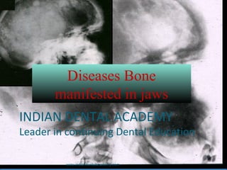
  • 1. Diseases Bone manifested in jaws INDIAN DENTAL ACADEMY Leader in continuing Dental Education www.indiandentalacademy.com
  • 2. 1. FIBROUS DYSPLASIA 2. OSTEITIS DEFORMANS (PAGET’S DISEASE) 3. CHERUBISM 4. CLEIDOCRANIAL DYSPLASIA 5. OSTEOPETROSIS 6. CEMENTO OSSEOUS DYSPLASIA 7. CEMENTO OSSIFYING FIBROMA 8. ACHONDROPLASIA INDEX www.indiandentalacademy.com
  • 3. FIBROUS DYSPLASIA •It is a fibo-osseous lesion in which normal medullary bone is gradually replaced by an abnormal fibrous C.T. proliferation. •The mesenchymal tissue contains various amounts of osteoid & osseous material that presumably arises through metaplasia. Etiology: •Unknown •Many workers believe --- --it is a non-neoplastic, hamartomatous growth resulting from deranged mesenchymal cell activity --another hypothesis suggests that it is an abnormal reaction of bone to a localized traumatic episode www.indiandentalacademy.com
  • 4. FIBROUS DYSPLASIA Clinical features:Clinical features: --Seen early in lifeSeen early in life -It can affect single bone or several to many bones.-It can affect single bone or several to many bones. M O N O S T O T I C F D - a f f e c t in g s in g le b o n e J a f f e -L ic h t e n s t e in t y p e - m u lt ip le b o n e le s io n s - s k in p ig m e n t a tio n ( c a f e a u la it m a c u le s ) M c C u n e - A lb r ig h t s t y p e - m u lt ip le b o n e le s io n s - s k in p ig m e n t a tio n ( c a f e a u la it m a c u le s ) - v a r o iu s t y p e s o f e n d o c r in e d is o r d e rs P O L Y O S T O T I C F D - a f f e c t in g m a n y b o n e s F I B R O U S D Y S P L A S IA www.indiandentalacademy.com
  • 5. MONOSTOTIC F.D. •It is much more common than Polyostotic type (80% of cases) •Jaws are most commonly affected sites •1st or 2nd decade of life •M = F •Slow growing, painless swelling of the affected area (usually unilateral) •Teeth involved in the lesion usually remain firm but may be displaced by bony mass • Although the mand lesions are truly Monostotic, maxillary lesions often involve adjacent bones such as zygoma, sphenoid, occiput & are not strictly monostotic. Termed as -- Craniofacial fibrous dysplasia FIBROUS DYSPLASIA www.indiandentalacademy.com
  • 6. POLYOSTOTIC F.D. •1st or 2nd decade of life (early in life) • It is less common than Monostotic type (20% of cases) •Clinical picture is dominated by symptoms related to long bone lesions. Jaws are less commonly affected. •deformity, bowing or thickening of long bones is evident •Pathological fractures with resulting pain & deformity is very common •Leg length discrepancy is noted as a result of involvement of upper portion of femur (Hockey stick deformity) • Café au lait (Coffee in milk) macules are pale brown well defined pigmentation with irregular outline, generally unilateral. --present on neck, trunk, thigh & also on oral mucosa. FIBROUS DYSPLASIA www.indiandentalacademy.com
  • 7. FIBROUS DYSPLASIA McCune-Albrights syndrome •Sexual precocity is the most common endocrine manifestation •Menstrual bleeding may occur during the first few months of life •Breast development may be seen in the first few years of life •Variety of other endocrine disturbances are seen -related to pituitary, thyroid, parathyroid etc…. www.indiandentalacademy.com
  • 8. FIBROUS DYSPLASIA Intra-oral appearance of the bony swelling which has remained virtually unchanged after five years (Next slide) www.indiandentalacademy.com
  • 9. Five years later FIBROUS DYSPLASIA www.indiandentalacademy.com
  • 11. FIBROUS DYSPLASIA Radiography: •Margins of the lesion are poorly defined -- lesion appears to blend into surrounding normal bone without evidence of circumscribed border •Lesions may be radiolucent or densely radiopaque •Characteristic X-ray picture – radiopacity of numerous poorly calcified bony trabeculae imparting a“Ground glass appearance” or “Orange peel appearance” •Skull X-ray – max sinus is radiopaque -- base of the skull shows increased density (involved bones are sphenoid, roof of orbit frontal bones & occiput) www.indiandentalacademy.com
  • 14. Histopath: •Typical microscopic feature of FD shows irregularly shaped trabeculae of immature (woven) bone in a cellular, loosely arranged fibrous stroma. •Lesional bone fuses directly to normal bone at the periphery of the lesion, so that NO capsule or line of demarcation is present •Bony trabeculae often assume curvilinear shapes, which have been described as “CHINESE LETTER” arrangement. (₣ ₤ ₪ ŧ Ħ ¥ コゴ ) •Bony trabeculae are considered to arise by metaplasia & are not surrounded by osteoblasts. •Jaw lesions might undergo maturation but long bone lesions do not show any maturation even in older aged individuals FIBROUS DYSPLASIA www.indiandentalacademy.com
  • 17. FIBROUS DYSPLASIA Treatment: •Difficult •Cosmetic reshaping after certain age (after the growth has stopped) •Radiation therapy is CONTRAINDICATED, as it carries the risk of development of post-radiation Osteosarcoma www.indiandentalacademy.com
  • 18. PAGET’S DISEASE (Osteitis Deformans) PD is characterized by abnormal & anarchic resorption & deposition of bone, resulting in distortion & weakening of the affected bones Disease principally affects older individuals (rarely below 40 yrs of age) Etiology: •Unknown •Inflammatory, genetic & endocrine factors may be contributing agents •Slow virus infection has received considerable attention (unproven) Slow virus-is one which produces a disease with a prolonged incubation period. www.indiandentalacademy.com
  • 19. Clinical features: •Most cases are polyostotic •M>>F (2:1) •Geographic variation is seen [Britain>>USA, Africa & Asia very rare] {W>>B} •Relatively asymptomatic, but severe bone pain is a common complaint •Pelvis, skull & femur are the most commonly affected bones •Jaw involvement is seen in about 10% - 15% of cases •Max >> mand •Bones affected by PD becomes thickened, enlarged & weakened •Skull involvement leads to increase in circumference of head PAGET’S DISEASE www.indiandentalacademy.com
  • 20. C/F: •The bones are weakened -- Due to which bowing deformity is present ---- this gives a “Simian Stance” (Monkey-like) •The alveolar ridge tend to become grossly enlarged (symmetrically). •In severe cases lip closure is difficult or impossible •Dentulous patients C/O : spacing of teeth •Edentulous patients C/O: non-fitting dentures (as the jaw is enlarging) PAGET’S DISEASE •In extreme cases of jaw enlargement “Leontasis Ossea” (Lion-like) facial deformity is seen •Neurological complications, such as deafness or visual disturbances may result from bony encroachment on cranial nerves www.indiandentalacademy.com
  • 24. Radiography: •Depends on the stage of the disease (osteoclastic & osteoblastic) •Early stages reveal a decreased radiodensity– slight radiolucecy with alteration in trabecular pattern •In early stages – skull might show large circumscribed areas of radiolucency (osteoporosis circumscripta) PAGET’S DISEASE •During later stages of (osteoblastic) disease there is haphazardly formed patchy areas of radio-opacities (bone formation) – giving it a “Cotton wool” appearance •Dental changes : -- hypercementosis of roots, loss of lamina dura, obliteration of PDL space & resorption of roots www.indiandentalacademy.com
  • 27. Lab findings: •Important information is provided •Serum Calcium & Phosphate levels are within normal limits •Serum Alkaline phosphatase levels are markedly elevated Normal value in adult: 1.5 – 5.0 (Bodansky units) In Paget’s disease : 50 – 250 (Bodansky units) PAGET’S DISEASE Other units for measuring Alkaline phosphatase •King-Armstrong – Normal value = 5 -10 units •Phenol units -- Normal value = 1.0 – 3.5 units www.indiandentalacademy.com
  • 28. Histopath: •Depends on the stage of the disease (osteoclastic or osteoblastic) •In the active resorptive stages, numerous Osteoclasts surround the bone trabeculae •Simultaneously, osteoblastic activity is seen with formation of osteoid rims around bone trabeculae •The marrow is replaced by fibrous C.T. •Characteristic picture of PD is presence of basophilic reversal lines. These lines indicate the junction between resorptive & formative phases of the bone and results in a “jigsaw puzzle” or “mosaic bone” appearance •Few cases show multiple small acellular bony masses that fuse as the disease progresses. PAGET’S DISEASE www.indiandentalacademy.com
  • 32. Treatment: •No treatment is required for mild cases •Use of parathyroid hormone antagonist, such as calcitonin or biophosphonates can reduce bone turnover •Dental: - difficulty in extraction is encountered (hypercementosis) - edentulous patients may require periodic new & larger dentures PAGET’S DISEASE Complications: • Development of malignant tumor, usually an osteosarcoma is reported. • Other neurological complications. www.indiandentalacademy.com
  • 33. CLEIDOCRANIAL DYSPLASIA (Marie & Sainton’s syndrome) •It is a disease of unknown etiology which is often but not always hereditary. •The disease shows an autosomal dominant inheritance pattern. •Chiefly involved bones are skull & clavicles, although a wide variety of anomalies may be found in other bones. www.indiandentalacademy.com
  • 34. CLEIDOCRANIAL DYSPLASIA Clinical features: •Short stature & have large heads •Pronounced frontal & parietal bossing with underdeveloped or narrow paranasal sinuses is evident. •Ocular hypertelorism is frequently present. •In the skull the fontanels often remain open or at least exhibit delayed closing. •Sutures also may remain open & wormian bones are common. •Mid-facial skeleton tends to be hypoplastic, and this combined with normal mandibular growth results in a relative prognathism. •Patients neck is long, the shoulders are narrow & show marked drooping. www.indiandentalacademy.com
  • 35. •Patients show an unusual mobility of their shoulders because of the absence or hypoplasia of the clavicles. (in some instances patient approximates the shoulders in front of chest) •A disease known as Pycnodysostosis (Maroteaux-Lamy syndrome) has all features of CD, but in addition to this -- bones are dense & fragile -- partial agenesis of the phalanges of hand & feet (these features are absent in CC dysplasia) CLEIDOCRANIAL DYSPLASIA www.indiandentalacademy.com
  • 36. CLEIDOCRANIAL DYSPLASIA Oral manifestation: •High, narrow, arched palate. Sometimes cleft palate is present •Maxilla is underdeveloped •Mandibular prognathism is seen •Prolonged retention of deciduous teeth & subsequent delayed eruption of permanent teeth •Numerous unerupted supernumerary teeth are present •The roots of the teeth are often short & thinner than usual & may be deformed •Absence or decreased amount of cementum is seen. www.indiandentalacademy.com
  • 41. Histopath: •Absence or lack of secondary cementum. Treatment: •No specific treatment •Care of the oral conditions is important CLEIDOCRANIAL DYSPLASIA www.indiandentalacademy.com
  • 42. CHERUBISM (Familial FD of Jaws; Hereditary FD of Jaws) •Cherubism is a rare developmental disease involving jaws •Cherub meaning plump-cheeked little angels •Autosomal dominant pattern of inheritance Clinical features: •Occur in children ( mean age – 7yrs) but can be noticed as early as 1 yr of age. Milder cases may not be detected until the patients reaches 10-12 yrs of age. •A painless bilateral expansion of the posterior mandible is the most common early manifestation. •Lesion tend to involve ascending ramus & body of the mandible. www.indiandentalacademy.com
  • 43. •The bilateral bony expansion imparts a “chubby” facial appearance •Extensive maxillary involvement causes stretching of the skin of the upper face to expose the sclerae. This results in an “eye upturned to heaven” appearance •Extensive bone involvement causes a marked widening & distortion of alveoli. •All 4 quadrants may be simultaneously involved. •Palatal vault may be obliterated. •Premature exfoliation of deciduous teeth may occur as early as 3 yrs of age. •Developing teeth are often displaced & fail to erupt & may be malformed •Lymphadenopathy- Submand & Upper cervical CHERUBISM www.indiandentalacademy.com
  • 44. Lab findings: •Serum Alkaline phosphatase levels may be elevated •Whereas serum Calcium & Phosphate levels are within normal limits CHERUBISM www.indiandentalacademy.com
  • 47. Radiographic findings: •Multilocular expansile bilateral radiolucencies (rarely Unilocular) •The borders are distinct & divided by bony trabeculae giving a “Soap Bubble” appearance •Unerupted teeth are often displaced & appear to be floating in the cyst-like spaces www.indiandentalacademy.com
  • 49. Histopath: •Stroma is loosely arranged •Consists of vascular fibrous tissue containing variable numbers of multinucleated Giant cells •The Giant cells tend to be small & are usually focally aggregated •In few cases perivascular eosinophilic cuff-like deposits are evident •Foci of extravasated blood are commonly present •In long standing cases (resolving lesions) the tissue becomes more fibrous, the number of Giant cells decreases & new bone formation is seen. CHERUBISM www.indiandentalacademy.com
  • 53. Histology of cherubism is similar to giant cell granulomas. *** the association between Cherubism & Noonan-like syndrome have been reported; but they are two separate diseases CHERUBISM Treatment: •Usually self limiting & regressive •Cosmetic surgery for improving esthetics & function www.indiandentalacademy.com
  • 54. OSTEOPETROSIS (Marble Bone Disease; Albers-Schönberg Disease) •Osteopetrosis is a rare hereditary skeletal disorder characterized by a marked increase in the bone density resulting from a defect in bone remodeling. www.indiandentalacademy.com
  • 55. OSTEOPETROSIS Clinical features: Infantile: Initial signs are normocytic anemia & hepato-splenomegaly resulting from compensatory extramedullary hematopoiesis Increased susceptibility to infection is common Facial deformity develops in many of the children -- broad face; hypertelorism; snub nose; frontal bossing Skull : failure of resorption & remodeling of skull bones produces narrowing of the various foramina (blindness, deafness &facial paralysis Dental : Tooth eruption is delayed, Osteomyelitis is common complication of extraction www.indiandentalacademy.com
  • 56. Adult: Detected at a later age than the infantile type Asymptomatic in early life & progressively involves more bones It is not life threatening NO- anemia, hepato-slenomegaly, blindness, deafness Dental : Fracture & Osteomyelitis are common complications of extraction OSTEOPETROSIS www.indiandentalacademy.com
  • 57. OSTEOPETROSIS Radiography: •Increase in skeletal density with defects in the metaphyseal remodeling •Distinction between cancellous & cortical bone is lost www.indiandentalacademy.com
  • 61. OSTEOPETROSIS Histopath: •Number of osteoclasts is increased, but their function to resorb bone is altered. •Osteoclasts appear atypical. • defective resorption combined with continued bone formation & endochondral ossification is seen. •Several patterns of abnormal endosteal bone formation have been described: # tortuous lamellar trabeculae replacing the cancellous portion of the bone # globular amorphous bone deposition in marrow spaces # osteophytic bone formation www.indiandentalacademy.com
  • 63. Treatment: •No specific treatment •Both the adult and childhood forms of osteopetrosis may benefit from Actimmune injections. Actimmune (interferon gamma-1b) delays the progression of osteopetrosis because it causes an increase in bone resorption (breakdown of old bone tissue) and in red blood cell production. •Bone marrow transplant is the only complete cure available. Bone marrow transplant has many risks, but if it is successful it saves the life of a child who would otherwise die from complications of the disorder. •Other treatments include nutrition, prednisone (helps improve the blood cell count), and physical and occupational therapy. OSTEOPETROSIS www.indiandentalacademy.com
  • 64. CEMENTO OSSEOUS DYSPLASIAS Periapical cemental dysplasia Florid osseous dysplasia They are seperated on the basis of extent of involvement of jaws. Now, Periapical cemental dysplasia :- Replacement of normal cancellous bone with fibrous tissue and cementum-like material, abnormal bone or a mixture of the two. By definition the lesion is located near the apex of a tooth. www.indiandentalacademy.com
  • 65. Clinical Features PCD is a common bone dysplasia that typically occurs in middle age More often in females than in males. Teeth are vital, and usually has no history of pain or sensitivity. It is a incidental finding during a periapical or panoramic radiographic examination made for other purposes. The lesions can become quite large, causing a notable expansion of the alveolar process, and may continue to enlarge slowly. Early stage Mixed stage www.indiandentalacademy.com
  • 66. Radiographic Features Lies at the apex of a tooth commonly in mandibular anterior teeth, although any tooth can be involved. The lesion is multiple and bilateral, but occasionally a solitary If the involved teeth have been extracted, this lesion can still develop and in these cases the term cemental dysplasia may be more appropriate. Lesion is well defined with radiolucent border, surrounded by a band of sclerotic bone. The lesion may be irregularly shaped or may have an overall round or oval shape centered over the apex of the tooth. www.indiandentalacademy.com
  • 67. In the early stage - radiolucency at the apex, continuous with the periodontal ligament with loss of the lamina dura. In the mixed stage - radiopaque tissue appears in the radiolucent structure. (round, oval, or irregular shape composed of cementum or abnormal bone k/as cementicles). Mature stage - Totally radiopaque with a thin radiolucent margin can be seen at the periphery because this lesion matures from the center outward in most of the cases this radiolucent margin is not apparent, which makes the differential diagnosis more difficult. Rarely root resorption and bone expansion. Mature stage www.indiandentalacademy.com
  • 68. D/D :-  Periapical rarefying osteitis.  Cementoblastoma.  Odontomas.  Cemento-ossifying fibroma. Treatment is not required. www.indiandentalacademy.com
  • 69. FLORID OSSEOUS DYSPLASIA :- Florid osseous dysplasia (FOD) is a widespread form of PCD.  The lesion has a poor vascular supply, a condition that likely contributes to its susceptibility to infection. Clinical features similar to PCD Occasionally low-grade, intermittent, poorly localized pain in the affected bone, especially when a simple bone cyst has developed within the lesion. If the lesions become secondarily infected, features of osteomyelitis may develop, including mucosal ulceration, fistulous tracts with suppuration, and pain. www.indiandentalacademy.com
  • 70. Radiographic Features Lesions are bilateral and present in both jaws. When they are present in only one jaw, the mandible is the more common location. The epicenter is apical to the teeth, within the alveolar process and usually posterior to the cuspid. Lesions are well defined and has a sclerotic border that can vary in width, very similar to PCD.  The soft tissue capsule may not be apparent in mature lesions. www.indiandentalacademy.com
  • 71. Density of the internal structure can vary from an equal mixture of radiolucent and radiopaque regions to almost complete radiopacity.  Some radiolucent regions, which usually represent the development of a simple bone cyst. These cysts may enlarge with time, even beyond the boundary of the lesion into the surrounding normal bone, or may fill in with abnormal dysplastic cementoosseous tissue. Can displace the inferior alveolar nerve canal in an inferior direction and the floor of the antrum in a superior direction and can cause enlargement of the alveolar bone by displacement of the buccal and lingual cortical plates. www.indiandentalacademy.com
  • 73. D/D :- Paget's disease Chronic sclerosing osteomyelitis. Treatment No treatment is required. Maintenance of good oral hygiene to prevent secondary infection. www.indiandentalacademy.com
  • 74. ACHONDROPLASIA •It is a hereditary condition that is transmitted as an autosomal dominant characteristic •Disease begins in utero & may actually be diagnosed before parturition •80% are stillborn www.indiandentalacademy.com
  • 75. Clinical features: Person is Dwarf (1.4 mtrs) with short & thick muscular extremities. Brachycephalic skull & bowed legs are seen Short hands & stubby fingers Lumbar lordosis with prominent buttocks & protruding abdomen Many joints show limited movement, because of which the elbows cannot be straightened They are of Normal intelligence They have unusual strength & agility (some adopt the occupation of professional wrestlers) ACHONDROPLASIA www.indiandentalacademy.com
  • 76. Oral manifestations: Maxilla is retruded because of restriction of growth of the base of the skull This may produce relative mandibular prognathism Difference in jaw sizes – causes malocclusion Dentition is usually normal (sometimes teeth have altered shape) ACHONDROPLASIA Radiographic features: Except for the retrusion of the maxilla & malocclusion, there are no changes in the jaw bones Long bones are short, thick & mild clubbing may be seen Bones at the base of the skull fuse www.indiandentalacademy.com
  • 78. ACHONDROPLASIA Hsitopath: Basic defect appears as a retardation or even aplasia of the zone of calcification of endochondral growth Cartilage columns fail to calcify and are not replaced by bone Treatment: No treatment www.indiandentalacademy.com
  • 79. DOWN SYNDROME (Trisomy 21 syndrome; Mongolism) •DS is a disease associated with subnormal mentality in which an extremely wide variety of anomalies & functional disorders may occur (cranial & facial deformities) Etiology: •Many factors like --- advanced maternal age& uterine & placental abnormalities, have been regarded as causes of the disease, Recent concept: chromosomal abnormality www.indiandentalacademy.com
  • 80. Three forms of DS: 1. One in which there is a typical trisomy 21 with 47 chromosomes (95% cases) 2. Another is a translocation type, in which there appears to be 46 chromosomes, although the extra-chromosomal material of no 21 is translocated to another chromosome (3% cases) 3. Another results from chromosomal mosaicism (2% cases) DOWN SYNDROME www.indiandentalacademy.com
  • 81. Clinical features: •Flat face, large anterior fontanel, open sutures, small slanting eyes with epicanthal folds, eye defects (refractive errors, nystagmus, cataracts etc..); open mouth with frequent prognathism, sexual underdevelopment, cardiac abnormalities & hypermobility of the joints, Congenital heart diseases is commonly seen. •Oral manifestations: - hypoplasia of maxilla, sphenoid bones, ribs & pelvic bones - macroglossia with protrusion of tongue, fissured tongue, open mouth, frequent mouth breathing causes drying & cracking of lips, palatal width is decreased & bifid uvula & cleft palate are seen - teeth : delayed eruption of permanent dentition, dental caries, microdontia, enamel hypocalcification is seen, severe PDL destruction is seen DOWN SYNDROME www.indiandentalacademy.com
  • 83. TREACHER COLLINS SYNDROME (Mandibulo-facial syndrome) •TCS is primarily affects structures developing from 1st branchial arch, but also involves 2nd arch to a minor degree Etiology: •TCS is transmitted by an autosomal dominant mode of inheritance •Incidence : between 0.5 – 10 cases in 10,000 births www.indiandentalacademy.com
  • 84. Clinical features: • various degree of hypoplasia of max, mand, zygomatic process of temporal bone, external & middle ear •Notched or colobomas of the lower eyelid, •Lower eye-lashes are absent •Malformation of external ear •Hair growth is Tongue-shaped in the region of pre-auricular area (known as “Hair-lick”) •Macrostomia, high palate, cleft palate & malocclusion of teeth •Facial clefts & skeletal deformities are seen in few individuals •Characteristic appearance are described as being “Bird like” or “Fish- like” TREACHER COLLINS SYNDROME www.indiandentalacademy.com
  • 87. Treatment: •None Radiography: •Underdeveloped or complete agenesis of malar bones and also mandible are seen •Clefting may be seen •Paranasal sinuses are grossly underdeveloped TREACHER COLLINS SYNDROME www.indiandentalacademy.com
  • 88. MARFAN’S SYNDROME (Marfan-Achard syndrome) •Is a heritable disorder of connective tissue, characterized by abnormalities of the skeletal, cardiovascular, & ocular systems Etiology: •MS is transmitted by an autosomal dominant mode of inheritance •Incidence : between 0.5 – 1 case in 10,000 births •The Marfan gene is believed to produce a change in one of the proteins that provides strenght to a component of C.T. (probably collagen) www.indiandentalacademy.com
  • 89. Clinical features: • tall, slender stature with relatively long legs & arms, large hands with long fingers, & loose joints •The arms, legs & digits are disproportionately long compared with patients trunk •Chest deformities include a protrusion or indentation of breast bone. •Various degree of scoliosis is present •Face appears long & narrow •Oral finding: narrow, high-arched palate & dental crowding •Cardiac : Mitral valve disease, •Ocular : dislocation of lens (ectopia lentis); myopia MARFAN’S SYNDROME Treatment: •Annual medical examination & treatment of cardiac & ocular defects www.indiandentalacademy.com
  • 90. PIERRE ROBIN SYNDROME (Robin anomalad) •It is a isolated defect (non-genetic) Etiology: •Incidence : between 5.3 – 22 case in 100,000 births •Fetal malposition & interposition of the tongue between the palatal shelves- is considered the etiology of palatal deformity • Evidence suggests that primary defect may be due to metabolic growth disturbances of the max & mand. www.indiandentalacademy.com
  • 92. PIERRE ROBIN SYNDROME Clinical features: •Severe micrognathia & mandibular hypoplasia •“U” shaped cleft palate is common. (speech & feeding difficulty) •Glossoptosis is seen (due to retropositioning of genioglossus muscle because of retrognathic mandible) •Airway obstruction (particularly in supine position) www.indiandentalacademy.com
  • 93. PIERRE ROBIN SYNDROME Treatment: •Multiple surgical therapy. Micrognathia www.indiandentalacademy.com
  • 94. REFERENCES White and pharoah. Oral radiology principles and interpretation. Diseases of bone manifested in jaws. 485-515. 5th edition. Mosby publication. Neville and Damm. Oral and maxillofacial pathology. Bone pathology. Pp-533. 2nd edition. Saunders publication. www.indiandentalacademy.com
  • 95. Shafer and levy. Oral pathology. Diseases of bone and joints. Pp-674. 4th edition. Elsevier publication. Wood and Goaz. Differential diagnosis of oral and maxillofacial lesions. Bony lesions. 5th editon. Mosby publication. N. H. Sherman, V. M. Rao. Fibrous dysplasia of the facial bones and mandible. Skeletal Radiology. 1982; (8)2: 141-143. www.indiandentalacademy.com
  • 96. Victor Perez Teixeira, Rogério Aparecido Dedivitis. CHERUBISM: CASE REPORT AND LITERATURE REVIEW. Rev. de Clín. Pesq. Odontol. 2004;(1)1, Nilton Alves , Reinaldo de Oliveira. Cleidocranial Dysplasia - A Case Report. Int. J. Morphol. 2008,26(4):1065-1068. Saadettin Dağistan, Ümmühan Tozoğlu. Florid cemento-osseous dysplasia: A case report. Med Oral Patol Oral Cir Bucal 2007;12:E348-50. www.indiandentalacademy.com