1. Science of the Eye: Vision
How do we see?
Have you ever thought about how we see the world around us? What is the science behind this
sense? In this unit, we will discuss how vision works, how we see color and some of the diseases
that affect vision. Let’s start with the general concept of vision. We will start with a few questions
that will help you understand how you think vision works.
Unit Objectives:
1. Understand the molecular basis of vision
2. Recognize the complexity of the retinal network of neurons (Massachusetts Biology Frameworks 4.7)
3. Practice Science Inquiry Skills (SIS)
Comments for Teachers are in italics (i.e., different activities to perform in the classroom and directions on
how to perform these activities).
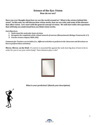
Mirror, Mirror, on the Wall: If a mirror is mounted flat against the wall, how big does it have to be in
order for you to see your entire body? Does distance play a role?
What is your prediction? (Sketch your description)
1
3.
The Digital Camera and the Eye:
You may have heard that the eye is like a camera. The digital camera is even a better analogy of how the
vertebrate eye works. A traditional camera uses a chemical process that “burns” the image on a thin film.
On the other hand, a digital camera uses a sensor that absorbs light and converts it to a signal more
analogous to what happens in our eyes.
Digital Camera Questions:
What is a digital camera?
Do you own a digital camera? If so, what kind (i.e., what’s the zoom, how many pixels, etc.)?
How do you think it works?
3
5.
In Living Color: Constructing Color Vision
Why is the sky blue? How can we distinguish between a purple flower and a yellow flower? Why are
there so many shades of gray? Color vision is essential to how we “see” our everyday world. It is
sometimes considered a construction project because it requires many components to function
completely. How we see color is still a subject of investigation for scientists. Scientists do not know all of
the details of color processing; that is, how we see or perceive colors. Have you ever looked at a picture
and thought that it did not exactly capture all of the contrast and hues of colors that you observed. Even
the most expensive camera does not have the same resolution as our visual system.
PreDiscussion Questions:
1. What are your favorite colors?
2. What organ(s) of the body do you think are responsible for color vision?
3. What is light and how is it useful?
4. Look at these two photographs. Describe three differences between the two photos.
5
6.
Discussion: Visible Light Spectrum
Most animals and plants can detect and respond to light. Animals detect a narrow region of the
electromagnetic radiation spectrum called the visible light region because it is visible to the human eye.
Light reflects from objects and is refracted by the cornea and the lens onto a photosensitive (light
detecting) region of the eye called the retina. Photosensitive cells in the retina can detect the visible light
region of the spectrum ranging from short wavelengths, starting at 390 nm, to longer wavelengths,
ending at 750 nm. How we see or perceive the colors of this visible light spectrum is the next topic of this
unit.
Diagram of the Electromagnetic Radiation Spectrum and the Visible Light Spectrum
Other animals are able to see outside of the visible light spectrum. For example, insects such as bees can
see in the ultraviolet region of the electromagnetic spectrum. If you look at an ultraviolet picture of a
flower, in some cases you can almost see a pattern reminiscent of a landing strip that directs bees to the
flower’s pollen (see picture below). We will see why bees can detect UV later in this unit.
6
7.
Marsh Marigold Flower – A. Visible Light (how humans see the flower) B. UV light (how bees see the flower)
(Image from http://www.eso.org/~rfosbury/home/natural_colour/biochromes/UV_flowers/nc_bio_flower_uv.html)
Discussion: Retina and Phototransduction
Introductory activities to introduce color vision in the classroom:
1. Where’s Waldo? Use a Where’s Waldo picture that is printed in color and in black and white. Give
half the class the color photo and half the class the black and white photo. Each student records how
long it takes to find Waldo. Average the time it takes students to find Waldo and create a
comparison chart (you can include in this analysis standard deviation and create bar graphs or other
graphs). (At the end of the activity, ask students what are some problems with this analysis, e.g.,
there is no real control, some people may have more experience with this type of puzzle than others,
which can affect the outcome.)
2. Obtain paint samples (swatches) from the store that are within the same color family and test
students’ ability to tell the different colors apart.
How the eye uses signals to communicate what we see?
PreDiscussion Questions:
1. Give an example of a signal. How do we use this signal?
2. Name one example of how signals are important in your everyday life?
3. What are some examples of signals in the cell or human body?
7
9.
The outer segment regions of the photoreceptors are membranous disc structures. Proteins that respond
to light are embedded in these structures.
Cartoon diagram of a rod and a cone
There are three major parts to the photoreceptors: the outer segment, the cell body, and the synaptic
terminal.
The outer segment regions of the photoreceptors are disk‐like structures made up of the cell membrane.
The proteins that respond to light are embedded in these structures.
The cell body is where the nucleus is located.
The synaptic terminal is where the neurotransmitter gets released.
9
10.
Under a high‐powered microscope (Scanning Electron Microscopy), the cones and rods look like the
following. (Only the outer segments are visible in this image.)
Image from http://www.chm.bris.ac.uk/webprojects2003/white/functional_anatomy_of_files/image004.jpg
Compare the sizes of the photoreceptor outer segments. Which one is bigger?
Photosensitive Proteins: Rhodopsin and Cone Opsins
Rhodopsin and the cone opsins are receptor proteins that span the membrane of disks in the outer
segments.
Rhodopsin and cone opsin are situated in the membrane of the discs in the photoreceptor outer
segments. Since the rod photoreceptors have larger outer segments, they have more opsin protein
(rhodopsin). This is part of the reason they can respond in lower light levels.
10
11.
The wavelengths of light that maximally initiate the response of the three types of cone
photoreceptors and rod photoreceptor
I can see in UV!! Some animals have an additional type of cone photoreceptor, which maximally
responds to UV light. This photoreceptor contains an opsin protein that responds to UV light (< 380 nm).
There is a huge amount of research ongoing that addresses how animals see in the UV and how seeing in
the UV affects their behavior. For example, in the picture of the flower shown above, bees are thought to
be able to see a contrast in the area of the flower’s pollen. It almost looks like a landing strip for the bee!
Phototransduction involves three steps:
1. In the dark, photoreceptors are constantly releasing neurotransmitter. They are depolarized or they
have a negative membrane potential (just like other neurons). The photoreceptors respond to light
through the rhodopsin (rod) and cone opsin (cones) proteins.
2. The absorption of light initiates a conformational change in the protein. This change sets off a cascade
of events (a signal) that closes Na+ ion channels in the cell. The Na+ ion channels are gated by binding
cGMP (cyclic GMP, a molecule in the cell). “Gated ion‐channel” means that the channel can only open if
something is bound to it. The signal initiated by light breaks up the cGMP molecule (an enzyme is
activated that breaks down cGMP to GMP) so that it no longer can bind to the Na+ ion channel. As a
result, the channel closes. Closing the channel prevents Na+ molecules from coming in the cell, which
makes the photoreceptor more negative or hyperpolarized (membrane potential).
3. The change in membrane potential reduces the release of a signal (neurotransmitter) in the synaptic
vesicle from the synaptic terminal. Fewer signals are then transmitted to the next layer of cells, the
bipolar cells.
Light is acting as a signal to turn down the amount of neurotransmitter that gets released from the cell.
The next cell, the bipolar cell, is a neuron that responds to both stimuli, lights on and lights off. There are
bipolar cells that are called ON‐bipolar cells that respond (send a signal) when the lights are ON and there
are OFF‐bipolar cells that respond (send a signal) when the lights are OFF. Also there are horizontal cells
that connect to photoreceptors and receive input from them. Although much is known about these
pathways, researchers are still uncovering information of how all of these neurons coordinate with one
another to pass along information about what we see to our brains.
11
12.
A diagram representing Phototransduction
It may seem strange that the photoreceptor releases less transmitter upon light and that the next level of
neurons, the bipolar cells, can respond to the less transmitter. One way to think about it is that, like other
neurons, the photoreceptors release neurotransmitter when depolarized (membrane potential is slightly
negative). Dark in this case does not have to be complete darkness. It can be a shadow that passes over
your eye, a change in light intensity.
Activity: What can you see in the dark?
This exercise demonstrates how the two types of photoreceptors in the eye, the rods and cones, adapt in dim
light conditions.
PreActivity Questions:
1. When you walk into a dark room, can you immediately see objects around you?
2. Are you able to distinguish colors in dim light?
3. Do you think that you can see certain colors in the dark? If so, which colors?
12
13. 4. When you walk out of a dark theatre after a movie, what can you see?
For this activity, students will first separate balls that have the same shape but are in three different colors.
You can do this with any object that feels the same but has different color or the writing on the object is
different. Have students place a pirate’s patch on one eye. Ask students to separate the balls into categories
in bright light. Mix up the balls again and turn off the lights. Immediately ask the students to separate the
balls in the dim light. This can be a quantitative exercise by counting the number of errors per group and
making charts with averages and standard deviations. Then have students switch the patch to the other
eye, repeat the sorting of the objects and count the number of errors among student groups.
Other classroom activities:
1. You can try to separate cups of water with different concentrations of food coloring and ask students to
sort the cups of water by increasing degree of color.
2. You can try magazine pictures or different colors of construction paper.
3. You can test variables, such as time, (e.g., how long does it take for the rod photoreceptors to adapt to dim
light?)
4. After being in the dark for several minutes, turn on the light and have students stare at different colored
items. Then, determine how long it takes for them to distinguish the colors. (This activity is testing how long
it takes cone photoreceptors to adapt to bright light.)
Is it difficult to distinguish color in the dark?
The cone photoreceptors are responsible for color vision. A certain amount of light energy (threshold)
has to be present for the cone photoreceptors to respond. However, in dark conditions, in humans and
other animals, the cone photoreceptors do not receive enough light to give a response.
Light Adaptation Activity: What is the minimum amount of light (threshold) required for the cone
photoreceptors to respond? Students can measure how much light is required for the cone receptors to
respond by performing the aforementioned activities and increasing the level of light. The amount of light
can be measured in Watts by a basic light meter (check with your school’s physics lab).
Why does it take a long time to adapt in dim light?
The rod photoreceptors are the photoreceptors responsible for vision in dim light. A single photon of
light is enough energy for the rod photoreceptors to respond. In bright light, the rod photoreceptors are
saturated with light and are bleached, which means that the visual pigment has converted to another
form. In this form, it can no longer respond to light and has to go through a set of reactions to return to
its original form. When you first turn off the lights, the rod photoreceptors are still saturated and it takes
a few minutes for the pigment molecules that respond to light to regenerate. It usually takes 20 ‐25
minutes to complete dark adaptation of the rod photoreceptors. When you turn the lights on, the rods
again will saturate (the visual pigment can no longer respond to light), but the light is now at a threshold
in which cone photoreceptors can respond.
13
15.
Crosssection of the retina
Central and Peripheral Vision Activity:
Hold this image paper a few inches from your face. Then, stare at the black cross in this picture for 10 –
20 seconds.
What did you observe?
What happened to the dots after staring at the cross for 10 ‐20 seconds?
15
17.
Procedure:
Before the Procedure, predict where you think you would see color or shapes on your giant
protractor.
1. Using the cups as the handles, hold the poster board base up to your face and put your nose in the
center hole.
2. Place a pushpin directly in front of your eyes.
3. Stare at the pushpin, while another person in your group slowly moves the index card with the
colored shape inward toward the pushpin.
4. When you see the color or the shape, let the person in your group know.
5. Make a mark at that point.
6. Using the protractor and ruler, measure the angle at which you recognized the shape and color.
For more advanced classrooms: Carrots are good for your eyes?
Rhodopsin and the cone opsins are located in the photoreceptor outer segment disks. A small
molecule called retinal binds to the rhodopsin and cone opsin proteins. The absorption of light causes a
change in the retinal molecule. Retinal changes conformation from its cis form to its trans form. This
change is like a domino effect in the photoreceptor cell. When retinal changes, rhodopsin also changes,
leading to a cascade of events. The last event is that the Na+ channels close and the cell becomes more
negative because now Na+ ions which are positive cannot flow into the cell. This change in membrane
potential causes a reduced amount of neurotransmitter to be released.
Carrots contain β ‐carotene, a molecule that the body uses to make retinal. We do not produce β‐
carotene, therefore we must obtain it from our diets. Besides carrots, olives are another good source of
β‐carotene. However, don’t worry too much that you’ll need to eat like a rabbit. As long as you are
eating sensibly, your body will make enough retinal for proper vision. However, in countries where
vitamin A deficiency due to lack of nutritious foods is a serious issue, vision problems, such as night
blindness, do occur.
17
18.
Colored Shadows: (Adapted from Exploratopia, from the Exploratorium)
The basic principle of this exercise is to remind students that there is a difference between subtractive and
additive color mixing. They probably learned this in elementary school or middle school. However, we will
know relate this to color vision.
Preactivity Questions:
What color to you get when you mix all colors together?
Procedure:
Using flashlights or lamps with color filters, arrange each light so that they converge on a whilte surface.
Try to make the room as dark as possible.
Observe the colors that you see once you begin to overlap the light.
Now, place your hand in front of the light.
Explorations Questions:
1. How many shadows do you see?
2. What are the colors of the shadows?
3. What are the colors of the shadows when they overlap?
4. Now, block one of the bulbs or turn off the light.
18
19. 5. How does this change the colors that you see in the shadows?
Other Activities for the classroom:
1. If you have a light source or a lamp in your class, you can look at the effects of light when you use
different color filters.
2. Using three flashlights you can place blue, red, and green color filters in front of each flashlights and
project it on a white board or white poster board.
Discussion: How do we detect different colors?
If you mix red, green, and blue light, you will equally activate all three cone photoreceptors
resulting in the perception of white color. White is an additive effect of all three colors, which activate all
three cone photoreceptors. Other colors are perceived by mixing the proper ratio from the red, green,
and blue light. The contributions of red, green, and blue cones largely determine the colors that we
perceive. The contribution of all three cones is processed in part by a direct path from photoreceptors to
bipolar cells to ganglion cells. Other retinal neurons influence the flow of this information indirectly. The
horizontal cells receive input from photoreceptors and influence the surrounding bipolar cells. Amacrine
cells contribute to the signal by receiving input from bipolar cells and sending signals to ganglion, bipolar,
and other amacrine cells. In a sense, the horizontal and amacrine cells form a web of information
contributing to the overall signal sent to the ganglion cells. Basically, information from 126 million
photoreceptors has to be funneled into 1 million ganglion cells to be sent to the brain. The ganglion cells
then pass on the message to the brain via its axons that make up the optic nerve (see picture below).
19
20.
How information flows through the retina (Image modified from Purves et al. (Ed.),
"Neuroscience", Sinauer Asociates Inc. Publisher)
Retinal Processing: the Flow of Information
Picture the retina as a train station to think about how it processes information. Passengers get to the
train station via different mechanisms; for example, some walked, some took the bus, some drove, or
some rode their bike. Next, passengers board the train to get to the next station. In this analogy, the
input of the photoreceptors, bipolar, amacrine, and horizontal cells come together to activate the
ganglion cells (the train). The train traveling to the next station is analogous to the output from the
ganglion cells to the brain. The axons of the ganglion cells make up the optic nerve, which travels from
the retina (one station) to the brain (next station).
Activity: What a weird American Flag!
20
21.
What are the colors represented in this version of the American Flag?
Stare at the white cross in the center of this image of the American Flag for 1 minute. Then move
your eyes to this blank box.
Do you see anything? If so, what colors do you see?
Discussion IV: Why does this happen?
So far we have discussed the initial stages of color vision, specifically how light can be converted
(transduced) into an electrical signal to be sent eventually to the brain. However, there is more to color
vision than the processing from the cone and rod photoreceptors. The signal gets processed in the retina
by other neurons and it gets processed in the brain. That is why we are often fooled by optical illusions!!
The ganglion cells respond to all of the input from multiple cone photoreceptors. Part of the retinal
processing of color is described by the color‐opponent theory.
21
22.
The ColorOpponent Theory (For more advanced classrooms):
Have you ever seen a color that is red‐green or blue‐yellow?
Certain colors are not seen in combination. These are called opponents, or antagonists. White and
Black are also opponents in the sense of light illumination (the measure of light and dark or whiteness
and blackness). The theory is that the information passed down to the some of the ganglion cells gets
sorted into three different channels before it goes to your brain. A channel represents a field of ganglion
cells or a group of cells responding together. Let’s say you are watching TV and you can only switch to
three different channels. However, those three different channels have all the shows categorized into
three different topics, sports, news, and reality TV.
In the retina, information about color and light intensity is sorted into the three channels. One
channel is devoted to information from the group of ganglion cells that responded to red and green light.
One channel is devoted to information from the group of ganglion cells that responded to blue and yellow
light. One channel is devoted the group of ganglion cells that respond to light intensity measured by the
degree of blackness or whiteness. Specific types of ganglion cells gather these three different processes
together (light versus dark, red versus green, yellow versus blue) to send to the brain. The ganglion cells
that respond in this fashion are called color‐opponent cells. These ganglion cells are sensitive to
differences in the wavelength of light. For each of the channels, if both types of stimuli stimulate the
ganglion cells, only one output gets read: red or green, blue or yellow, and light or dark. For example, if a
concentric field of ganglion cells is stimulated by red light (via mainly red cone photoreceptors) and
green light (via mainly green cone photoreceptors), the center ganglion cell might have a direct pathway
from the red photoreceptor cell while the surrounding ganglion cells are receiving signals mainly from
green photoreceptors. The channel from this field of cells will cancel each other out and the ganglion cell
will not fire (send an electrical pulse). The same would happen if the center ganglion cell is illuminated
by green light and the surrounding cells are illuminated by red light. The same is true for blue and yellow
light responses. Thus, you never get both responses from both red and green light or blue and yellow
light sending their signal at the same time. That is why you never see colors that are red‐green or blue‐
yellow.
Analogy: Imagine you have entered an arm wresting contest. In this scenario, you are the input from a
red light stimulus and your opponent is the input from a green light stimulus. Your first opponent is
someone who is stronger than you so he or she wins the match. In this case, the ganglion cell fires in
22
24.
Questions that can be investigated in classroom activities:
1. Repeat the exercise by altering the time that you stare at the boxes.
How does the time you stare at the box make a difference in the color that you see?
2. Repeat exercise this time with your left eye closed, then open your left eye and stare at the box
with your left eye open but right eye closed. How does this make a difference in the color that you
see? What is your explanation for your results?
3. Make your own afterimage using opponent colors.
Research Focus: A “Bionic” Eye?
Note: I would like to include a comparison of the digital camera and eye first in this section and maybe
incorporate an activity with cameras, etc?
Questions:
Understanding what you know now about the retina, what would happen if you no longer had functional
photoreceptors?
Looking at the pictures below, describe how the vision of an individual with retinitis pigmentosa is
affected? An individual with age‐related macular regeneration? (Hint: What part of your vision is
affected, central or peripheral?)
24
25.
Images Courtesy of the National Eye Institute (http://www.nei.nih.gov/health/examples/)
Have you ever heard of the TV shows, “The Six Million Dollar Man” and “The Bionic Woman”? The
shows were about two individuals who were in terrible accidents and had parts of their bodies replaced
with prosthetic limbs. However these prosthetic limbs actually improved their ability to run, hear, and
see. For example, the six million dollar man had prosthetic legs that allowed him to run at the speed of 60
mph (miles per hour)! Sounds like science fiction, right? Well, maybe not! What if you could implant a
prosthetic retina in someone to restore eyesight? Last year, researchers at multiple labs funded by the
US Department of Energy performed clinical trials in which they implanted an artificial retina or what is
being called a “bionic” eye in blind individuals. This “bionic” eye will not give the blind individual better
vision than non‐blind individuals, but it is still a remarkable advance in the field of vision science.
There are two major retinal diseases, Retinitis Pigmentosa and Age‐related Macular Degeneration,
which can cause blindness due to loss of photoreceptor function (see pictures above). Remember the
photoreceptors are a part of the retina and respond to light sending electrochemical signals to the rest of
the retina and brain. In these diseases, the photoreceptors degenerate, but in many cases, other retinal
neurons, such as the bipolar, horizontal, amacrine, and ganglion cells are still present and functional. One
of these diseases causes photoreceptors primarily in the fovea to be destroyed.
What is the function of the fovea? Which disease affects the photoreceptors of the fovea?
There are no known cures for these retinal diseases. Millions of people have lost their sight due
to these diseases. Two million Americans have Age‐related Macular Degeneration; while hundreds of
thousands of people suffer from Retinitis Pigmentosa. Engineers and scientists are now trying to
improve this device to assist millions of people who have lost their sight due to these retinal diseases.
How does it work?
If the retina contains photoreceptors that are not functional, how is vision affected? What could you do to
simulate other retina cells?
An external miniature digital camera is placed on a pair of sunglasses worn by the patient. The digital
camera gathers the image and transmits the information to a miniprocessor. This miniprocessor
25
27.
(Image courtesy of California Institute of Technology, http://artificialretina.energy.gov)
Further Discussion Questions:
Do you think that with the “bionic” eye implant, individuals will be able to see colors?
If not, what do you think would be necessary for them to see colors?
You are the scientist working for the artificial retina project. What are some things you would like to
know before making the implant available to any blind patient?
If you knew someone who was thinking about receiving the “bionic eye” implant, what would you tell him
or her? Would you have any concerns?
27