
Recommended
More Related Content
What's hot
What's hot (20)
Similar to Prenatal development
Similar to Prenatal development (20)
Recently uploaded
Recently uploaded (20)
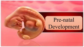
Prenatal development
- 2. Human Life begins at Conception Conception The union of a male sperm cell and a female egg cell to form a new organism.
- 3. Pre-natal development is divided into three (3) periods: 1. Germinal 2. Embryonic 3. Fetal.
- 4. 1. Germinal Period (First 2 weeks after conception) – This includes the: a) creation of zygote. b) continued cell division and c) the attachment of the zygote to the uterine wall.
- 5. The following are the details of development during this period: a. 24 to 30 hours after fertilization-the male (sperm) and the female (egg) chromosome unite. b. 36 hours-the fertilized ovum, zygote. divides into two (2) cells. c. 48 hours (2days) – 2 cells become 4 cells d. 72 hours (3days) – 4 cells become a small compact ball of 16-32 cells
- 6. f. 96 hours (4days) – hollow ball of 64-128 cells g. 4-5 days – inner cell mass (blastocyst) still free in the uterus h. 6-7 days – blastocyst attaches to the wall of uterus i. 11-15 days – blastocyst invades into uterine wall and becomes implanted in it (implantation)
- 7. The Blastocyst, the inner layer of cells that develops during the germinal period, develops later into the embryo. The trophoblast, the outer layer of cells that develops also during the germinal period, later provides nutrition and support for the embryo
- 8. 2. Embryonic Period (2-8 weeks after conception) In this stage, the name of the mass cells, Zygote, become embryo. The following developments take place: a) Cell differentiation intensifies b) Life-support system for the embryo develop and c) Organs appear
- 9. The embryo’s endoderm, the inner layer of cells develops into the digestive and respiratory systems. The outer layer of cells is divided into two parts:the ectoderm and the mesoderm. The ectoderm is the outermost layer which becomes the nervous system, sensory receptors (eyes, ears, nose) and skin parts (nails, hair).
- 10. The mesoderm is the middle layer which becomes circulatory, skeletal, muscular, excretory and reproductive systems. This process of organ formation during the first two months of pre-natal development is called organogenesis. The life-support system of the embryo are: placenta, the umbilical cord, and the amnion.
- 11. The placenta is a life-support system that consists of a disk-shaped group of tissues in which small blood vessels from the mother and the offspring intertwine but do not join. The umbilical cord contains arteriesand one vein that connects the baby to the placenta. The amnion is a bag or an envelope that contains a clear fluid in which a developing embryo floats
- 12. Fetal Period (2 to 7 months after conception) – Growth and development continue dramatically during this period. The details of the developmental process are as follows
- 13. a. 3 months after conception Fetus is about 3 inches long and weighs about 1 ounce; fetus has become active, moves its arms and legs, opens and closes its mouth, and moves its head; the face, forehead, eyelids. Nose and chin can now be distinguished and also the upper arms, lower arms, hands and lower limbs; the genital can now be identified as male or female.
- 14. b. 4 months after conception – fetus is about 6 inches long and weighs 4 – 7 ounces; growth spurt occurs in the body’s lower parts; pre-natal reflexes are stronger, mother feels arm and leg movements for the first time. c. 5 months after conception – fetus is about 12 inches long and weighs close to a pound; structures of the skin (fingernails, toenails) have formed; fetus is more active.
- 15. d. 6 months after conception – fetus is about 14 inches long and weighs one and half pound; eyes and eyelids are completely formed; fine layer of head covers the head; grasping reflex is present and irregular movements occur. e. 7 months after conception – fetus is about 16 long and weighs 3 pounds. f. 8 and 9 months after conception – fetus grows longer and gains substantial weight, about 4 pounds.
- 16. Teratology and Hazards to Pre-natal Development Teratology is the field that investigates the causes of congenital (birth) defects. A teratogen is that which causes birth defects. I comes from the Greek word “tera” which means “monster”.
- 17. Below are clusters of hazards to pre-natal development: 1. Prescription and nonprescription drugs – These include prescription as well as non-prescription drugs. Antibiotic is an example of a prescription drug that can be harmful. Examples of nonprescription drugs are diet pills, aspirin and coffee. 2. Psychoactive drugs – These includes nicotine, caffeine and illegal drugs such as marijuana, cocaine and heroin. Heavy drinking by pregnant women results to the so-called fetal alcohol syndrome (FAS) which is a cluster of abnormalities that appears in the children of mothers who drink alcohol heavily during pregnancy.
- 18. 3. Environmental hazards – These include radiation in jobsites and X-rays, environmental pollutants, toxic wastes and prolonged exposure to heat in saunas and bath tubs. 4. Other maternal factors such as Rubella (German Measles), syphilis, genital herpes, AIDS, nutrition, high anxiety and stress, age (too early or too late, beyond 30) Rubella (German measles) in 1964 – 65 resulted in 30,000 pre-natal and neonatal (newborn) deaths and more than 20, 000 affected infants were born with malformations, including mental retardation, blindness, and deafness and heart problems A mother can infect her child in three ways: 1) during gestation across the placenta, 2) during delivery through contact with maternal blood or fluids, and 3) postpartum (after birth) through breast-feeding.
- 19. Folic acid is necessary for pregnant mothers. Folic acid can be reduced the risk of having a baby with a serious birth defect of the brain and spinal cord, called the ‘neural tube’. A baby with spina bifida, the most common neural tube defect is born with a spine that is not closed. The exposed nerves are damaged, leaving the child with varying degrees of paralysis and sometimes mental retardation. A baby with Down syndrome rarely is born to mother an under age 30 but the risk increases after the mother reaches 30. By age 40, the probability is slightly over 1 in 100, and by age 50 it is almost 1 in 10. The risk is also higher before age 18.
- 20. 5. Paternal factors – Fathers’ exposure to lead, radiation, certain pesticides and petrochemicals may cause abnormalities in sperm that lead to miscarriage or diseases such as childhood cancer. As in the case of older mothers, older fathers also may place their offspring at risk for certain defects.