1. Neuro-Oncology 13(4):410 – 416, 2011.
doi:10.1093/neuonc/noq205
Advance Access publication February 22, 2011
N E U RO - O N CO LO GY
Temozolomide in the treatment of children
with newly diagnosed diffuse intrinsic
pontine gliomas: a report from the Children’s
Oncology Group
Kenneth J. Cohen, Richard L. Heideman, Tianni Zhou, Emiko J. Holmes,
Robert S. Lavey, Eric Bouffet, and Ian F. Pollack
An open-label phase II study (ACNS0126) testing the
efficacy of chemoradiotherapy with temozolomide
(TMZ) followed by adjuvant TMZ was conducted by
the Children’s Oncology Group. During the period
from July 6, 2004 through September 6, 2005, 63 children with newly diagnosed diffuse intrinsic pontine
glioma (DIPG) were enrolled in the study. All patients
received TMZ at a dosage of 90 mg/m2/day for 42
days to a dose of 59.4 Gy. Four weeks following
irradiation, TMZ was given at a dosage of 200 mg/
m2/day for 5 days every 28 days, for a total of 10
cycles. The primary objective of the statistical analysis
was to determine whether the current treatment produced a 1-year event-free survival (EFS) rate higher
than the historical baseline of 21.9% observed in
CCG-9941. The mean 1-year EFS (+ standard devi+
ation) was 14% + 4.5%, compared with 21.9% + 5%
for CCG-9941. The P value of the test of comparison
of 1-year EFS, based on a 1-sided, 1-sample test of
proportions, was .96. There was no evidence that temozolomide produced a 1-year EFS rate higher than
21.9%. The mean 1-year OS (+ standard deviation)
+
was 40% + 6.5%, compared with 32% + 6% for
CCG-9941. The median time to death was 9.6
months. Chemoradiotherapy with TMZ followed by
Received September 23, 2010; accepted December 2, 2010.
Correspondence Author: Kenneth J. Cohen, MD, MBA, The Sidney
Kimmel Comprehensive Cancer Center at Johns Hopkins, CMSC-800;
600 North Wolfe Street, Baltimore, MD 21287 (kcohen@jhmi.edu).
adjuvant TMZ is not more effective than previously
reported regimens for the treatment of children with
DIPG.
Keywords: temozolomide, DIPG, chemoradiotherapy.
T
he management of children with the diagnosis of a
diffuse intrinsic pontine glioma (DIPG) remains
one of the great challenges within pediatric oncology. Children often present with relatively modest
neurologic findings only to have MRI reveal classic
imaging features of an enlarged pons, with minimal contrast enhancement after gadolinium administration,
diffuse T2/FLAIR signal, and engulfment of the basilar
artery.1 Biopsy of this lesion, although rarely obtained,
demonstrates a diffuse, infiltrating astrocytoma World
Health Organization (WHO) grades 2, 3, or 4.2
Regardless of histologic grade, with classic imaging
features, death is anticipated with most children dying
of their disease within 6 months to 2 years from the
time of initial diagnosis.3,4
The mainstay of treatment for DIPG remains radiation therapy (XRT), which is essentially palliative,
delaying the inevitable progression of disease for a
period of some months.5 In an effort to alter the
dismal prognosis anticipated for most children, numerous studies have been undertaken in the hopes of
improving the outcome for these children.6 Recent strategies have included neoadjuvant chemotherapy prior
# The Author(s) 2011. Published by Oxford University Press on behalf of the Society for Neuro-Oncology.
All rights reserved. For permissions, please e-mail: journals.permissions@oup.com.
Downloaded from http://neuro-oncology.oxfordjournals.org/ by guest on July 20, 2013
The Sidney Kimmel Comprehensive Cancer Center at Johns Hopkins, Baltimore, Maryland (K.J.C.);
Department of Pediatrics, University of New Mexico School of Medicine, Albuquerque, New Mexico (R.L.H.);
Department of Preventive Medicine, University of Southern California, Los Angeles, California (T.Z.);
Operation Office, Children’s Oncology Group, Arcadia, California (T.Z., E.J.H.); Department of Radiation
Oncology, H. Lee Moffitt Cancer Center, Tampa, Florida (R.S.L.); Hospital for Sick Children, Toronto,
Ontario (E.B.); Department of Neurological Surgery, University of Pittsburgh, Pittsburgh, Pennsylvania (I.F.P.)
2. Cohen et al.: Temozolomide in the treatment of pediatric DIPG
Subjects with other brain stem tumors, including exophytic brain stem gliomas, cervicomedullary junction
tumors, and focal low-grade gliomas of the midbrain
or brain stem, were not eligible. Patients and/or their
parents or legal guardians were required to sign institutional review board approved consent prior to
enrollment.
Pathology
Pathology was not required for participation on the trial
if imaging features were consistent with a DIPG. In cases
in which pathologic tests were performed, the institutional pathologist had to confirm that the tumor was
an infiltrating astrocytoma of WHO grade 2– 4.
Treatment
All subjects enrolled in the study received the same
planned therapy. Within 42 days after diagnosis, subjects started radiation therapy with the dose to the planning target volume being 59.4 Gy given in 33 fractions of
1.8 Gy delivered once daily, 5 days per week. The planning target volume encompassed the gross tumor volume
on MRI scan with a 1.3 –1.5-cm 3-dimensional margin.
TMZ was administered at a dosage of 90 mg/m2/day
beginning within 5 days after the start of radiation
therapy and continued uninterrupted for a total of
42 days. Four weeks after the completion of radiation
therapy, maintenance therapy was initiated (200 mg/
m2/day for 5 days every 28 days for a total of 10
cycles). Each cycle of TMZ commenced when there
had been adequate hematologic recovery from the
prior cycle of treatment. All children received
Pneumocystis jiroveci pneumomia prophylaxis throughout treatment.
Patients and Methods
Response evaluation
Eligibility
Children were eligible for the study provided they had
undergone gadolinium-enhanced MRI with imaging
features consistent with the diagnosis of DIPG. Biopsy
of the lesion was neither required nor recommended
unless the treating physician believed that imaging features were not entirely consistent with the diagnosis of
DIPG. MRI had to demonstrate that at least two-thirds
of the tumor was situated in the pons and that the
origin of the tumor was clearly within the pons.
Eligibility criteria included that cases be newly diagnosed and that the patients were 3 –21 years of age,
had received no prior therapy other than corticosteroids,
and had a Lansky/Karnofsky performance status ≥50,
life expectancy of ≥ 2 months, and adequate hematologic, renal, and hepatic function. Exclusion criteria
included the inability to begin treatment within
42 days from the time of diagnosis, DIPG presenting as
a second malignancy, or metastatic spread of tumor.
Evaluation of response to chemoradiotherapy and adjuvant chemotherapy was based on institutional assessment of the change in size of the tumor using the
maximal 2-dimensional cross-sectional tumor measurements on T2 or FLAIR sequences. Response was characterized as a complete response (ie, disappearance of all
abnormal signal within the brain stem and return to
normal size of the brain stem), partial response (ie, ≥
50% decrease in the sum of the products of the 2 perpendicular diameters of the target lesion), stable disease
(ie, neither sufficient decrease in the products of the 2
perpendicular diameters of the target lesion to qualify
for a partial response, nor sufficient increase in the
target lesion to quality as progressive disease), or progressive disease (ie, a ≥25% increase in the crosssectional area of the largest 2 diameters of the target
lesion). Patients were removed from protocol therapy
upon evidence of clinical and/or radiologic progressive
disease.
NEURO-ONCOLOGY
†
APRIL 2011
411
Downloaded from http://neuro-oncology.oxfordjournals.org/ by guest on July 20, 2013
to XRT,7 – 9 chemoradiotherapy (often followed by
adjuvant therapy),10 – 17 adjuvant therapy alone,18 highdose chemotherapy with stem cell rescue,19,20 hyperfractionated radiation therapy,9,21 anti-hypoxic treatment
during radiation therapy,22,23 and salvage therapy at
the time of tumor progression.24,25 The previous
Children’s Cancer Group treatment protocol for DIPG
(CCG 9941) that serves as the historical control for
the present study consisted of 3 cycles of induction
multiple-agent chemotherapy followed by radiation
therapy given twice daily to a total dose of 7200 cGy.9
None of these approaches has proven satisfactory,
leaving once-daily XRT alone as the only routinely
recommended “standard of-care.”
Temozolomide (TMZ) is an orally bioavailable
prodrug that is metabolized at physiologic pH to its
active metabolite monomethyl 5-triazeno imidazole carboxamide. The primary mechanism of action is methylation at the O6 position of guanine with a minor
contribution at the N7 position. TMZ is lipophilic facilitating gastrointestinal absorption and CNS penetration.
TMZ is widely utilized in the treatment of high-grade
gliomas (HGGs), including anaplastic astrocytoma
(AA) and glioblastoma (GBM). In the United States,
TMZ is approved for the treatment of newly diagnosed
GBM in adults concomitantly with radiotherapy and
then as maintenance therapy. It is also approved for
the treatment of adults with refractory AA following
progression on a nitrosourea/procarbazine regimen.
Given its apparent activity in the treatment of patients
with infiltrating astrocytomas, the Children’s Oncology
Group (COG) undertook a study (ACNS0126) to determine the utility of TMZ in the treatment of children with
infiltrating astrocytomas. Stratum A was restricted to
children with a diagnosis of AA, GBM, or gliosarcoma;
stratum B was restricted to children with DIPG, the
results of which are reported here.
3. Cohen et al.: Temozolomide in the treatment of pediatric DIPG
Statistical methods
The primary study endpoint for treatment efficacy was
time to treatment failure, measured from the start of
treatment, from which event-free survival (EFS) percentage was computed. Failure events included tumor progression, tumor recurrence, or death due to any cause.
The secondary efficacy endpoint was time to death due
to any cause, from which overall survival (OS) was computed. The primary objective of the statistical analysis
was to determine whether the current treatment produced a 1-year EFS rate higher than the historical baseline of 21.9%, which is the maximum likelihood
estimates derived from an exponential model during
the first year from study CCG-9941. Both ACNS0126
and CCG 9941 were assumed to have exponential distribution. The test of comparison is based on a 1-sided,
1-sample test of proportions,
ˆ
p−p
ˆ ˆ
V p
ˆ
where p is the estimated proportion of patients who are
ˆ ˆ
event-free at 1 year, p ¼ 0.219, and V(p) is the estimated
ˆ
asymptotic variance of p. Because censoring in this
disease in the first year of follow-up is minimal, the
test statistic is based on the simple estimate of the proportion of patients who are event free at 1 year. The estiˆ
ˆ
mated variance in this case is p(12p)/n, where n is the
total sample size. The test was performed after the last
enrolled patient has been followed for a minimum of 1
year. The criteria for significance was the upper 90th
percentile of the standard normal distribution, so that
the test was performed at a nominal 10% type I error
level.
Results
Patient characteristics
During the period from July 6, 2004 through September
6, 2005, 63 children were registered on ACNS0126 BSG
stratum. Three children were ineligible because their
tumors were coded as being not centered in the pons
(did not have DIPG) and two children were ineligible
because they failed to meet other eligibility requirements
(one had RT at a non-COG institution and the other did
not begin treatment in the required time frame). Patient
characteristics for the 58 eligible patients are shown in
Table 1. The median age for the 58 patients is 7.7
years (range, 3.3 –16.2 years). Forty-eight percent were
male; 57% were white and 29% were black. Three children had biopsies of their tumor at presentation, with 1
tumor being AA and 2 being GBM.
Therapies administered, response, and toxicity
For the 58 eligible children, 49 (84%) completed the
prescribed chemoradiotherapy and went on to receive
412
NEURO-ONCOLOGY
†
APRIL 2011
Characteristic
Sex
Frequency
Percentage
48.3
Primary site
30
51.7
,5 years
5– 10
13
32
22.4
55.2
12
20.7
≥15 years
White
1
33
1.7
56.9
Black
Race
28
10 –15
Age
Male
Female
17
29.3
8
3
13.8
5.2
55
94.8
Other/unknown
Midbrain
Pons
adjuvant chemotherapy. Of the 9 patients who went
off protocol therapy before maintenance, 7 progressed,
1 was withdrawn by the parents who declined further
treatment secondary to significant changes in mental
and motor status, and 1 died. The responses of the
49 remaining patients at the end of chemoradiotherapy
included 18 with partial response, 20 with stable
disease, and 1 with progressive disease who did not
discontinue protocol therapy. Ten other patients were
not evaluated at that time but continued to undergo
treatment; their responses in the next course in
which they were evaluated included 2 with partial
responses, 5 with stable diseases, and 3 with progressive disease. The median number of adjuvant maintenance courses was 2 (range, 1– 10). Only 2 patients
completed all 10 courses of maintenance, and 1 of
them progressed during the last course.
There was a break in the course of radiation therapy
ranging from 1 to 7 days in 9 patients due to unstable
medical condition precluding delivery of XRT.
Protocol-directed dose modification occurred during
the first course of chemotherapy due to toxicity for
7 patients (3 cases of thrombocytopenia and 1 each of
pneumonia, tachypnea, increased intracranial pressure/
unresponsiveness, and radiation-induced edema). Two
patients had an allergic reaction to TMZ during the
first course. Over all courses of treatment, there were
20 times (17 patients) when the drug was modified due
to toxicity; 11 modifications were due to low platelet
counts.
Targeted toxicities are shown in Table 2 for the
chemoradiotherapy course and the cumulative rates
over all the maintenance courses (with number of
patients ranging from 49 in course 1 to only 2 after
course 7). Hematologic toxicities predominated. One
child was removed from study therapy because of
prolonged thrombocytopenia. Nontargeted toxicities
were rare, with the majority being neurologic
toxicities with unlikely attribution to TMZ; during chemoradiotherapy, the nontargeted toxicities included
ataxia (n ¼ 3), depressed level of consciousness
(n ¼ 3), and speech impairment (n ¼ 3).
Downloaded from http://neuro-oncology.oxfordjournals.org/ by guest on July 20, 2013
Z=
Table 1. Characteristics of patients with diffuse intrinsic pontine
glioma.
4. Cohen et al.: Temozolomide in the treatment of pediatric DIPG
Table 2. Summary of targeted toxicities for diffuse intrinsic
pontine glioma stratum
Toxicity
Maintenance
(n 5 49)
0
8
Anemia
Leukocytes (total white
blood cell count)
5
10
13
35
Lymphopenia
Neutrophils/granulocytes
38
10
39
47
Thrombocytopenia
9
34
Nausea
Vomiting
3
3
8
7
Alanine aminotransferase
level
Febrile neutropenia (fever
of unknown origin)
0
4
0
4
Infection with grade 3 or
4 neutropenia
Infection with unknown
ANC
2
5
5
0
Infection without
neutropenia
9
8
12
17
35
46
5
0
7
3
Allergic reaction/
hypersensitivity
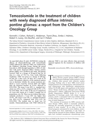
Fig. 1. ACNS0126 DIPG Event-Free and Overall Survival.
Neuropathy
Cranial
Motor
Neurology
Sensory
Seizures
Data are percentage of grade 3 or 4 toxicities for chemoradiotherapy
and maintenance courses.
Outcome
Figure 1 demonstrates the EFS and OS for all eligible
patients with DIPG. Figure 2 presents an event-free
survival plot for the 58 eligible DIPG patients on
ACNS0126 and compares them with a similar cohort
of 63 patients from the historical control study
CCG-9941. The primary objective of the statistical
analysis was to determine whether the current treatment
produced a 1-year EFS rate higher than the historical
baseline of 21.9%. On ACNS0126, 46 patients progressed and an additional 4 patients died with presumptive progression in the absence of an MRI within 1 year.
Fourteen percent (8 of 58) of patients were event-free at
1 year. The 1-sided P value of the test of comparison
based on a 1-sided, 1-sample test of proportions was
.96. Therefore, we failed to collect evidence to show
that the 1-year EFS rate on the current treatment is
higher than the historical baseline of 21.9% at the .1
significance level.
The mean Kaplan-Meier estimate (+standard deviation) of 1-year EFS was 14% + 4.5% for ACNS0126,
compared with 21.9% + 5% for CCG-9941; the mean
1-year OS (+standard deviation) was 40% + 6.5%
for ACNS0126, compared with 32% + 6% for
CCG-9941. The median times to progression and
Fig. 2. Event-Free Survival Comparison of ACNS0126 and
CCG-9941.
death were 6.1 and 9.6 months, respectively, for patients
with DIPG in ACNS0126. The mean 2-year EFS and OS
(+standard deviation) were 1.7% + 1.7% and 3.6% +
2.5%, respectively. The only patient not known to have
died was lost to follow-up after 25 months.
Discussion
Results of this study demonstrated no apparent survival
benefit associated with the addition of TMZ to conventional radiation therapy among children with DIPG. In
this study and in CCG-9941, the majority of children
died with disease progression within 1 year of diagnosis.
This finding is particularly disappointing given the
encouraging results seen with a similar chemoradiotherapy and maintenance regimen using TMZ in adult
patients with supratentorial HGG.26 Two smaller pediatric trials27,28 recently reported similarly poor outcomes, albeit with slightly reduced doses of TMZ. The
addition of chemoradiotherapy in the current study
appeared to offer no therapeutic advantage, with a
dismal outcome similar to that seen using maintenance
TMZ following radiation therapy alone.29 Seven children progressed following chemoradiotherapy prior to
any courses of maintenance therapy. One explanation
could have been pseudoprogression, although all 7
NEURO-ONCOLOGY
†
APRIL 2011
413
Downloaded from http://neuro-oncology.oxfordjournals.org/ by guest on July 20, 2013
Chemoradiotherapy
(n 5 58)
5. Cohen et al.: Temozolomide in the treatment of pediatric DIPG
414
NEURO-ONCOLOGY
†
APRIL 2011
regulatory requirements are met for such banking.
Alternatively, if the specimen were used to influence
the plan of care for the child undergoing a biopsy, then
the prospect of direct benefit could be argued, allowing
for classification of the research as more than minimal
risk but with the prospect of direct benefit.
Alternatives have been recently reported, including the
use of so-called “warm autopsy” specimens, in which
pontine tissue is acquired as soon after the death of the
child as possible. Whether such tissue reflects the pretreatment biology of a DIPG is unclear, but potential
molecular targets are being identified by these
methods. Another possibility would be to use specimens
obtained from children who present with a classic bithalamic infiltrating astrocytoma, a lesion type that is commonly biopsied. These lesions tend to image in a manner
quite similar to that of a DIPG, and their natural history
is equally unfavorable.38
Numerous strategies for the treatment of DIPG are
being studied in both the preclinical and early clinical
setting. Chemotherapeutics that show promise against
supratentorial AA and GBM are often proposed as
rational candidates for study in children with DIPG.
Exploiting this approach has been ineffective in the
past, but perhaps newer agents will provide greater
benefit. Molecularly targeted therapies hold some conceptual promise, but the utility of these agents is, as
yet, unproven in the setting of a DIPG, in part because
of the lack of available tissue to analyze. Local delivery
strategies (eg, convection-enhanced delivery) are being
explored on the presumption that the current failure of
systemic therapy is related to inefficient delivery of
drug to the target tissue.
Despite anecdotal reports of efficacy, the data are sufficiently poor that there appears to be little justification
for the continued use of TMZ in this patient population,
even in combination therapy. Currently, the only
therapy remains palliative up-front XRT. Other treatments are best explored in the setting of early phase
clinical trials.
Acknowledgments
Presented in part at The 12th Annual Meeting of the
Society for Neuro-Oncology, Dallas, Texas.
Conflict of interest statement. None declared.
Funding
The Chair’s Grant (U10 CA98543-08) and Statistics and
Data Center (U10 CA98413-08) of the Children’s
Oncology Group from the National Cancer Institute,
National Institutes of Health.
Downloaded from http://neuro-oncology.oxfordjournals.org/ by guest on July 20, 2013
children were dead of disease wtihin 1 year after diagnosis, suggesting that this phenomenon, if present, did not
alter the natural history of this tumor.
It is unclear why TMZ was ineffective in this patient
population. One possible explanation is that DIPG
contains an active, presumably unmethylated,
O6-methylguanine DNA methyltransferase (MGMT)
promoter, as is often seen in supratentorial HGGs.
MGMT activity would be anticipated to rapidly remove
methyl and alkyl groups from the O6 position of
guanine abrogating the cytotoxic impact of TMZ. This
hypothesis is speculative, because a biopsy of the pons
was not required for enrollment on this stratum. A
small number of children underwent biopy, presumably
when the treating physician felt that the imaging features
at the time of presentation were not classic for a diagnosis
of DIPG. However, these specimens were not tested for
MGMT expression. Agents that inhibit MGMT activity,
such as O6-benzylguanine,30 are being studied as potential therapeutic strategies to enhance the sensitivity of
tumor cells to TMZ exposure.31 A major concern with
this approach has been marked bone marrow suppression
caused by the alkylating agent.32
A second possible explanation is that, despite the
central nervous system penetration of TMZ, it fails to
adequately reach the target tissue. At presentation,
most DIPGs show limited or no contrast enhancement
after the administration of gadolinium, raising the possibility that the blood-brain barrier is relatively intact in
that region. Whether TMZ effectively penetrates the
pons is unknown.
Many practitioners have suggested that improvements in the outcome for children diagnosed with a
DIPG will require tissue acquisition so that appropriate
molecular analyses can be undertaken in an effort to
better understand the biology of this tumor. Central to
this perspective is the presumption that the biology of
DIPG is fundamentally different from that of other infiltrating astrocytomas, such as supratentorial AA and
GBM, for which numerous specimens are available for
analysis. Small series have recently suggested that there
are fundamental biological differences in DIPG versus
HGG.33, 34 However, biopsy of the brain stem is a contentious issue because such a procedure poses some surgical risk to the child, although in capable hands, this
appears to be a relatively low-risk procedure35, 36 with
limited, or no likelihood of direct benefit.37 In selected
cases, an upfront biopsy is justified for clinical purposes
when imaging features are not typical for the diagnosis.
One recent study reported that, if biopsies are obtained
from newly diagnosed children with presumed DIPG,
an alternate histology will occasionally be identified.
Whether this finding justifies the routine use of biopsies
in all children is doubtful. Currently, if a biopsy is clinically indicated, every effort should be made by the
practitioner to bank such specimens after histologic
confirmation of the diagnosis, provided appropriate
6. Cohen et al.: Temozolomide in the treatment of pediatric DIPG
References
19. Dunkel IJ, Garvin JH, Jr., Goldman S, et al. High dose chemotherapy
with autologous bone marrow rescue for children with diffuse pontine
Schumacher M, Schulte-Monting J, Stoeter P, et al. Magnetic resonance
brain
imaging compared with biopsy in the diagnosis of brainstem diseases of
2.
Donaldson SS, Laningham F, Fisher PG. Advances toward an understanding of brainstem gliomas. J Clin Oncol. 2006;24(8):1266 –1272.
1.
1998;37(1):67 –73.
childhood:
a
multicenter
review.
J
Neurosurg.
2007;106(2
Suppl):111 – 119.
stem
tumors.
Children’s
Cancer
Group.
J
Neurooncol.
20. Bouffet E, Raquin M, Doz F, et al. Radiotherapy followed by high dose
busulfan and thiotepa: a prospective assessment of high dose che-
4.
Hargrave D, Bartels U, Bouffet E. Diffuse brainstem glioma in children:
motherapy in children with diffuse pontine gliomas. Cancer.
critical review of clinical trials. Lancet Oncol. 2006;7(3):241 –248.
3.
2000;88(3):685 –692.
Laigle-Donadey F, Doz F, Delattre JY. Brainstem gliomas in children and
21. Mandell LR, Kadota R, Freeman C, et al. There is no role for hyperfrac-
adults. Curr Opin Oncol. 2008;20(6):662 –667.
6.
tionated radiotherapy in the management of children with newly diag-
Langmoen IA, Lundar T, Storm-Mathisen I, et al. Management of pedi-
nosed diffuse intrinsic brainstem tumors: results of a Pediatric Oncology
atric pontine gliomas. Childs Nerv Syst. 1991;7(1):13 –15.
5.
Group phase III trial comparing conventional vs. hyperfractionated
Frazier JL, Lee J, Thomale UW, et al. Treatment of diffuse intrinsic brainstem
radiotherapy [see comments]. Int J Radiat Oncol Biol Phys.
gliomas: failed approaches and future strategies. J Neurosurg Pediatr.
2009;3(4):259–269.
Kretschmar CS, Tarbell NJ, Barnes PD, et al. Pre-irradiation chemotherapy and hyperfractionated radiation therapy 66 Gy for children with
brain stem tumors. A phase II study of the Pediatric Oncology Group,
ation therapy in children with high-risk brainstem gliomas. Pediatr
Blood Cancer. 2008; 50(2):397– 399.
23. Marcus KJ, Dutton SC, Barnes P, et al. A phase I trial of etanidazole
Protocol 8833. Cancer 1993;72(4):1404 –1413.
8.
and
Doz F, Neuenschwander S, Bouffet E, et al. Carboplatin before and
brainstem glioma. Int J Radiat Oncol Biol Phys. 2003;55(5):
during radiation therapy for the treatment of malignant brain stem
tumours: a study by the Societe Francaise d’Oncologie Pediatrique.
Eur J Cancer. 2002;38(6):815 –819.
hyperfractionated
radiotherapy
in
children
with
diffuse
p. 1182 –1185.
24. Fouladi M, Nicholson HS, Zhou T, et al. A phase II study of the farnesyl
transferase inhibitor, tipifarnib, in children with recurrent or progressive
Jennings MT, Sposto R, Boyett JM, et al. Preradiation chemotherapy in
high-grade glioma, medulloblastoma/primitive neuroectodermal tumor,
primary high-risk brainstem tumors: phase II study CCG-9941 of the
9.
or brainstem glioma: a Children’s Oncology Group study. Cancer.
Children’s Cancer Group. J Clin Oncol. 2002;20(16):3431– 3437.
2007;110(11):2535–2541.
10. Jakacki RI, Jamison C, Mathews VP, et al. Dose-intensification of procar-
25. Pollack IF, Jakacki RI, Blaney SM, et al. Phase I trial of imatinib in chil-
bazine, CCNU (lomustine), vincristine (PCV) with peripheral blood stem
dren with newly diagnosed brainstem and recurrent malignant gliomas:
cell support in young patients with gliomas. Med Pediatr Oncol.
a
1998;31(6):483 –490.
2007;9(2):145 –160.
11. Packer RJ, Krailo M, Mehta M, et al. Phase 1 study of concurrent
Pediatric
Brain
Tumor
Consortium
report.
Neuro
Oncol.
26. Stupp R, Mason WP, van den Bent MJ, et al. Radiotherapy plus conco-
RMP-7 and carboplatin with radiotherapy for children with newly diag-
mitant and adjuvant temozolomide for glioblastoma. N Engl J Med.
nosed brainstem gliomas. Cancer. 2005;104(6):1281 –1287.
2005;352(10):987 –996.
12. Walter AW, Gajjar A, Ochs JS, et al. Carboplatin and etoposide with
27. Jalali R, Raut N, Arora B, et al. Prospective evaluation of radiotherapy
hyperfractionated radiotherapy in children with newly diagnosed
with concurrent and adjuvant temozolomide in children with newly
diffuse pontine gliomas: a phase I/II study. Med Pediatr Oncol.
diagnosed diffuse intrinsic pontine glioma. Int J Radiat Oncol Biol
1998;30(1):28 –33.
13. Allen J, Siffert J, Donahue B, et al. A phase I/II study of carboplatin
Phys. 77(1):113 –118.
28. Chiang KL, Chang KP, Lee YY, et al. Role of temozolomide in the
combined with hyperfractionated radiotherapy for brainstem gliomas.
treatment
Cancer. 1999; 86(6):1064 –1069.
children: experience at a single institution. Childs Nerv Syst.
14. Bernier-Chastagner V, Grill J, Doz F, et al. Topotecan as a radiosensitizer
of
newly
diagnosed
diffuse
brainstem
glioma
in
26(8):1035 –1041.
in the treatment of children with malignant diffuse brainstem gliomas:
29. Broniscer A, Iacono L, Chintagumpala M, et al. Role of temozolomide
results of a French Society of Paediatric Oncology Phase II Study.
after radiotherapy for newly diagnosed diffuse brainstem glioma in chil-
Cancer. 2005;104(12):2792 –2797.
dren: results of a multiinstitutional study (SJHG-98). Cancer.
15. Turner CD, Chi S, Marcus KJ, et al. Phase II study of thalidomide and
radiation in children with newly diagnosed brain stem gliomas and glioblastoma multiforme. J Neurooncol. 2007;82(1):95 –101.
16. Wagner S, Warmuth-Metz M, Emser A, et al. Treatment options in
childhood pontine gliomas. J Neurooncol. 2006;. 79(3):281 –287.
17. Korones DN, Fisher PG, Kretschmar C, et al. Treatment of children with diffuse
intrinsic brain stem glioma with radiotherapy, vincristine and oral VP-16: a
Children’s Oncology Group phase II study. Pediatr Blood Cancer.
2008;50(2):227–230.
18. Jenkin RD, Boesel C, Ertel I, et al. Brain-stem tumors in childhood: a prospective randomized trial of irradiation with and without adjuvant
CCNU, VCR, and prednisone. A report of the Childrens Cancer Study
Group. J Neurosurg. 1987;66(2):227 –233.
2005;103(1):133 –139.
30. Bobola MS, Silber JR, Ellenbogen RG, et al. O6-methylguanine-DNA
methyltransferase, O6-benzylguanine, and resistance to clinical alkylators in pediatric primary brain tumor cell lines. Clin Cancer Res.
2005;11(7):2747 –2755.
31. Broniscer A, Gururangan S, MacDonald TJ, et al. Phase I trial of singledose
temozolomide
and
continuous
administration
of
o6-benzylguanine in children with brain tumors: a pediatric brain
tumor
consortium
report.
Clin
Cancer
Res.
2007;13(22
Pt
1):6712 –6718.
32. Quinn JA, Desjardins A, Weingart J, et al. Phase I trial of temozolomide
plus O6-benzylguanine for patients with recurrent or progressive malignant glioma. J Clin Oncol. 2005;23(28):7178–7187.
NEURO-ONCOLOGY
†
APRIL 2011
415
Downloaded from http://neuro-oncology.oxfordjournals.org/ by guest on July 20, 2013
7.
1999;43(5):959 –964.
22. Aquino-Parsons C, Hukin J, Green A. Concurrent carbogen and radi-
7. Cohen et al.: Temozolomide in the treatment of pediatric DIPG
33. Paugh BS, Qu C, Jones C, et al. Integrated molecular genetic profiling of
36. Roujeau T, Machado G, Garnett MR, et al. Stereotactic biopsy of
pediatric high-grade gliomas reveals key differences with the adult
diffuse pontine lesions in children. J Neurosurg. 2007;107(1 Suppl):
disease. J Clin Oncol. 2010;28(18):3061 –3068.
1 – 4.
34. Zarghooni M, Bartels U, Lee E, et al. Whole-genome profiling of pedi-
37. Leach PA, Estlin EJ, Coope DJ, et al. Diffuse brainstem gliomas in chil-
atric diffuse intrinsic pontine gliomas highlights platelet-derived growth
dren: should we or shouldn’t we biopsy? Br J Neurosurg.
factor receptor alpha and poly (ADP-ribose) polymerase as potential
therapeutic targets. J Clin Oncol. 2010;28(8):1337 –1344.
35. Cartmill M, Punt J. Diffuse brain stem glioma: a review of stereotactic
biopsies. Childs Nerv Syst. 1999;15(5):235 – 237; discussion 238.
2008;22(5):619 –624.
38. Reardon DA, Gajjar A, Sanford RA, et al. Bithalamic involvement predicts poor outcome among children with thalamic glial tumors.
Pediatr Neurosurg. 1998;29(1):29 –35.
Downloaded from http://neuro-oncology.oxfordjournals.org/ by guest on July 20, 2013
416
NEURO-ONCOLOGY
†
APRIL 2011