1. III. Introduction
Congestive heart failure
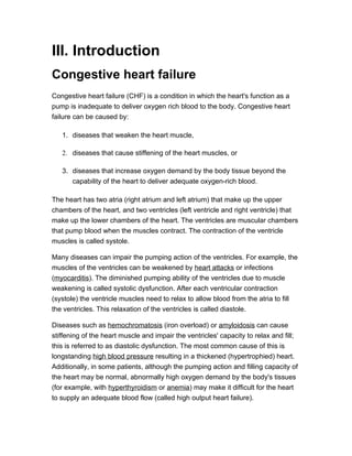
Congestive heart failure (CHF) is a condition in which the heart's function as a
pump is inadequate to deliver oxygen rich blood to the body. Congestive heart
failure can be caused by:
1. diseases that weaken the heart muscle,
2. diseases that cause stiffening of the heart muscles, or
3. diseases that increase oxygen demand by the body tissue beyond the
capability of the heart to deliver adequate oxygen-rich blood.
The heart has two atria (right atrium and left atrium) that make up the upper
chambers of the heart, and two ventricles (left ventricle and right ventricle) that
make up the lower chambers of the heart. The ventricles are muscular chambers
that pump blood when the muscles contract. The contraction of the ventricle
muscles is called systole.
Many diseases can impair the pumping action of the ventricles. For example, the
muscles of the ventricles can be weakened by heart attacks or infections
(myocarditis). The diminished pumping ability of the ventricles due to muscle
weakening is called systolic dysfunction. After each ventricular contraction
(systole) the ventricle muscles need to relax to allow blood from the atria to fill
the ventricles. This relaxation of the ventricles is called diastole.
Diseases such as hemochromatosis (iron overload) or amyloidosis can cause
stiffening of the heart muscle and impair the ventricles' capacity to relax and fill;
this is referred to as diastolic dysfunction. The most common cause of this is
longstanding high blood pressure resulting in a thickened (hypertrophied) heart.
Additionally, in some patients, although the pumping action and filling capacity of
the heart may be normal, abnormally high oxygen demand by the body's tissues
(for example, with hyperthyroidism or anemia) may make it difficult for the heart
to supply an adequate blood flow (called high output heart failure).
2. In some individuals one or more of these factors can be present to cause
congestive heart failure. The remainder of this article will focus primarily on
congestive heart failure that is due to heart muscle weakness, systolic
dysfunction.
Congestive heart failure can affect many organs of the body. For example:
• The weakened heart muscles may not be able to supply enough blood to the
kidneys, which then begin to lose their normal ability to excrete salt (sodium)
and water. This diminished kidney function can cause the body to retain more
fluid.
• The lungs may become congested with fluid (pulmonary edema) and the
person's ability to exercise is decreased.
• Fluid may likewise accumulate in the liver, thereby impairing its ability to rid
the body of toxins and produce essential proteins.
• The intestines may become less efficient in absorbing nutrients and
medicines.
• Fluid also may accumulate in the extremities, resulting in edema (swelling) of
the ankles and feet.
Eventually, untreated, worsening congestive heart failure will affect virtually every
organ in the body.
3. Picture of the heart and valves, left and right ventricles, left and right atria
Despite the paucity of epidemiologic work on congestive heart failure (CHF), the
salient features of the natural course of cardiac failure are understood. The
estimated 1983 incidence of CHF in the United States was 214,000 men and
184,000 women. The estimate of prevalence was 2.3 million persons, with a
remarkable increase with advancing age and higher rates in men than women at
all ages. Overt heart disease plus age are the principal determinants of the
incidence of CHF. Nearly 90% of patients with CHF have systemic hypertension
or coronary heart disease, or both, as the antecedent underlying condition.
Diabetes mellitus increases the risk of CHF at all ages, particularly in women and
those treated with insulin. The prognosis after diagnosis of CHF is grim and is
related to the degree of myocardial dysfunction. The challenge is to develop
more effective drugs not only for the management of overt CHF, but also for the
prevention of its progression
4. Heart failure is the clinical endpoint for a number of diseases resulting in
myocardial dysfunction. Ischemic cardiomyopathies due to coronary artery
disease and dilated cardiomyopathies, either idiopathic or familial, make up most
cases. Many other diseases can also lead to end-stage heart failure. These
include valvular, congenital, metabolic, and inflammatory disorders.
In terms of the pathology, cardiac remodeling is characterized by myocytic
hypertrophy, chamber dilatation, and changes in the composition of the matrix.
The fibrotic heart ultimately becomes somewhat spherical, and, subsequently, it
loses efficiency as a pump. Therefore, an important goal is to identify and
favorably intervene before terminal myocardial remodeling begins.
IV. Structural Organization
Anatomical back ground
The heart is a muscular pump that contains four chambers: right atrium, left
atrium, right ventricle and left ventricle. The two small atria make up the top of
5. the heart, and the two large ventricles make up the bottom of the heart. The right
atrium pumps blood to the right ventricle, and the left atrium pumps blood to the
left ventricle. A wall, called the septum, separates the right atrium and right
ventricle, from the left atrium and left ventricle.
Blood flows through the heart in the following manner:
• The right atrium receives oxygen-poor blood from the body, and then
pumps the blood through the tricuspid valve and into the right ventricle.
• The right ventricle pumps the blood through the pulmonic valve and to the
lungs, where it picks up more oxygen.
• The left atrium receives the oxygen-rich blood from the lungs, and then
pumps the blood through the mitral valve and into the left ventricle.
• The left ventricle pumps blood through the aortic valve and to the rest of
the body.
• The blood supplies oxygen to the body and the cycle starts again.
Anatomy examples:
• Normal circulation through the heart
• The human heart
• Cross-section of heart at the level of the atria
• Cross-section of heart at the level of the ventricles
• The heart sits inside the pericardium
• The heart valves
Coronary Arteries
The coronary arteries supply oxygen to the heart muscle.
6. The heart has three main coronary arteries:
• Right coronary artery: supplies the right ventricle
• Left coronary artery: supplies the left ventricle
• Posterior circumflex artery: supplies the posterior aspect of both ventricles
Anatomy examples:
• Coronary angiogram
• Front view of the heart and coronary arteries
• Back view of the heart and coronary arteries
Cardiac Conduction System
An electrical impulse stimulates the muscle fibers in the heart to contract. The
impulse spreads through the heart in a very organized manner, so that the atria
contract first, followed by the ventricles.
The electrical impulse proceeds in the following manner:
• The electrical impulse originates at the sinoatrial (SA) node, which is
located in the wall of the right atrium.
o The SA node is the heart's natural pacemaker: it regulates the heart rate.
• The impulse proceeds through the atria, stimulating them to contract.
• After the atria are stimulated to contract, the atrioventricular (AV) node
slows the electrical impulse before it proceeds to the ventricles. This pause
allows the ventricles to fill with blood before they contract.
o The AV node is located between the atria and the ventricles.
• After the pause, the impulse then proceeds through the ventricles,
stimulating them to contract.
7. Anatomy examples:
• The cardiac conduction system
• EKG showing electrical activity of heart
.
V. Confimatory test
Diagnosis
Physicians can often make a preliminary diagnosis of heart failure with only a medical
history and careful physical examination. An English study suggests, in fact, that the
8. condition may be under-diagnosed. The following signs along with a history of heart
disease strongly suggest heart failure:
• Enlarged heart.
• Irregular heart sounds.
• Abnormal sounds in the lungs.
• Swelling or tenderness of the liver.
• Fluid retention.
• Elevation of pressure in the veins of the neck.
Confirming these findings definitely or determining the severity of the condition,
however, is difficult. Further tests are usually needed.
Laboratory
Both blood and urine tests are used to check for malfunctions of the liver and kidneys and
to detect signs of diabetes.
Blood tests can also be used to evaluate the following:
• Cholesterol and lipid levels.
• Anemia.
• Thyroid disease.
Urine tests can also be used to assess:
• Albumin. The presence of this protein in the urine is usually a sign of kidney disease,
but even tiny amounts (microalbumin) signal an increased risk for heart failure in
people with and without diabetes.
VI. Sign, Symptoms and complication
Signs and Symptoms
Irrespective of the etiology, the first manifestation of congestive heart failure
(CHF) is usually tachycardia. An obvious exception to this finding occurs in
congestive heart failure due to a primary bradyarrhythmia or complete heart
9. block. As the severity of congestive heart failure increases, signs of venous
congestion usually ensue. Left-sided heart failure is generally associated with
signs of pulmonary venous congestion, whereas right-sided heart failure is
associated with signs of systemic venous congestion. Marked failure of either
ventricle, however, can affect the function of the other, leading to systemic and
pulmonary venous congestion. Later stages of congestive heart failure are
characterized by signs and symptoms of low cardiac output. Generally,
congestive heart failure with normal cardiac output is called compensated
congestive heart failure, and congestive heart failure with inadequate cardiac
output is considered decompensated.
Signs of congestive heart failure vary with the age of the child.2 Signs of
pulmonary venous congestion in an infant generally include tachypnea,
respiratory distress (retractions), grunting, and difficulty with feeding. Often,
children with congestive heart failure have diaphoresis during feedings, which is
possibly related to a catecholamine surge that occurs when they are challenged
with eating while in respiratory distress.
Right-sided venous congestion is characterized by hepatosplenomegaly and,
less frequently, edema or ascites. Jugular venous distention is not a reliable
indicator of systemic venous congestion in infants because the jugular veins are
difficult to observe. Also, the distance from the right atrium to the angle of the jaw
may be no more than 8-10 cm, even when the individual is sitting upright.
Uncompensated congestive heart failure in an infant primarily manifests as
a failure to thrive. In severe cases, failure to thrive may be followed by signs of
renal and hepatic failure.
In older children, left-sided venous congestion causes tachypnea, respiratory
distress, and wheezing (cardiac asthma). Right-sided congestion may result in
hepatosplenomegaly, jugular venous distention, edema, ascites, and/or pleural
effusions. Uncompensated congestive heart failure in older children may have
fatigue or lower-than-usual energy levels. Patients may complain of cool
extremities, exercise intolerance, dizziness, or syncope.
Clinical findings may include hypotension, cool extremities with poor peripheral
perfusion, a thready pulse, and decreased urine output. Chemical evidence of
renal and liver dysfunction may be present, as well as a diminished level of
10. consciousness. Children with uncompensated congestive heart failure,
particularly older children, generally have a lower cardiac output than what most
experienced clinicians would estimate on the basis of the clinical signs.
Signs and symptoms of congestive heart failure include the following:
• Tachycardia
• Venous congestion
o Right-sided
Hepatomegaly
Ascites
Pleural effusion
Edema
Jugular venous distension
o Left-sided
Tachypnea
Retractions
Nasal flaring or grunting
Rales
Pulmonary edema
• Low cardiac output
o Fatigue or low energy
o Pallor
o Sweating
o Cool extremities
o Poor growth
o Dizziness
o Altered consciousness
o Syncope
VII. Treatment
Surgical
11. Heart transplantation
When progressive end-stage heart failure occurs despite maximal medical
therapy, the criterion standard for therapy has been heart transplantation. Since
Christiaan Barnard performed the first orthotopic heart transplantation in 1967,
the world has seen tremendous advancement in the field of cardiac
transplantation.
Compared with patients who receive only medical therapy, transplant recipients
have fewer rehospitalizations, marked functional improvements, enhanced
quality of life, more gainful employment, and longer lives with 50% surviving to 10
years.11 Heart transplantation is associated with a 1-year survival rate of 83%,
which decreases in a linear manner by approximately 3.4% per year. Careful
selection of donors and recipients, as well as efforts to minimize potential
perioperative dangers (ischemic times, pulmonary hypertension, mechanical
support, cardiogenic shock), are critical to ensure good outcomes.
The single greatest advancement in ensuring long-term function of the allograft is
the development of immunologic modulators. Pioneered by Dr Norman Shumway
at Stanford University, steroids and antipurine metabolites including azathioprine
and mycophenolate mofetil (MMF) have been widely used.
Central to the current immunosuppression regimens are calcineurin inhibitors,
cyclosporine, and tacrolimus. These drugs inhibit cellular pathways responsible
for the production of interleukin (IL)-2 and subsequent T-cell activation. They
inhibit the nuclear translocation of cytoplasmic factors needed to bind to the IL-2
gene promoter. Immunosuppressive regimens have evolved from cyclosporin to
a predominant use of tacrolimus.
Triple drug therapy consisting of steroids, calcineurin inhibitors, and MMF has
become standard initial immunotherapy after heart transplantation.12 Additional
agents, such as antithymocyte globulin, rapamycin, and IL-2 receptor
antagonists, also have important roles in modern immunosuppression protocols.
The Achilles heel in the long-term success of heart transplantation is the
development of coronary graft atherosclerosis, the cardiac version of chronic
rejection. Coronary graft atherosclerosis is uniquely different from typical
12. coronary artery disease in that it is diffuse and usually not amenable to
revascularization. Furthermore, although heart transplantation is a feasible
solution for patients with end-stage heart disease, its use is limited by an
inadequate donor supply.
In the United States, fewer than 2500 heart transplantation procedures are
performed each year.13 Each year, an estimated 10-20% of patients die while
awaiting a heart transplant. Of the 5 million people with heart failure,
approximately 30,000-100,000 have such advanced disease that they would
benefit from transplantation or mechanical circulatory support.14 This disparity
between the number of patients needing transplants and the availability of heart
donors has refocused efforts to find other ways to support the severely failing
heart.
VIII. Reference
http://www.medicinenet.com/congestive_heart_failure/article.htm#1whatis
http://adam.about.com/reports/000013_5.htm
http://www.medicinenet.com/congestive_heart_failure/page2.htm
http://www.ncbi.nlm.nih.gov/pubmed/3966408
http://emedicine.medscape.com/article/901307-overview