13 peripheral venous duplex ct mri in dvt dr ahmed esawy

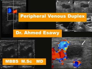
VENOUS DULEX DEEP VENOUS THROMBOSIS DVT Acute DEEP VENOUS THROMBOSIS 1-low echogenicity(intraluminal material producing low void ,incomplete color fill in) 2-venous distention 3-loss of compressibility 4-free floating thrombus 5-doppler signal abnormality a-flow augmentation absent or decreased proximal to thrombosed segment b-distally flow is continous rather phasic 6- Static valve leafets 7- Absent or abnormal flow on spectral or colour Doppler 8-Loss of spontaneous fow and respiratory variation 9- Increased flow in collateral channels subacute DEEP VENOUS THROMBOSIS 1-increased echogenicity similar to muscle 2-decreased thrombosed size 3-reduced vein size 4-adherence of thrombus 5-resumption of flow(doppler signal may remain abnormal) chronic DVT Ultrasound duplex signs 1-echogenic intraluminal material(more than adjacent muscle) a-focal or diffuse thickening of vein wall b-vein reduced to echogenic cord 2-valve abnormality a-reflux►varicosities b-persistent venous distention 3-doppler flow abnormalities a-lack of spontaneity b-lack of phasicity c-absence of valsalva response d-or absence of augmentation Acute Occlusive DEEP VENOUS THROMBOSIS PARTIAL THROMBOSIS ACUTE DEEP VENOUS THROMBOSIS Partial thrombosis of inferior cava vein partially occlusive thrombus Spectral Doppler In DEEP VENOUS THROMBOSIS AUGMENTATION IN ACUTE DEEP VENOUS THROMBOSIS LEG CALF VEINS THROMBOSIS IVC thrombosis Subclavian and Internal Jugular Veins DVT superficial venous system thrombosis thrombophlebitis Compression ultrasound venous thrombosis Chronic DVT Follow-up evaluation Post thrombotic syndrome Wall thickening , rigidity ,irregularity Persistent occlusion Collaterals Valvular damage, incompetency (reflux) Loss of phasicity Superficial varicose veins less easily compressed Recanalization Tips & Tricks in DVT DVT PITFALLS DIFFERENTIAL DIAGNOSIS Swollen / edematous / fatty legs Non-occlusive thrombus Segmental calf vein thrombus Segmental iliac vein thrombus Pregnant patients Leg swelling 1-suboptimal irregularity 2-mistaken identity 3-incorrect description of age of thrombus 4-chronic thrombotic residua 5-improper use of color duplex image 6-poor venous distention of reduced flow 7-venous stasis Popliteal cysts Haematoma Superficial thrombophlebitis Iliac nodes/pelvic masses Arteriovenous fistula Lymphoedema Hyperperfusion syndrome Muscle tears