More Related Content
Similar to Research poster 2015 (20)
Research poster 2015
- 1. RESEARCH POSTER PRESENTATION DESIGN © 2015
www.PosterPresentations.com
Astrocytes are glial cells located in the central nervous system (CNS) that
are known to secrete various neurotrophic & apoptotic factors.
Experiments conducted using wild type astrocyte conditioned media
(WTACM) on undifferentiated SH-SY5Y cells suggest that a neurotrophic
factor present in the media protected the cells preferentially when
compared to differentiated SH-SY5Y cells. It is unclear whether this factor
was astrocyte-secreted or a component of fetal bovine serum (FBS), which
is used to supplement the growth of the cells. To further investigate the
source of these factors, undifferentiated cells were treated with media from
astrocytes grown with FBS either present in or absent from the media.
While FBS clearly contributed neurotrophic factors which affected the
undifferentiated SH-SY5Y cells, experimental results indicated the
presence of an astrocyte-secreted factor that induced cell death.
Fractionation experiments were conducted to narrow down the size of the
factor and the results suggest the presence of neurotrophic factors that
weigh below 50kDa and between 50 and 100kDa as well as a cell-death
inducing factor that weighs between 50 and 100kDa. Future research aims
to identify the nature of these astrocyte-derived neurotrophic factors and
cell-death inducing factor.
Abstract
Introduction
❖ To make the >50kDa and >100kDa retentate media:
➢ 12mL of WTACM without serum was placed into the Amicon
Ultra-15 centrifugal filter unit tube with an ultracel-50 membrane
and the Amicon Ultra-15 centrifugal filter unit tube with an
ultracel-100 membrane.
➢ The tubes were centrifuged at 5,000rpm for 30 minutes at 23°C.
➢ The filter from each tube was removed and 5mL of DMEM F-12
was added to it.
➢ After mixing the DMEM with the retentate, the resulting media
from each filter was then transferred to a 14mL centrifuge tube.
❖ The media left in the Amicon tube and was considered the <50kDa and
<100kDa filtrate media.
Ultra-fractionation of media
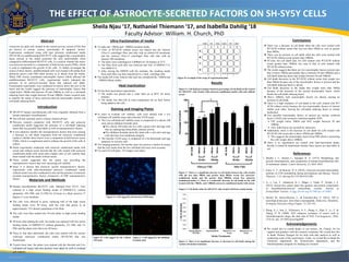
Figure 1: There is a significant increase in cell death between the cells treated
with the less than 50kDa and greater than 50kDa serum free astrocyte-
conditioned media and the greater than 100kDa serum free astrocyte
conditioned media (*). There is a noticeable decrease in cell death in the cells
treated with the >50kDa and >100kDa astrocyte-conditioned media with serum.
Results
Conclusions
❖ There was a decrease in cell death when the cells were treated with
WTACM without serum that was less than 50kDa as well as greater
than 50kDa.
❖ There was an increase in cell death when the cells were treated with
WTACM without serum greater than 100kDa.
❖ Of note, the cell death ratio for cells treated with WTACM without
serum greater than 100kDa was near to that of cells treated with
WTACM without serum.
❖ The results suggest that there are two neurotrophic factors present (one
that is below 50kDa and another that is between 50 and 100kDa) and a
cell-death inducing factor that weighs between 50 and 100kDa.
❖ Cell death decreases in the WTACM without serum that weighs less
than 50kDa because one of the neurotrophic factors is present and the
cell-death inducing factor is absent.
❖ Cell death decreases in the media that weighs more than 50kDa
because of the presence of the second neurotrophic factor, which
blocks the cell-death inducing factor.
❖ Above 100kDa, both neurotrophic factors are absent, causing an
increase in cell death.
❖ There is a high incidence of cell death in the cells treated with WT
ACM without serum because the two neurotrophic factors of interest
inhibit each other, leaving the cell-death inducing factor to remain
unchecked.
❖ Two possible neurotrophic factors of interest are nuclear erythroid
factor 2 (Nrf2) and vasoactive intestinal peptide (VIP).
➢ VIP weighs below 50kDa and Nrf2 weighs between 50 and
100kDa.
➢ Also, both factors are secreted by astrocytes.
❖ Additionaly, there is the decrease in cell death for cells treated with
WTACM with serum that is above 50kDa and 100kDa.
➢ This suggests the neurotrophic factor from FBS which is protceting
the cells weighs above 100kDa.
❖ There is no significance are treated with heat-inactivated media,
thereby it cannot be determined whether these factors are heat labile or
not.
References
1. Biedler J. L., Helson L., Spengler B. A. (1973). Morphology and
growth, tumorigenicity, and cytogenetics of human neuroblastoma cells
in continuous culture. Cancer Research, 33, 2643–2652.
1. Jones, E. V., & Bouvier, D. S. (2014). Astrocyte-secreted matricellular
proteins in CNS remodelling during development and disease. Neural
Plasticity, 1-12. doi.org/10.1155/2014/321209
1. Li, J., Lee, Y., Johansson, H. J., Mäger, I., Vader, P., Nordin, J. Z.
(2015). Serum-free culture alters the quantity and protein composition
of neuroblastoma-derived extracellular vesicles. Journal of
Extracellular Vesicles, 4. http://dx.doi.org/10.3402/jev.v4.26883
1. Morell, M., Souza-Moreira, L., & Gonzalez-Rey, E. (2012). VIP in
neurological diseases: more than a neuropeptide. Endocrine, Metabolic,
& Immune Disorders-Drug Tragets, 12, 323-332.
1. Wang, X. J., Sun, Z., Villeneuve, N. F., Zhang, S., Zhao, F., Li, Y...&
Zhang, D. D. (2008). Nrf2 enhances resistance of cancer cells to
chemotherapeutic drugs, the dark side of Nrf2. Carcinogenesis, 29(6),
1235-43. doi: 10.1093/carcin/bgn095.
Acknowledgements
❖ We would like to extend thanks to our mentor, Dr. Church, for his
support and guidance with the research condcuted. We would also like
to thank Thomas Naragon for his help with data analysis as well as
conducting some of the experiments. Lastly, we would like to thank the
Chemistry department, the Neuroscience department, and the
Interdisciplinary program for funding our research.
❖ SH-SY5Y human neuroblastoma cells were originally obtained from a
human metastatic neuroblastoma1.
❖ The cell line currently used is a thrice cloned cell line1.
❖ Previous research conducted with SH-SY5Y cells and astrocyte
conditioned media suggested the presence of a cell-death inducing
factor that was possibly heat labile as well as neuroprotective factors.
❖ It was unknown whether the neuroprotective factors that were causing
a decrease in cell death originated from the astrocyte conditioned
media or whether these factors were a component of fetal bovine serum
(FBS), which is a supplement used to enhance the growth of the cells in
culture.
❖ Initial experiments conducted with astrocyte conditioned media with
serum and without serum showed that the cells treated with astrocyte
conditioned media with serum had a higher ratio of cell viability than
those treated with the media without serum.
❖ These results suggested that the serum was providing the
neuroprotective factors that were increasing cell viability.
❖ Since it is known that astrocyte secrete neuroprotective factors,
experiments with ultra-fractionated astrocyte conditioned media
without serum were also conducted to rule out the presence of astrocyte
secreted neuroprotective factors irrespective of FBS neuroprotective
factors.
Faculty Advisor: William. H. Church, PhD
Sheila Njau ‘17, Nathaniel Thiemann ‘17, and Isabella Dahilig ‘18
THE EFFECT OF ASTROCYTE-SECRETED FACTORS ON SH-SY5Y CELLS
Heat Inactivation
Materials and Methods
❖ Human neuroblastoma SH-SY5Y cells, obtained from ATCC, were
cultured in a high serum feeding media of DMEM-F12 without
glutamine, 10% FBS, and 1% PSG for 24 hours in a flask stored at 37
degrees C in an incubator.
❖ The cells were allowed to grow, replacing half of the high serum
feeding media every 48 hours, until the cells had grown to an
approximately 75% density population in the flask.
❖ The cells were then seeded into 24-well plates in high serum feeding
media.
❖ 48 hours after plating the cells, the media was replaced with low serum
feeding media of DMEM-F12 without glutamine, 2% FBS, and 1%
PSG and the plates were fed every 48 hours.
❖ Three or four days afterwards, the cells were treated with the various
wild-type astroicyte conditioned media (WTACM) that was
fractionated.
❖ Twenty-fours later, the plates were stained with the Hoechst and Live
cell/dead cell assays and nine pictures were taken by well to evaluate
cell viability.
Staining and Imaging Plates
❖ For the heat inactivation experiments:
➢ The media was placed into a water bath set at 60°C for thirty
minutes.
➢ The media was then left at room temperature for an hour before
being added to the cells.
❖ In order to evaluate cell viability, cells were stained with a live
cell/dead cell viability assay and a hoechst 33258 assay:
➢ The Live cell/dead cell viability assay is composed of a calcein AM
stain and an ethidium bromide stain.
■The calcein AM stains the live cells a green color and tags cells
that are undergoing intracellular esterase activity.
■The ethidium bromide stains the dead cells a red color and tags
cells that have lost membrane integrity.
➢ The hoechst 33258 assay is composed of a bis-benzimide and tags
apoptotic or necrotic cells.
❖ For imaging purposes, the hoechst stain was used as a marker to ensure
that the cell counts from the live cell/dead cell assay were accurate.
❖ For each 24-well plate, 216 images were taken.
Figure B: Cells tagged by the Calcein
AM stain.
Figure C: Cells tagged by the ethidium
bromide stain
Figure A: Cells tagged by the hoechst 33258 stain.
Figure D: Example of the setup of experimental plate.
Figure 2: There is no significant increase or decrease in cell death among the
various cell media treatments.
Figure 2: Cell death ratios for SH-SY5Y cells treated with heat treated media
Figure 2: Cell death percentages based on percentage of cell death in the control
for SH-SY5Y cells treated with astrocyte-conditioned media with and without
serum