How to do quick user assign in kanban in Odoo 17 ERP
skin and derivatives.pdf
1. I R Y
c ABIRTAlIKA aAh nPxu AAA OKA CIRDCOR HWUALIE RMA ORIT EAR PDM
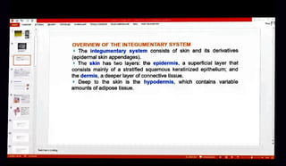
oVERVIEw OF THE INTEGUMENTARY SYSTEM
The integumentary system consists of skin and its derivatives
(epidermal skin appendages)
The skin has two layers: the epidermis, a superficial layer that
consists mainly of a stratified squamous keratinized epithelium; and
the dermis, a deeper layer of connective tissue.
Deep to the skin is the hypodermis, which contains variable
amounts of adipose tissue.
aMeToe KCAaay
2. A 1 A BCTAIKA
MA nNma vALAA OKAI CIAPROB AiE
M OMIT REAOCR PC
EPDERMIS
The epidermis is composed primarily of keratinocytes (85%) that undergo diferentiation to form stratified
squamous keratinized epithelium.
Five distinct layersofepidemis(strata) can
bedistinguished.
The stratum basale is a single layer of smal, mitotically active basal cells that are attached by
hemidesmosomes tounderlying connectivetissueandby desmosomesto eachother.
The stratum spinosum contains
several layers oflargerkeratinocytes that are
attachedto each other by
desmosomes located at the ends of their eytoplasmie processes containing intermediate filaments (keratin
ilaments)
The stratum granulosum is a
distinct layer of
latlened keratinocytesfilled with keratohyalingranules
(contain precursors to filagerin, which aggregates keratin filaments and lamellarbodies containing lipids,
which, when secreted, are
responsible for theformation ofthe
epidermal waterbarrier
The stratum lucidum, which is limited to
thick skin and
consideredasubdivision ofthe stratum comeum
In the light microscope, it often has a refractile appearance and may stain poorly. This highily reractile layer
contains eosinophilic cells in which theproucess of keratinization is well advanced. The micleus and
cytoplasmic organelles become disnuptedand disappearasthecell eradually fnllswithkeratin.
The stratum corneum is the most superficial layerof terminally differentiated squamous cells(withno
nuclei) that are entirely filled with keratin filaments. These cells are constantly desquamating from the skin
surface.
Total epidermal tumover time takes approximately 47 days.
w*23
3. 0AA RABIABTABKA
MJAI IEROaM AAALIR
nOKAJC a PEuEHAAAIME B TOT READER
CELLS OF THE EPIDERMIS
There are four diferent types of cells in the epidermis:
Keratinocytes are highly speciallzed eplthellal cells designed to perform a very
specitficfunction: separation ofthe organism from Its external environment. They account
for 85% of the cells in the epidermis.
Melanocytes are the pigment-producing cells of the epidermis. They account for
approximately 5% ofthe cells In the epidemis. Melanocyles synthesize melanin plgment
in melanosomes and during the
process of pigment donation, melanocytes transfer
them into adjacent keratinocytes. The transferred plgment accumulates above nuclei of
keratinocytes to protect nuclear DNA from ultraviolet (Uv) radiallon and damage.
Langerhans' cells are the antigen-presenting cells involved in signaling in the
immune system. They account for approximately 2%% to 5% of the cells in the epidermis.
Also, It possesses characteristic, tennis racquet-shaped Birbeck granules. They represent
relatlvely small vesicles, which appear as rods with a bulbous expansion at their end.
Merkel's cells are the sensitive mechanoreceptor cells assoclated with sensory nerve
endings. They account for approximately 69% to 10% of the cells in the epidermis.
JaMeTKH K CanAy
-
4. OAA FAADIIABCTABKA DA raxOu MAA OKAICAO PELEVOMM MOTLADUK
Melanocytes (5% of cells in epidemis) reside in the stratum basale and have long processes that
extend belween keratinocytes into the stratum spinosum.
Melanocytes synthesize melanin pigment in melanosomes and during the process of pigment
donation, melanocytes transfer them into adjacent keratinocytes. The transfered pigment
accumulates above nuclel of keratinocytes to protect nuclear DNA from ultraviolet (UV) radiation and
damage. Diagram of the epidernmls
This diagram shows a melanocyte interacting with
everal cells of the strutum basale and the stratum
Langerhans ne melunocyle has
long dendritic processes
cel that contain accumulated melanosomes and extend
between the cells of the epidermis.
melanocyte The Langerhans' cell is a dendritic cell often confused
with a melanocyte but is actually part of the
mononuclear phagocytolic system and tunctions
as an
keratinocyteantigen-presenting cell of
the immune systemin the
initiation of cutaneous hypersensitivity reactions
(contact allergic demmatitis). Also, it possesses
characteristic, tennis racquet-shaped Birbeck granues.
melanoeye They represent relatively s1mall vesicles, which appear
as rods with a bulbous expansion at their end.
SaMetkH KChaAay
5. A l AADIAR BCTABKA MIA n O AAT nioKAC4 OWAT EM T RIAHRPE
Schematic diagram of keratinocytes in the epldermis.
The keratinocytes in this figure retlect different stages in
the life cycle of the cell as it passes from the basal layer to
EKTI
t h e skin surface, where it becomes desquamated.
The baal cell begins to
synthesize intermediate (keratin)
filaments; these are grouped into bundles and are seen in
the light inicroscope as
tonofibrils.
The cell enters the spínous layer, where the synthesis of
inlermediate lilaments conlinues. In the upper purt of the
spinous layer. the cells begin to produce keratohyalin
granules containing intermediate ilament-associated
dmde Proteinsand glycolipid-containing lamellar bodies.
H-75 Within the granular layer, the cell discharges lamellar
bodies that contribule to formution of the water barrier of
the epidermis; the remainder of the cell cytoplasm contains
numerouskeratonyalin granules Ihal, in close association
with tonofilaments, fom the cell envelope.
The
surface cells are keratinized; they contain a
thickcell
envelope und bundles of tonofilanents in
amla bodes
besal cel
mals fase
boeomes
pecialized
atrix.
6. DERMIS
The dermis is composed of wo
layers.
The papillary layer is superficial
and consists of loose connective
tissue (collagen I and l1) that contains
extensive plexus ofblood, lymphatic
vessels, and sensory nerve endings.
The reticular layer is deeper and is
composed of dense irregular
connective tissue containing type
Enepic Epidermis
ws Derm
collagen, elastic fi bers, and larger
blood vessels.
The epiderma-dermal junction has
numerous finger-like connective tissue
protrusions called dermal papillae
that correspond to
similar epidermal
profrusions (epidermal ridges).
Dermal papillae contain nerve
endings and a network of blood and
Iymphatic capillaries.
p l v
Tenan pu
rtern rtines ef swrst l n
MPTM KaAny
7. M ABI 1AM TADRA
A
iPIXULIu A.LAA DNAT CAAPERM uEHOPOEA
DA FOMIT READER POI
This specimen obtained from the skin of the sole of the
Toot (numan) showS epidermis (Epi) containing the
extremely thick stratum corneum (So). Remaining9
layers of the epldermis (except for the stratum lucidum,
which Is not present on this slide) -
that is,
the stratum basale (SB),
SC Epi
the stratum spinosum (SS), and
the stralum granulosum (SGr) - are clearly visible in this
routine H&E preparauon.
The duct of a sweat gland (D) can be seen on the jeft
as it traverses the dermis (Derm) and further spirals
through the epidermis. At the sites where the ducts of
the sweat
gland enter
the epldermis, note the epiderma
downgrowths known as interpapilary pegs
The dermis contains papllae, protrusions of connective
tissue that le between the interpapillary pegs. Note also
the greater cellulanty of the papilary layer (PL) and that
ne collagentibers oftherelicular layer (RL) are thicker
than those of the papilary layer.
SGr
Derm
aMeIKM K
8. Baca
GAiI ABIA BCTAIKA MJAN rPXOAM
AA noSACC
TEE A MA FONT IACR PEV
SENSORY NERVE RECEPTORS OF SKIN
ne eidermiS contains ree nerve endings, which delect fine touch, heat, cold, and pain.
n 8ddition, et ealed nerve endinas, such as Pacinian corpuscles to detect pressure and VIDrations,
Meieen
wn a nerve ending) is a
sensitive mechanoreceptor.
Meissner's corpuscles to detect light touch, and Ruffini's corpuscies to delect skin stretch and torque.
erhafs cel
ensitve echauoe Diagram of the sensory receptors in
paln, Dae touch
wnperstare hEa, t skin.
nd er fbe
Free nerve endings:
a. Epldermal free
endings.
D.
Merkers corpuscles conlaining Merkel's
cellsand disc receptors of afferent
myelinated nerve fibe
Encapsulated nerve endings:
c. Pacinian corpuscle located In the deep
e tbe
nen
Ocinian corpAIsce
(vibrationded
layer of deep dermis and hypodermis.
d. Krause's end bulb serves as cold
b e b (celd
wmted eprot
receptor.
e. Melssner's corpuscle in dermal papilla.
. Ruffini's corpuscle in deep layers of the
dermis.
Raña'h corpwee (shin stertch and torque)
slebsners cotpuscie (ught toach)
SMeTat EAMay
9. DA
r m a m i a n
TAUIKA A imu A*MUALA OKACiAPA0a HbO06AIEBMA FORIT READER PO
Epidermal Skin Appendages
Skin appendages are derived from downgrowths of epidermal epithelium
during development. They include the following:
Hair follicles and theirproduct, hair
Sebaceous glands and their product, sebum
Merocrine sweat glands and their product, sweat
Apocrine sweat glands and their mixed product containing a form ofsweat
with a high concentration ofcarbohydrates, lipids, and proteins
JaMeTKMK CAaRAyy
10. 73 x
Bata
A BTAIKA 1A nEPOROB AoALA OxAJ CAPIOH uEHIPOHA, IE A FOXiT READMR PM
EPIDERMAL SKIN APPENDAGES eboh
Hairs and hair follicles are present over
almost the entire body.
The hair follicle contains a reservoir of
epidermal stem cells (follicular bulge) that
are responsible for differentiation into hair-
forming matrix cells.
Hair is formed by the differentiation of
matrix cells in the inferior segment of the
air follicle (bulb) to form the medulla, cotex
(80% of hair mass), and cuticle of a hair shaft.
The hair shaft is surrounded by the internal
and external root sheath. The internal root
epidermis-
dermis-
sebaceous
eccrine external root shealn
apocrine
9land
arrectorpilmuscle
sensory
nerve
ending
gassy membrane
cuticle
OUar
Duige
sheath has three layers of cells: Henle's
layer, Huxley's layer, and the internal root
sheath cuticle. The external root sheath is
nair ortex
hair m trix
blood vessel demal papilla
continuous with the epidermis.
Sateete k CMaiay
11. DAR FAABAR BCTABKA AMIA nEPEAOM AIMAta hoNAcINNOB
amOBAHME B
FOTREADERPD
air
haft
Diagram showing a hair follicle.
Note the cell layers that form the hair shaft and
the surrounding external and internal root
sheaths.
The sebaceous gland consists of the secretory
portion and a short duct that empties into the
infundibulum, theupper part of the
hair folicle.
The arreclor pili muscle accompanies the
sebaceous gland; contraction of this smooth
muscle assistsin gland secretion and discharges
the sebum into the infundibulum ot the halr
epidermis-
dermis-
sebaceous
Jland
eccrine-
glana
external root sheath
follicle.
ocrine
land
arrectorpili muscle
internal root sheath
glassy membrane
cuticle
nhaircortox
medula
Projection of the external root sheath near
insertion of the arrector pil muscle forms the
follicular bulge that contalns epidermal stem
cells.
Nerve endings (yelow) surround the follicular
bulge with nearby insertion of arrector pil muscle.
The apocrine sweat gland also empties into the
Infundibulum.
sensory
andinn
follicular
bulge
hair matrix
Notethat eccrine sweat glands are Independent
structures and are nol associated directy witn the
blood vessel demal papilla
hair follicle.
JaMet K CAaHIy
12. 2
Halr
follicleand pathwaysof epidermal
stem cell migration. This dlagram shows the
location and migration pathways of epidermal
stem cells that reside In the follicular bulge. Under
normal
conditions, epidermal stem cells migrale
upward to
the sebaceous gland and downward to
intera root
internal external
hair
shat sheain sheath
reach the halr matrix in the bulb of the follicle
nair shaft (black arrows). Hair matrix is fomed by
dmerenuaung ceIS nat migrate along the external
root sheath from the follicular bulge. As the
differentiation progresses, cells leave
the matrix
they form cell layers that differentiate into the hair
shaft containing
atri
sebaceouUS
gland
follicular
bulge
(7) medula,
(2) hair cortex, and
(3) hair cuticle,
and the internal root sheath containing
(4) intemal root sheath cuticle,
(5) Huxley's layer, ana
(6) Henle s layer.
During injury of the epldermis, the epldermal stem
cells migrate from the follicular bulge toward the
skin surface (red arrow) and
participate in the
Initial resurfacing of damaged epidermis.
hair matrix
cells
nal
papilla
aMeteChamay
13. i riti -
S TAB-ABCALKA
MIAH EPCXOM AMMAIR nOKAI CIAPROB PELEOONAIME B
ORITREADRPO
Internal Root Sheath
Hair Structure
Sheath cuticle-
Huxtey's layer-
Henle's layer-
Bulb (at the base)
Papilla(areolarCT)
Root (belowthe surface)
Shaft (above the surface)
Cuticle(singlelayerof hard,
Extenal root sheath-
Glassy membrane
Root hair pexus
Hair sh
keratinized cells)
Cortex (keratinized,pigmented)
Medula (soft keratin)
Myoepithela
cell
Dark,
cel
Hair papIla
Clear
eTMK ChaMay
14. AA AB A TAIKA AVH Mai AMMALUR noKAJ CAAMAO8 PELUEWOA IME HaA FORT READR PDF
4
Hair
25
6
Lanugo- type of hair on the body of a fetus or newborn baby, on
the ody of an adult except on the palms, of the hands, the soles
of the feet and the parts where long hair grows.
Terminal hair(long)- head, beard, pubic hair, axillary space.
Bristly hair-eyebrows, eyelashes, external auditory tube,
vestibule of the nasal cavity.
31
2
33
15. DAMA AABA BCTABKA A nEPEAQAM AvMAIR OACAAS P OA aREAR
The intemal
root sheath is
amultlayered cellularcovering
that surounds the deep part of the halr. The internal root
shealth has three
layers
Henle's layer consists of an outer single layer of
cuboidal cells. These cells are in direct contact with the
outermost part of the hair follicle, which represents a
downgrowth of the epidermis and is designated the
external root sheath.
Huxley's layer consists of a single or double layer of
flattened cells that form the middle plate of the internal
root sheath.
The internal root sheath cuticle consists of
squamous cells whose outer free surface faces the halr
shaft.
JaMeTMK ChaMiy
16. A AIA BCTAKA
OA nEPXu AMALIR noKA CAPAOB PELInaMOBAIME M FONT HEADCR PDF o
EPIDERMAL SKIN APPENDAGES
Sebaceous glands produce sebum that coats the hair and skin surface. Sebum is produced by
holocrine secretion and is discharged via pilosebaceous canal into the hair follicle.
Apocrine sweat glands secrete protein-rich sweat into the hair follicles, but they are restricted to
specific regions of the body (axillae, perineum). Axillary and perineal regions, circumanal regions; labia
majora; areolae of breast)
Apocrine sweat glands are coiled tubular glands with wide lumen.
Their secretory parts contain
myoepithelial cells, the contraction of which is responsible for expression of sweat.
Eccrine (merocrine) sweatglandsare not related to hair folicies. They produce sweat that is
similarin compositionto an uitrailtrate of bloodinkidney
ECCrine sweat
glands play a major role in temperature regulation through the cooling that results from
the evaporation of water from sweat on the body surface. Thelr secretory parts also contain
myoepithelial cells.
Nails are plates of keratinized cells resting on nail beds
containing hardkeratinthatisformedina
nail root at the proximal part of the nail. Keratinocytes proliferate there and differentiate to form hard
keratin.
As the nail plate grows, it moves over the nail bed with edges covered by skin folds.
uE-
17. BAR ABHABCTABKA A EPXOJ AALMA nOKAJ GIARON PEL VWKAiE DAA FOT READER PDE
Free edge
Nail body
Lunula.
Nail body-or
perinychium,hard
keratinized cells of
Cuticle
Nail root stratum corneum
Stratum
corneum
Nail root- underneath
skin
Nail root
Eponychiumn
Nail body
Lunula-regionof
thickened stratum
basale, area of nail
growth
.Cuticle-or
eponychium, a narrow
band of epidermis
around nail margin
Janaetkn x CAsay