Bending this way that way forwards and backwards
•Download as PPTX, PDF•
2 likes•587 views

A presentation given at the South West UK radiographers study day in 2014. The overview was in vivo measurement of spinal biomechanics using quantitative fluoroscopy. The take home message is that flexion extension radiographs of the lumbar spine are unreliable due to measurement error and intra subject variability.

Recommended
Recommended
More Related Content
What's hot
What's hot (20)
Viewers also liked
Similar to Bending this way that way forwards and backwards
Similar to Bending this way that way forwards and backwards (20)
Recently uploaded
Recently uploaded (20)
Bending this way that way forwards and backwards
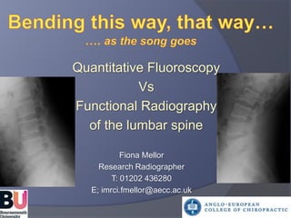
- 1. Quantitative Fluoroscopy Vs Functional Radiography of the lumbar spine Fiona Mellor Research Radiographer T: 01202 436280 E; imrci.fmellor@aecc.ac.uk
- 2. Learning outcomes Why measure intervertebral motion? Sources of errors and variation in flex/ext (functional) radiographs A new adaptation of fluoroscopy (quantitative fluoroscopy - QF) Comparison of radiation dose Novel uses for QF/other studies
- 4. Diagnostic categories of back pain (CSAG 1994) 1% Serious pathology 4% Nerve root compression 95% ‘Simple’ (Non-specific) backache - Chemical - Central sensitization - Mechanical (Instability)
- 5. Why measure intervertebral motion? Diagnosis Treatment Disability Research
- 6. Motion Subsystems (Panjabi 1992) Passive Active Motor Control
- 8. Back…… Wellcome film library. London
- 9. …via the present Intra and inter subject variation Intra and inter examiner error Positioning Definition of normal
- 11. …. to the future
- 12. Passive Quantitative Fluoroscopy Acquisition Image Analysis Output
- 13. Image analysis
- 16. PhD Hypothesis: There is a higher prevalence of abnormal mid lumbar inter-vertebral motion patterns in patients with mechanical LBP compared to controls QF passive motion 40 Patients (mechanical CNSLBP) & 40 healthy volunteers Coronal and sagittal Global range 40o Each direction (Lt Rt, flx, ext) Funded by the NIHR Clinical Academic Training Fellowship
- 17. Results
- 18. ‘Abnormal motion patterns’ Maximum rotation p <0.05 Left L4/5 pts < controls Right L3/4 pts > controls
- 19. Reference intervals A definition of ‘abnormal ‘ is those whose rotation falls beyond that achieved by 95% of the healthy population Hyper-mobility: p<0.05 Right L3/4 and Flexion L4/5 Hypo-mobility; p<0.05 Left and Right
- 20. Continuous motion patterns: Reference intervals Hyper mobility: Left L3/4 and Flexion L3/4 Hypo-mobility; Left L3/4 and L4/5. Right L4/5 and Flexion
- 21. Variation is still a problem! - How to account for the variation - How to measure the co-dependency of segments
- 24. …. The future of inter-vertebral measurements
- 25. Radiation dose
- 26. Radiation dose
- 27. Conclusions QF is more responsive than functional radiography with a similar radiation dose The coronal plane should be considered ‘Non Specific’ back pain = further subgrouping
- 28. Implications for clinical practice
- 29. QF research at AECC i. Characteristics of kinematics in healthy adults and their reproducibility over time ii. Effect of muscle interaction in healthy adults (surface electromyography) iii. Effects of manipulation of the cervical spine and patient reported outcomes iv. Relationship between prosthetic fit and intervertebral motion
- 31. Uncertainties: Healthy Passive Vs Active motion Subtle differences detected by QF
- 32. Healthy recumbent passive flexion Inter-vertebral angle (o) Time (15 frames = 1 second)
- 33. Healthy weight-bearing flexion Time (15 frames = 1 second) Inter-vertebral angle (o)
- 35. Cervical spine rotation in a patient with whiplash Flexion
- 36. Muscle activity: weight bearing flexion (sEMG)
- 37. Prosthetic fit and inter-vertebral motion
- 38. Summary Functional views of cervical and lumbar spine could be replaced with QF Further sub-grouping of non specific neck and back pain Further analysis of existing data and economic analysis
- 39. Fiona Mellor E: imrci.fmellor@aecc.ac.uk Acknowledgements: National Institute of Health. Clinical Academic Training Fellowship. Bournemouth University Santander travel award. Anglo-European College of Chiropractic. Bournemouth . UK Orthokinematics. Texas USA Professor Alan Breen and the team at IMRCI. Bournemouth. UK Professor Nat Ordway and the team at SUNY. Syracuse. USA
- 40. Bibliography Breen, A., Muggleton, J. and Mellor, F., 2006. An objective spinal motion imaging assessment (OSMIA): reliability, accuracy and exposure data. BMC Musculoskeletal Disorders, 7 (1), 1-10. Breen, A. C., Teyhen, D. S., Mellor, F. E., Breen, A. C., Wong, K. and Deitz, A., 2012. Measurement of inter-vertebral motion using quantitative fluoroscopy: Report of an international forum and proposal for use in the assessment of degenerative disc disease in the lumbar spine. Advances in Orthopaedics, 1-10. Deitz, A. K., Mellor, F.E., Teyhan, D.S., Panjabi, M.M., Wong, K.W.M., 2010. Kinematics of the Aging Spine: A Review of Past Knowledge and Survey of Recent Developments, with a Focus on Patient-Management Implications for the Clinical Practitioner. Yue, Guyer, Johnson, Khoo & Hochschuler (eds) In: Yue, J. L., Guyer, R. D., Johnson, P. J., Khoo, L. T., and Hochschuler, S. H., eds. The Comprehensive Treatment of the Aging Spine: Minimally Invasive and Advanced Techniques. Elsevier. Mellor, F., Breen, A., 2009. Objective assessment of spinal motion: the future? Imaging and Oncology, 3, 34-41. Mellor, F. E. and Breen, A. C., 2014. Discrimination of biomechanical back pain patient subgroups from continous inter-vertebral motion data: a protocol. Bone & Joint Journal Orthopaedic Proceedings Supplement, 96-B (SUPP 4), 5. Mellor, F. E., Muggleton, J. M., Bagust, J., Mason, W. M. A., Thomas, P. W. and Breen, A. C., 2009. Midlumbar lateral flexion stability measured in healthy volunteers by in-vivo fluoroscopy. Spine, 34 (22), E811-E817. Mellor, F. E., Thomas, P. and Breen, A., . 2014a. Moving Back: the radiation dose received from lumbar spine quantitative fluoroscopy compared to lumbar spine radiographs with suggestions for further dose reduction. Radiography, In print. Mellor, F. E., Thomas, P., Thompson, P. and Breen, A., 2014b. Proportional lumbar spine inter-vertebral motion patterns: a comparison of patients with chronic, non-specific low back pain and healthy controls European Spine Journal, epub ahead of print (March). Panjabi, M., Abumi, K., Duranceau, J. and Oxland, T., 1989. Spinal Stability and Intersegmental Muscle Forces: A Biomechanical Model. Spine, 14 (2), 194-200. Panjabi, M. M., 1992. The stabilising system of the spine - Part 2: Neutral zone and instability hypothesis. Journal of Spinal Disorders, 5 (4), 390-397.
- 41. The Neutral Zone Theory Neutral zone Flexion Extension Range of motion Force 2Kg