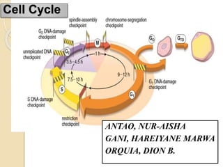
2. Phases and Checkpoints within the
Cell Cycle
The cell cycle represents a self-
regulated sequence of events that
controls cell growth and cell division.
to produce two daughter cells, each
containing chromosomes identical to
those of the parental cell.
3. Two Principal Phases:
Interphase – representing continuous
growth of the cell
G1 (gap1) phase
S (synthesis) phase
G2 (gap2) phase
M Phase (Mitosis) - characterized by the
partition of the genome
4.
5. Checkpoints - monitor and modulate the
progression of cells through the cell cycle
in response to intracellular or
environmental signals.
6. G1 Phase
The cell gathers nutrients and synthesizes RNA
and proteins necessary for DNA synthesis and
chromosome replication.
Two checkpoints:
1) the G1 DNA-damage checkpoint, which
monitors the integrity of DNA
2) the restriction point, which is sensitive to the
size of the cell, the state of the cell’s
physiologic processes, and its interactions
with extracellular matrix
7. S Phase
DNA is replicated
The DNA of the cell is doubled during
the S phase, and new chromatids are
formed that will become obvious at
prophase or metaphase of the mitotic
division.
S DNA-damage checkpoint - monitors
quality of replicating DNA.
8. G2 Phase
Cell prepares for cell division
The cell examines its replicated DNA in
preparation for cell division. This is a period of
cell growth and reorganization of cytoplasmic
organelles before entering the mitotic cycle.
Two checkpoints monitor DNA quality:
1. G2 DNA-damage checkpoint – detects DNA
damage
2. Unreplicated-DNA checkpoint - prevents the
progression of the cell into the M phase before
DNA synthesis is complete
9. M Phase (Mitosis)
Mitosis nearly always includes:
Karyokinesis (division of the nucleus)
and Cytokinesis (division of the
cytoplasm)
Separation of two identical daughter
cells concludes the M phase
10. M Phase
Two checkpoints:
1. Spindle-assembly checkpoint - prevents
premature entry into anaphase
2. Chromosome-segregation checkpoint -
prevents the process of cytokinesis until
all of the chromosomes have been
correctly separated.
11.
12. Mitosis
Mitosis is a process of chromosome
segregation and nuclear division followed by
cell division that produces two daughter cells
with the same chromosome number and DNA
content as the parent cell.
mitosis - equal partitioning of replicated
chromosomes and their genes into two
identical groups.
Includes: Karyokinesis (division of the nucleus)
Cytokinesis (division of the
cytoplasm)
13. Mitosis consists of 4 main phases:
1) Prophase
2) Metaphase
3) Anaphase
4) Telophase
14.
15. Imprint cytology from a breast cancer, stained with H&E. After
diagnosis, the specimens were distained, Feulgen stain, relocated and
analyzed for DNA content. Abnormal DNA content recorded with
microphotometry: 6.1 c prophase CDF (a)
16.
17. Kinetochore - a highly specialized
protein complex which appears on each
chromatid opposite to the centromere
and allows it to attach to a spindle fiber
on a chromosome.
20. Imprint cytology from a breast cancer, stained with H&E. After
diagnosis, the specimens were destained, Feulgen stain, relocated and
analysed for DNA content. Abnormal DNA content recorded with
microphotometry: 7.2 c metaphase CDF (b)
21. Mitotic spindle in metaphase. Using indirect
immunofluorescence techniques, the mitotic spindle in
a Xenopus XL-177 cell was labelled with an antibody
against α-tubulin conjugated with fluorescein (green).
22. H&E (HP). This micrograph of a malignant tumour of the skin
contains an abnormal mitotic figure A. The cell is in metaphase,
but rather than a metaphase plate with two sets of chromatids and
two spindles, the cell has produced four sets of chromatids and
four spindles, a quadripolar mitosis.
23.
24. Imprint cytology from a breast cancer, stained with H&E. After diagnosis,
the specimens were destained, Feulgen stain, relocated and analysed for
DNA content. Abnormal DNA content recorded with microphotometry: 5.2
c anaphase CDF (c)
29. The series of micrographs shown in the
next slides illustrate the mitotic process
in actively dividing immature blood
cells from a smear preparation of
human bone marrow using Giemsa
Stain.
32. Defects of mitosis result in various nuclear
abnormalities, namely, micronuclei,
binucleation, broken egg appearance,
pyknotic nuclei, and increased numbers of
and/or abnormal mitotic figures.
Mitotic activity remains restricted to somatic
stem cells that eventually repair injuries, and
to committed stem cells that substitute for
tissue turnover.
33. The following are the criteria that characterize
aberrations from regular mitotic activity in the
soma:
Dislocated divisions with relentless persistency
Multipolar anaphase distortion
Centromere defects and chromosome disaggregation
resulting in multiple mitotic figures
Spindle defects- Aberrant cellular divisions
Genome instability (Failures in check points and
apoptotic system) resulting in proliferation and aberrant
chromosome division figures (CDFs)
Chromosome mutations- Acquisition of successive
mutations leading to tumour initiation or syndrome
manifestations
Interphase aneuploidy
Chromosome division figures- Pathologic mitosis with
aberrant DNA content.