
Recommended
Recommended
More Related Content
What's hot
What's hot (20)
Similar to Cvd signs and symptoms
Similar to Cvd signs and symptoms (20)
Recently uploaded
Recently uploaded (20)
Cvd signs and symptoms
- 1. Signs and symptoms of CVD
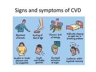
- 2. Symptoms Cardinal symptoms – Dyspnoea – Chest pain/ tightness – Edema – Palpitation – Cyanosis – Syncope – Fatigue – Hemoptysis
- 3. Dyspnoea • Unpleasant subjective awareness of breathing • Breathlessness inappropriate to the level of physical activity
- 4. Dyspnoea • Classified according to NYHA (New York Heart Association):- 1. Class I • No limitation and symptoms during ordinary activity 2. Class II • Symptoms with ordinary activity, slight limitation in physical activity
- 5. Dyspnoea 3. Class III • Symptoms with less than ordinary activity, marked limitation of activity 4. Class IV • Symptoms even at rest, unable to carry out any activity without symptoms
- 6. Systemic causes Acute dysspnoea Chronic exertional dyspnoea Cardiovascular system Acute pulmonary edema (acute LVF) Chronic congestive cardiac failure Myocardial ischemia Acquired valvular heart diseases Coronary artery disease Hypertensive heart disease Cardiomyopathies Respiratory system Acute severe asthma Acute exacerbation of COPD Pneumothorax Pneumonia Pulmonary embolus Acute respiratory distress syndrome Inhaled foreign body Lobar collapse Laryngeal edema e.g anaphylaxis COPD Chronic asthma Chronic pulmonary thromboembolism Bronchial carcinoma Interstitial lung diseases Lymphatic carcinomatosis Large pleural effusion
- 7. Acute dyspnoea Chronic dyspnoea Others Metabolic acidosis (diabetic ketoacidosis,uremia,salicylate overdose, ethelyne glycol poisoning ) Hyperventilation Severe anemia Obesity Myasthenia gravis Guillain-Barre syndrome Ankylosing spondylitis Kyphoscoliosis Anxiety
- 8. Mechanism of dyspnoea in heart disease • Left heart dysfunction – Increased pulmonary venous pressure • Transudation of fluid into the interstitial space – Thickening of vessel wall (perivascular cells and fibers) • Causes decreased compliance of lungs – Increased airway resistance – Both lead to increased work of breathing leading to dyspnoea
- 9. Orthopnoea • Dyspnoea on lying down/ in recumbent position • Relieved on sitting up or elevation of head
- 10. Orthopnea • Mechanism – Increased venous return – Fluid returns to the vascular system – Failing heart is unable to cope with the extra volume of fluid / blood delivered to it • Results in increase in pulmonary venous and capillary pressure leading to pulmonary interstitial edema and decreased airway resistance – Elevated diaphragm leads to decreased vital capacity of the lung
- 11. Paroxysmal nocturnal dyspnoea • Episodes of dyspnoea which occurs at night and awakens patient after 2-4 hours of sleep and takes 10-30 minutes for recovery after assuming upright postion
- 12. Mechanism • Similar to orthopnoea • Fall in PaO2 and decreased sympathetic support to left ventricular function during sleep
- 13. Causes of PND • Ischemic heart disease • Aortic valve disease • Hypertension • Cardiomegaly • Atrial fibrillation • Rarely in mitral disease • Earliest symptom of Left ventricular failure
- 14. Dyspnoea • Trepopnea – Dyspnoea in left or right lateral decubitus position • Platypnoea – Dyspnoea in upright position – Left atrial thrombus – Left atrial tumors- myxomas – Pulmonary arterio-venous fistula
- 15. Chest pain • Also called angina pectoris • Typical chest pain – Central/ substernal/ retrosternal chest pain – Increased on exertion – Relieved on rest, or by nitrates
- 16. Systems Causes Cardiovascular Coronary artery disease Myocardial ischemia, MI Spasm or narrowing of coronary arteries Aortic dissection Aortic aneurysm Myocarditis Pericarditis MVP Aortic stenosis, Aortic regurgitation Esophageal Esophagitis Esophageal spasm Mallory- Weiss syndrome Lungs/ pleura Brochospasm Pulmonary infarct Pneumonia
- 17. Systems Causes Musculoskeletal Osteoarthritis Rib fracture Costochondritis (Tietze’s syndrome) Intercostal muscle injury Neurological Prolapsed intervertebral disc Herpes zoster Thoracic outlet syndrome
- 18. Edema • Accumulation of fluid in the interstitial spaces • Occurs especially in heart failure • Leads to defective systolic emptying by the heart or impaired relaxation – Accumulation of blood in the heart and venous circulation
- 19. Mechanism • Due to the failing heart, the circulation in the kidney also decreases resulting in ischemic kidney – Secretion of renin – Renin- Angiotensin system is activated – Secretion of aldosterone • Sodium and water retention – Due to aldosterone and ADH secretion – The excretion of sodium from the kidney is reduced when heart is not working efficiently. So sodium and water retention occurs
- 20. Mechanism • Results in increase in plasma volume and hydrostatic pressure in the capillaries, veins and lymphatics – Leads to transudation of the fluid into interstitial spaces – Results in EDEMA
- 21. Palpitation • Awareness of ones own heartbeat • Different kinds of palpitation patient might experience – Fast and regular e.g. Paroxysmal tachycardia – Fast and irregular e.g. atrial fibrillation – Fleeting and repetitive e.g. premature ectopics – Fleeting and heavy heart beat • Increased stroke volume • Physical exertion, anxiety, emotion, fear, anemia, aortic, mitral regurgitation, patent ductus arteriosus, VSD, ASD
- 22. Cyanosis • Normally, the arterial blood is 95% oxygen saturated whereas venous blood is 70% saturated. The capillary blood is somewhere in between • Oxygen in the blood is attached to the hemoglobin. • Hemoglobin content is 15gm%
- 23. Cyanosis • In the capillaries, out of 15gm% of Hb – If 5gm% or more of Hb is devoid of oxygen (reduced hemoglobin), – It leads to bluish discoloration of skin and mucous membrane leading to CYANOSIS
- 24. Cyanosis • Central – When oxygen saturation is very low – Conjunctiva, palate, tongue, inner surface of the lips and cheeks and outer lips – Causes • Congenital heart disease with right to left shunt • Arterio-venous fistula of the lung • Diseases of the lung e.g. COPD with corpulmonale • Congestive heart failure with severe lung congestion
- 25. Cyanosis • Peripheral – Arterial oxygen saturation is normal – Tissues extract excess of oxygen from the blood and increase in the amount of reduced hemoglobin in the blood – Low cardiac output e.g. congestive heart failure – Exposure to excess cold – Fingers, earlobes, nose, cheeks, outer lips
- 26. Syncope/ presyncope • Transient loss of consciousness resulting from sudden and momentary reduction or stoppage of blood supply to the brain. • May or may not be preceded by feeling of faintness or lightheadedness
- 27. Causes • Cardiac causes – Ventricular systole – Ventricular fibrillation – Paroxysmal tachycardia – Hypertrophic obstructive cardiomyopathy – Aortic stenosis – Massive pulmonary embolism – Cardiac compression due to hemopericardium – Cardiogenic shock
- 28. Syncope • Non cardiac causes – Epilepsy – Head injury – Burns – Drugs – Pain, emotion and fear – Cerebrovascular accidents – Severe cough for prolonged periods
- 29. Fatigue • Manifestation of cardiac failure • Heart doesn’t pump enough blood to meet the oxygen demand – Leading to FATIGUE
- 30. Cough • Left heart failure – venous pressure falls – fluid goes into the bronchioles – peri-bronchial edema – irritates the bronchi • Sudden transudation of fluid in the alveoli – pt tries to cough it out • ACE inhibitors used in several cardiac diseases
- 31. Hemoptysis • Causes – Mitral stenosis • Pressure in veins and venules is high due to rise in LA pressure, if this pressure increases further, after physical exertion, pulmonary venules rupture and cause hemoptysis • Recurrent bronchitis- hemoptysis • Acute pulmonary edema – pink frothy sputum • Pulmonary infarction – hemoptysis
- 32. Causes • Left ventricular failure/ acute pulmonary edema • Arterio-venous aneurysm and syphilitic or mycotic aneurysm
- 33. Recurrent bronchitis • Recurrent respiratory infections may occur in patients with heart disease • Those with large amount of blood going to the lungs due to left to right shunt e.g. ASD, VSD, PDA • Those with pulmonary congestion e.g. LVF, MS • Chronic corpulmonale due to COPD
- 35. Systemic embolus • Thrombus or clot in the left side of the heart • May get detached and reach different parts of the body • Embolism
- 36. Signs • General examination including appearance • Vitals – Pulse – BP – Temperature and respiratory rate if relevant • Examination of the JVP • Proper examination of the cardiovascular system
- 37. Appearance • Indicators of increase in accumulation of lipid in the blood – Yellowish deposits along the eyelids – Xanthelasma – Over the skin and extensor surfaces – Xanthomas – Around the rim of cornea – corneal arcus – BMI
- 38. Appearance • Anemia - pallor • Clubbing • Splinter hemorrhages • Osler and Janeway nodules
- 39. Character of the pulse • Normal pulse – Percussion wave followed by a dicrotic notch which coincides with aortic valve closure. This is followed by dicrotic wave – We can only feel the percussion wave
- 40. Pulse • Plateau pulse – Anacrotic pulse/ Pulsus parvus et tardus – Slow rising pulse – Seen in AS • Pulsus bisferiens – Twice beating pulse – Two systolic peaks – Seen in AS+AR
- 41. Pulse • Bounding pulse – Sudden rise and sudden fall of the pulse wave – Pulse amplitude is also high – Bounding character felt best on lifting arm high up – Also known as Water Hammer pulse/ Corrigan’s pulse – Found in • AR High fever • Thyrotoxicosis Pregnancy • Severe anemia Chronic corpulmonale • PDA Carbon dioxide retention • AV fistula
- 42. Pulse • Bounding pulse – Consists of closed glass tube with little water inside it, the rest of the tube is vaccum – If it is turned upside down, water from one end of the tube suddenly falls to the other end and strikes suddenly at the finger.
- 43. Pulse • Dicrotic pulse – Twice beating pulse – After the percussion wave, a second wave is felt – dicrotic wave – Occurs in diastole – Found in • Severe congestive cardiac failure • Cardiac tamponade • Typhoid fever
- 44. Pulse • Pulsus alternans – Large wave and small wave are alternating each other – LVF
- 45. Pulse • Pulsus bigeminus – Pulse is felt in pairs when one normal beat is alternating with a premature ventricular contraction – Two beats are felt followed by a long pause – Digitalis toxixity
- 46. Pulse • Pulsus paradoxus – During deep inspiration, the pulse becomes feeble or disappears – During expiration, the pulse is felt again – Accentuation of the normal phenomenon – Normally there should be a fall of SBP by 10mm Hg during inspiration – In pulsus paradoxus, it falls > 10mmHg
- 48. Thank you