SlideShare a Scribd company logo
1 of 2
EFFECTS OF INFRARED LASER EXPOSURE IN A CELLULAR MODEL OF WOUND HEALING
                             Mark D. Skopin and Scott C. Molitor, Department of Bioengineering, University of Toledo, Toledo OH

      To investigate the efficacy of low-level exposure to 980 nm laser                 Images were acquired using a Quantix 57 scientific grade digital CCD camera
light, we have measured cell growth rates following wound induction using               (Roper Scientific) using custom made software designed to run under Matlab
an in vitro model of wound healing. A small pipette was used to                         (Mathworks). To assess cell growth, an automated analysis routine was developed
                                                                                        to measure the area of the imaged field that was covered by cells. This procedure
mechanically induce a wound in fibroblast cell cultures, which were then                utilized the Matlab edge detection function edge( ) with the Canny algorithm option to
imaged at specific time intervals following wound induction and exposure                detect lines of pixels along cell edges. Once edges were detected, the Matlab
to various doses of laser light. Our results show that exposure to low and              function imclose( ) was utilized to fill in the gaps between edge lines using circular
medium intensity laser light significantly accelerate cell growth; high                 structuring elements approximately one cell width across (12 - 15 pixels). The result
intensity laser light negated the beneficial effects of laser exposure on cell          of this analysis was the detection of pixels in regions covered by cells where edges
growth.      Further experiments demonstrated that cell growth was                      were in proximity, whereas regions such as the wound that had little or no cell
accelerated over a wide range of exposure durations using medium                        coverage were left undetected. Cell coverage area was then quantified at each time
                                                                                        interval following wound induction and laser exposure by adding the number of
intensity laser light with no marked reduction in cell growth at the longest            detected pixels and comparing this to the coverage area immediately following
exposure durations. Our results confirm clinical observations that low-                 wound induction. For each laser exposure dose and time interval following wound
level exposure to 980 nm laser light can accelerate healing of superficial              induction, images were acquired from five different culture dishes to provide
wounds.       However, these results also demonstrate the need for                      repeated data samples for statistical analysis.
appropriate supervision of laser therapy sessions to prevent overexposure               Statistical analysis
to laser light that may reverse increases in cell growth rates observed in                     To separate the effects of recovery time and laser exposure dose, a two-factor
response to lower levels of laser exposure.                                             analysis of variance (ANOVA) was performed on the cell coverage area data
                                                                                        obtained from images acquired at different post-exposure elapsed times and from
Introduction                                                                            different laser exposure doses. Without any laser exposure, a complete re-growth of
                                                                                        fibroblast cells into the wound region will occur over the 48 hour period during which
      Photobiotherapy is the clinical application of light for healing
                                                                                        cells were imaged. Therefore, a statistically significant increase in cell coverage
decubitus ulcers (bedsores) and other superficial wounds. Previous                      area will occur over the course of the experiment. However, the two-factor ANOVA
studies have demonstrated significant clinical value for the use of low-                procedure allows for the separation of two experiment factors, in this case elapsed
level laser light to accelerate the healing of superficial wounds (Mester et            time and laser exposure dose. Therefore, statistically significant effects of laser
al., 1985). Although the cellular mechanisms of this accelerated wound                  exposure dose can be compared to the effects of elapsed time, and can provide an
healing are not known, a recent study has demonstrated that low-level                   estimate of the acceleration of cell growth by the various laser exposures. The
light from a 633 nm HeNe laser accelerates cell growth in a cellular model              statistical software package Minitab 14 (Minitab Inc.) was utilized to perform the two-
                                                                                        factor ANOVA; data was entered as three columns with elapsed time, exposure dose
of wound healing and improves cellular metabolism in a dose-dependent
                                                                                        and percent increase in cell coverage area from images immediately taken after
manner (Hawkins and Abrahamse, 2006).                                                   wound induction.
      The use of infrared (IR) light may have significant advantages
compared to visible light for clinical applications. In particular, the longer
wavelength light minimizes scatter produced by superficial layers of the
skin, and allows for a penetration of the light into deeper layers of skin
that are most active during wound healing processes. In addition, IR light
produces heating of deeper skin layers, promoting increased blood flow
and to further accelerate healing processes (Dierickx, 2006).
      Although IR light has demonstrated clinical value, the effects of IR
light at the cellular level have not been examined. To determine whether
IR can improve cell growth and recovery, we have utilized a clinical IR
laser in a cellular model of wound healing. Our results show that limited
doses of IR light can increase the rate of cell growth within hours of light            Figure 1: cell culture model of wound healing. A sterile pipette approximately 1 mm
                                                                                        in diameter is used to induce a wound in a monolayer of fibroblast cells plated onto
exposure.                                                                               35 mm culture dishes (far left image). Fibroblast cells grow back into the wound
                                                                                        region within 8 hours (middle image) and the wound is completely overgrown within
Methods                                                                                 24 hours. Scale bar: 250 μm. The algorithm used to calculate cell coverage area
Cell culture                                                                            found that 64.9%, 76.7% and 97.7% of the imaged area was covered with cells in
       Fetal human skin fibroblast cells (cell line CCD-1070SK, American Type           the three images displayed above.
Culture Collection) were grown in Dulbecco’s modification of Eagle’s medium
(DMEM) supplemented with 1 mM L-glutamine, 1% penicillin-streptomycin and 3%            Results
fetal bovine serum. The cultures were incubated at 37 ºC with 95% O2 - 5% CO2 at              Our results demonstrate that exposure to light from a 980 nm laser
85% humidity. Cells were trypsinized using a 0.25% (w/v) trypsin and 0.03% EDTA
solution in DMEM and seeded into sterile 35 mm polystyrene culture dishes at a
                                                                                        can enhance cell growth rates in an in vitro wound model. A range of
density of 7.0 x 10⁴ cells per cm2. Cells were incubated overnight to allow the cells   exposure doses was investigated by varying laser output power over a
to recover from trypsinization and to adhere to the bottom of the culture dishes.       fixed exposure duration, or by varying exposure duration at a fixed laser
Wound healing model                                                                     output power. Figure 2 shows the results of the first experiment in which
       Culture dishes with plated cells were removed from the incubator, placed on      laser output power was varied from 1.5 - 7.5 W to produce an exposure
                                                                                                                    2
the stage of an inverted microscope equipped with relief contrast optics (IX-71,        level of 26 - 120 mW/cm over a two minute exposure, resulting in
                                                                                                                               2
Olympus) and visualized using a 4x, 0.13 numerical aperture objective. To simulate      exposure doses from 3.1 - 14.4 J/cm . Regardless of exposure level,
a wound, confluent monolayers were scratched with a sterile pipette approximately 1     significant cell recovery was observed within three hours of wound
mm in diameter (figure 1). Following wound induction, the output of a 7.5W, 980 nm      induction; however, exposure to moderate levels of laser light (26 - 97
laser used for clinical applications (VTR 75, Avicenna Laser Technologies) was                   2
                                                                                        mW/cm ) appeared to enhance cell growth at all time intervals relative to
focused on a spot 12.5 mm in diameter centered on the wound with the visible red
aiming beam disabled. Cell growth into the wound region at time intervals up to 48      control experiments in which no laser exposure was applied (figure 2, top
hours post-exposure was compared to control dishes in which no laser light was          panel). These results were confirmed by the results of a two-factor
used. To attenuate the laser output and focus the light on a smaller spot, a 3 mm       ANOVA (figure 2, lower right), which shows that significant increases in
                                                                                                                                                                 2
fiber approximately 1.5 m in length was coupled to the laser output and directed        cell growth were observed with two minute exposures to 26 - 73 mW/cm
                                                                                                                  2
toward the center of the 35 mm culture dish approximately 10 mm above the dish          (p < 0.01) and 97 mW/cm (p < 0.05). These results also show that the
surface. Two different sets of experiments were performed: the first compared           beneficial effects of laser exposure are negated by over-exposure:
different exposure intensities over the same exposure time, the second compared                                            2
                                                                                        fibroblasts exposed to 120 mW/cm of laser light for two minutes did not
different exposure times at the same exposure intensity. For the first set of
experiments, the laser output was varied from 1.5 - 7.5 W to produce exposure           show any significant increase in growth rates relative to control
densities 26 - 120 mW/cm2 for 2 minutes, resulting in exposure doses from 3.1 - 14.4    experiments.
J/cm2. For the second set of experiments, the laser output was fixed at 4.5 W or 73           The two-factor ANOVA analysis also provided a measure of how
mW/cm2 and the exposure times were varied from 20 sec to 15 min, resulting in           much cell growth was accelerated by laser exposure. Over the first eight
exposure doses from 1.5 - 66 J/cm2.                                                     hours following wound induction, the average cell coverage increased
Image acquisition and analysis                                                          linearly by approximately 1.7% per hour (figure 2, lower left); the growth
      To assess the growth of cells back into the wound region, images were             rate begins to slow before 24 hours, when cells across the wound margin
acquired at hourly intervals up to 8 hours after wound induction and laser exposure,    begin to contact each other and completely fill the area previously devoid
and then at 24 and 48 hours post exposure. To maintain a controlled environment,        of cells. When compared to the mean cell growth measured at various
culture dishes were returned to the incubator between image acquisition sessions.
time intervals following wound induction, the 4 - 6% increase in cell
                                       2
growth produced by 49 - 73 mW/cm of laser exposure over a two minute
period represented an acceleration of wound healing by approximately 2.5
- 3.5 hours within the first eight hours of healing. This represents a
sizeable acceleration in cell growth considering that the wounds from our
in vitro model were nearly completed healed within 24 hours following
wound induction. Despite the significant increases in cell growth across
various time intervals and exposure levels, the two-factor ANOVA analysis
did not find any significant interaction between elapsed time and exposure
level. In other words, the various exposure levels showed consistent
effects across all time intervals following wound induction, and there were
no exposure levels whose effects were only observed at a particular time
interval or subset of time intervals following wound induction.


                                                                                       Figure 3: Top panel: cell growth in wound model as a function of time elapsed from
                                                                                       wound induction and laser exposure dose. Vertical bars show % change in cell
                                                                                       coverage area averaged across five experiments in which cells were not exposed to
                                                                                       laser light, or in which cells were exposed to 73 mW/cm2 of light during exposures
                                                                                       that varied from 20 sec to 15 min to give exposure doses from 1.5 - 65.7 J/cm2.
                                                                                       Error bars show S.E.M across five experiments. Bottom left: confidence intervals
                                                                                       from the two-factor ANOVA analysis demonstrate a significant increase in cell
                                                                                       coverage area observed as early as 3 hours after wound induction regardless of
                                                                                       laser exposure (* p < 0.05 for 3 hours, ** p < 0.01 for 4 hours and beyond). Bottom
                                                                                       right: confidence intervals from the two-factor ANOVA analysis demonstrate a
                                                                                       significant increase in cell coverage area for moderate laser exposure doses when
                                                                                       compared to no laser exposure (** p < 0.01 for 1.5 - 8.8 J/cm2). No significant
                                                                                       increase in cell coverage was observed at the second highest dose (21.9 J/cm2), and
                                                                                       a significant decrease in cell coverage was observed at the highest exposure dose (*
                                                                                       p < 0.05 for 65.7 J/cm2).
Figure 2: Top panel: cell growth in wound model as a function of time elapsed from
wound induction and laser exposure intensity. Vertical bars show % change in cell      Conclusions
coverage area averaged across five experiments in which cells were not exposed to             Our results confirm the clinical observation that low-level exposure to
laser light, or in which cells were exposed to 26 - 120 mW/cm2 of light during a two   980 nm of laser light can accelerate cell growth in a wound healing model.
minute exposure by varying the laser power from 1.5 - 7.5 W to give exposure doses     Because our measurements were obtained from an in vitro cell culture
from 3.1 - 14.4 J/cm2. Error bars show S.E.M across five experiments. Bottom left:     model, these results also suggest that the mechanisms involved in the
confidence intervals from the two-factor ANOVA analysis demonstrate a significant      acceleration of cell growth following laser exposure are cellular or
increase in cell coverage area observed as early as 3 hours after wound induction
regardless of laser exposure (** p < 0.01). Horizontal bars show 95% confidence
                                                                                       molecular in nature. The hypothesis that IR light accelerates healing
intervals with midline at mean value; error bars show 99% confidence intervals.        processes by heating skin and promoting increased blood flow (Dierickx,
Bottom right: confidence intervals from the two-factor ANOVA analysis demonstrate      2006) could not explain the increased cell growth rates in an in vitro cell
a significant increase in cell coverage area for low and moderate levels of laser      culture model. Our measurements suggested that IR exposure produced
exposure when compared to no laser exposure (** p < 0.01 for 26 - 73 mW/cm2; * p       temperature increases less than 2 ºC, and the use of a controlled
< 0.05 for 97 mW/cm2). No significant increase in cell coverage was observed at the    incubation environment between image acquisition intervals further
highest exposure level (120 mW/cm2).                                                   minimizes the temperature variability in our experiments. Previous
                                                                                       researchers have suggested that light exposure increases ATP levels by
      Figure 3 shows the results of the second experiment in which                     altering the energetic state of light-sensitive cytochromes within the inner
exposure durations were varied from 20 sec - 15 min at a constant laser                mitochondrial membrane that participate in oxidative phosphorylation
                                                                         2
output power of 4.5 W to produce an exposure level of 73 mW/cm ,                       (Karu et al., 1995); we are conducting further experiments to examine
                                                     2
resulting in exposure doses from 1.5 - 66 J/cm . As with changes in                    ATP levels following low-level exposure to 980 nm laser light.
exposure level, significant cell recovery was observed within three hours                     Our results also demonstrate the importance of appropriate
of wound induction regardless of exposure duration, and a wide range of                supervision of laser light exposure in a clinical setting. In particular, the
exposure durations appeared to enhance cell growth at all time intervals               average cell growth rates formed a non-monotonic function of laser
relative to control experiments in which no laser exposure was applied                 exposure levels (figure 2, lower right) and exposure doses (figure 3, lower
(figure 3, top panel). These results were confirmed by the results of a                right); with peak growth rates at moderate exposures, and reduced benefit
two-factor ANOVA (figure 3, lower right), which shows that significant                 at higher exposure intensities and doses. This suggests that excessive
                                                         2
increases in cell growth were observed with 73 mW/cm exposures having                  light exposure could have potentially damaging effects that negate any
                                                                          2
durations of 20 sec - 2 min to produce exposure doses of 1.5 - 8.8 J/cm                initial benefit of light exposure. Therefore the appropriate exposure levels
                                                               2
(p < 0.01). Note that a long exposure of 15 min (65.7 J/cm ) produced a                and durations must be selected in order to maximize cell growth rates.
significant decrease in cell growth (p < 0.05), suggesting that long
exposure to laser light at a moderate level can reverse the benefits of                References
lower exposure does. A comparison of these results to the mean cell
                                                                                       Dierickx CC (2006) The role of deep heating for noninvasive skin rejuvenation.
growth measured at various time intervals following wound induction                              Lasers Surg Med 38:799-807.
(figure 3, lower left) showed that the 6% increase in cell growth produced
                  2                                                                    Hawkins DH, Abrahamse H (2006) The role of laser fluence in cell viability,
by 73 mW/cm of laser exposure over a 20 sec - 2 minute period                                   proliferation, and membrane integrity of wounded human skin fibroblasts
represented an acceleration of wound healing by approximately two hours                         following helium-neon laser irradiation. Lasers Surg Med 38:74-83.
within the first eight hours of healing. Furthermore, the two-factor ANOVA
                                                                                       Karu T, Pyatibrat L, Kalendo G (1995) Irradiation with He-Ne laser increases ATP
analysis did not find any significant interaction between elapsed time and                       level in cells cultivated in vitro. J Photochem Photobiol B 27:219-223.
exposure duration despite the significant increases in cell growth across
                                                                                       Mester E, Mester AF, Mester A (1985) The biomedical effects of laser application.
various time intervals and various exposure durations.
                                                                                                 Lasers Surg Med 5:31-39.

More Related Content

What's hot

Vol.2, issue 2, no.3 (1)
Vol.2, issue 2, no.3 (1)Vol.2, issue 2, no.3 (1)
Vol.2, issue 2, no.3 (1)Satya Mamidi
 
Introduction to radiation therapy
Introduction to radiation therapyIntroduction to radiation therapy
Introduction to radiation therapyRad Tech
 
nanobiotechnology QD in bioimaging
nanobiotechnology QD in bioimagingnanobiotechnology QD in bioimaging
nanobiotechnology QD in bioimagingYong Zhang
 
Delsa Nano 0.6 nm - 30 microns
Delsa Nano 0.6 nm - 30 micronsDelsa Nano 0.6 nm - 30 microns
Delsa Nano 0.6 nm - 30 micronsadityahpatel
 
Imaging for Radiotherapy delivery and verification
Imaging for Radiotherapy delivery and verificationImaging for Radiotherapy delivery and verification
Imaging for Radiotherapy delivery and verificationMiami Cancer Institute
 
Quantum dots and application in medical science
Quantum dots and application in medical scienceQuantum dots and application in medical science
Quantum dots and application in medical sciencekeyhan *
 
Lasers in gynaecology
Lasers in gynaecologyLasers in gynaecology
Lasers in gynaecologySai Sashãnk
 
BMES_Abstract_Microscope_Comparison_Meyer
BMES_Abstract_Microscope_Comparison_MeyerBMES_Abstract_Microscope_Comparison_Meyer
BMES_Abstract_Microscope_Comparison_MeyerAndrew Meyer
 
Early stage detection of skin cancer via terahertz spectral profiling and 3D ...
Early stage detection of skin cancer via terahertz spectral profiling and 3D ...Early stage detection of skin cancer via terahertz spectral profiling and 3D ...
Early stage detection of skin cancer via terahertz spectral profiling and 3D ...Applied Research and Photonics, Inc.
 
Biomedical applications of quantum dots
Biomedical applications of quantum dotsBiomedical applications of quantum dots
Biomedical applications of quantum dotsANJUNITHIKURUP
 
Terahertz Spectroscopic Analysis and Multispectral Imaging of Epitaxially Gro...
Terahertz Spectroscopic Analysis and Multispectral Imaging of Epitaxially Gro...Terahertz Spectroscopic Analysis and Multispectral Imaging of Epitaxially Gro...
Terahertz Spectroscopic Analysis and Multispectral Imaging of Epitaxially Gro...Applied Research and Photonics, Inc.
 
Glimpse of clinical radiobiology course
Glimpse of clinical radiobiology courseGlimpse of clinical radiobiology course
Glimpse of clinical radiobiology courseManoj Gupta
 
LONG TEST IN STS.docx
LONG TEST IN STS.docxLONG TEST IN STS.docx
LONG TEST IN STS.docxkervin casuga
 
New Techniques in Radiotherapy
New Techniques in RadiotherapyNew Techniques in Radiotherapy
New Techniques in RadiotherapySantam Chakraborty
 
Determination ofRadiological Quality Parameters Using Optical Densitometer an...
Determination ofRadiological Quality Parameters Using Optical Densitometer an...Determination ofRadiological Quality Parameters Using Optical Densitometer an...
Determination ofRadiological Quality Parameters Using Optical Densitometer an...IOSR Journals
 
Laser therapy unit for physiotherapy
Laser therapy unit for physiotherapyLaser therapy unit for physiotherapy
Laser therapy unit for physiotherapyMedical Bazzar
 

What's hot (20)

Vol.2, issue 2, no.3 (1)
Vol.2, issue 2, no.3 (1)Vol.2, issue 2, no.3 (1)
Vol.2, issue 2, no.3 (1)
 
Introduction to radiation therapy
Introduction to radiation therapyIntroduction to radiation therapy
Introduction to radiation therapy
 
nanobiotechnology QD in bioimaging
nanobiotechnology QD in bioimagingnanobiotechnology QD in bioimaging
nanobiotechnology QD in bioimaging
 
Delsa Nano 0.6 nm - 30 microns
Delsa Nano 0.6 nm - 30 micronsDelsa Nano 0.6 nm - 30 microns
Delsa Nano 0.6 nm - 30 microns
 
Imaging for Radiotherapy delivery and verification
Imaging for Radiotherapy delivery and verificationImaging for Radiotherapy delivery and verification
Imaging for Radiotherapy delivery and verification
 
Quantum dots and application in medical science
Quantum dots and application in medical scienceQuantum dots and application in medical science
Quantum dots and application in medical science
 
Lasers in gynaecology
Lasers in gynaecologyLasers in gynaecology
Lasers in gynaecology
 
Ftir
FtirFtir
Ftir
 
BMES_Abstract_Microscope_Comparison_Meyer
BMES_Abstract_Microscope_Comparison_MeyerBMES_Abstract_Microscope_Comparison_Meyer
BMES_Abstract_Microscope_Comparison_Meyer
 
Early stage detection of skin cancer via terahertz spectral profiling and 3D ...
Early stage detection of skin cancer via terahertz spectral profiling and 3D ...Early stage detection of skin cancer via terahertz spectral profiling and 3D ...
Early stage detection of skin cancer via terahertz spectral profiling and 3D ...
 
Particle size analysis
Particle size analysisParticle size analysis
Particle size analysis
 
Biomedical applications of quantum dots
Biomedical applications of quantum dotsBiomedical applications of quantum dots
Biomedical applications of quantum dots
 
Terahertz Spectroscopic Analysis and Multispectral Imaging of Epitaxially Gro...
Terahertz Spectroscopic Analysis and Multispectral Imaging of Epitaxially Gro...Terahertz Spectroscopic Analysis and Multispectral Imaging of Epitaxially Gro...
Terahertz Spectroscopic Analysis and Multispectral Imaging of Epitaxially Gro...
 
Tour prime ok
Tour prime okTour prime ok
Tour prime ok
 
Glimpse of clinical radiobiology course
Glimpse of clinical radiobiology courseGlimpse of clinical radiobiology course
Glimpse of clinical radiobiology course
 
LONG TEST IN STS.docx
LONG TEST IN STS.docxLONG TEST IN STS.docx
LONG TEST IN STS.docx
 
New Techniques in Radiotherapy
New Techniques in RadiotherapyNew Techniques in Radiotherapy
New Techniques in Radiotherapy
 
Determination ofRadiological Quality Parameters Using Optical Densitometer an...
Determination ofRadiological Quality Parameters Using Optical Densitometer an...Determination ofRadiological Quality Parameters Using Optical Densitometer an...
Determination ofRadiological Quality Parameters Using Optical Densitometer an...
 
Laser therapy unit for physiotherapy
Laser therapy unit for physiotherapyLaser therapy unit for physiotherapy
Laser therapy unit for physiotherapy
 
Application Note: Angiogenesis
Application Note: AngiogenesisApplication Note: Angiogenesis
Application Note: Angiogenesis
 

Viewers also liked

Harian Pelita Jumat 16 November 2012 Halaman 1
Harian Pelita Jumat 16 November 2012 Halaman 1Harian Pelita Jumat 16 November 2012 Halaman 1
Harian Pelita Jumat 16 November 2012 Halaman 1Taruna Ikrar
 
Programa De Licenciatura En Lenguas Modernas[1]
Programa De Licenciatura En Lenguas Modernas[1]Programa De Licenciatura En Lenguas Modernas[1]
Programa De Licenciatura En Lenguas Modernas[1]adyuli
 
Elance final v puti l1 verbs (1) 1 out of 4
Elance final v puti l1 verbs (1) 1 out of 4Elance final v puti l1 verbs (1) 1 out of 4
Elance final v puti l1 verbs (1) 1 out of 4icehouse2460
 
Bullying the reality and affect in sports-1
Bullying  the reality and affect in sports-1Bullying  the reality and affect in sports-1
Bullying the reality and affect in sports-1ROBERT COLEMAN
 
Tonsil Mouth Gag Fiberoptic
Tonsil Mouth Gag FiberopticTonsil Mouth Gag Fiberoptic
Tonsil Mouth Gag FiberopticJames_DuCanto_MD
 
OrthoTexas: Physical Therapy Plano, TX
OrthoTexas: Physical Therapy Plano, TXOrthoTexas: Physical Therapy Plano, TX
OrthoTexas: Physical Therapy Plano, TXOrtho Texas
 
Controlling toxic substances 2012
Controlling toxic substances 2012Controlling toxic substances 2012
Controlling toxic substances 2012h-wilkinson
 
Bab2 eko koperasi
Bab2 eko koperasiBab2 eko koperasi
Bab2 eko koperasianjanifanny
 
Water purifier manufacturer in india
Water purifier manufacturer in indiaWater purifier manufacturer in india
Water purifier manufacturer in indiablucol1
 
The Bootz presentation for the Venture Lab Crash Course on Creativity
The Bootz presentation for the Venture Lab Crash Course on CreativityThe Bootz presentation for the Venture Lab Crash Course on Creativity
The Bootz presentation for the Venture Lab Crash Course on Creativitydonatop
 
Step by step email
Step by step emailStep by step email
Step by step emailjessblunt123
 
Daftar isi
Daftar isiDaftar isi
Daftar isikartoba
 

Viewers also liked (19)

Harian Pelita Jumat 16 November 2012 Halaman 1
Harian Pelita Jumat 16 November 2012 Halaman 1Harian Pelita Jumat 16 November 2012 Halaman 1
Harian Pelita Jumat 16 November 2012 Halaman 1
 
Programa De Licenciatura En Lenguas Modernas[1]
Programa De Licenciatura En Lenguas Modernas[1]Programa De Licenciatura En Lenguas Modernas[1]
Programa De Licenciatura En Lenguas Modernas[1]
 
Elance final v puti l1 verbs (1) 1 out of 4
Elance final v puti l1 verbs (1) 1 out of 4Elance final v puti l1 verbs (1) 1 out of 4
Elance final v puti l1 verbs (1) 1 out of 4
 
Bullying the reality and affect in sports-1
Bullying  the reality and affect in sports-1Bullying  the reality and affect in sports-1
Bullying the reality and affect in sports-1
 
Desktop power supply
Desktop power supplyDesktop power supply
Desktop power supply
 
Tonsil Mouth Gag Fiberoptic
Tonsil Mouth Gag FiberopticTonsil Mouth Gag Fiberoptic
Tonsil Mouth Gag Fiberoptic
 
Recipe
RecipeRecipe
Recipe
 
OrthoTexas: Physical Therapy Plano, TX
OrthoTexas: Physical Therapy Plano, TXOrthoTexas: Physical Therapy Plano, TX
OrthoTexas: Physical Therapy Plano, TX
 
Controlling toxic substances 2012
Controlling toxic substances 2012Controlling toxic substances 2012
Controlling toxic substances 2012
 
Bab2 eko koperasi
Bab2 eko koperasiBab2 eko koperasi
Bab2 eko koperasi
 
Water purifier manufacturer in india
Water purifier manufacturer in indiaWater purifier manufacturer in india
Water purifier manufacturer in india
 
Cloogy: a Rede Inteligente acessível a todos
Cloogy: a Rede Inteligente acessível a todosCloogy: a Rede Inteligente acessível a todos
Cloogy: a Rede Inteligente acessível a todos
 
RPL
RPLRPL
RPL
 
The Bootz presentation for the Venture Lab Crash Course on Creativity
The Bootz presentation for the Venture Lab Crash Course on CreativityThe Bootz presentation for the Venture Lab Crash Course on Creativity
The Bootz presentation for the Venture Lab Crash Course on Creativity
 
Class 2 vocabulary about company
Class 2 vocabulary about companyClass 2 vocabulary about company
Class 2 vocabulary about company
 
Step by step email
Step by step emailStep by step email
Step by step email
 
Daftar isi
Daftar isiDaftar isi
Daftar isi
 
Kekerasan remaja
Kekerasan remajaKekerasan remaja
Kekerasan remaja
 
Transport Layer
Transport LayerTransport Layer
Transport Layer
 

Similar to U of Toledo Wound Healing Study—AvicennaLaser.com

RecoveryRx® Accelerated healing and better pain relief
RecoveryRx® Accelerated healing and better pain reliefRecoveryRx® Accelerated healing and better pain relief
RecoveryRx® Accelerated healing and better pain reliefMax Poritzky
 
Study of Mitotic Index and DNA profile when exposure to He-Ne laser and UVC r...
Study of Mitotic Index and DNA profile when exposure to He-Ne laser and UVC r...Study of Mitotic Index and DNA profile when exposure to He-Ne laser and UVC r...
Study of Mitotic Index and DNA profile when exposure to He-Ne laser and UVC r...IOSR Journals
 
Analysis and Classification of Skin Lesions Using 3D Volume Reconstruction
Analysis and Classification of Skin Lesions Using 3D Volume ReconstructionAnalysis and Classification of Skin Lesions Using 3D Volume Reconstruction
Analysis and Classification of Skin Lesions Using 3D Volume ReconstructionIOSR Journals
 
Radiotherapy ppt. Types of radiation and chronic
Radiotherapy ppt. Types of radiation and chronicRadiotherapy ppt. Types of radiation and chronic
Radiotherapy ppt. Types of radiation and chronicneestom1998
 
simonsonISMRM2016final (1)
simonsonISMRM2016final (1)simonsonISMRM2016final (1)
simonsonISMRM2016final (1)Michael Simonson
 
Radiotherapy /certified fixed orthodontic courses by Indian dental academy
Radiotherapy /certified fixed orthodontic courses by Indian dental academy Radiotherapy /certified fixed orthodontic courses by Indian dental academy
Radiotherapy /certified fixed orthodontic courses by Indian dental academy Indian dental academy
 
NirvanaSensingGlove
NirvanaSensingGloveNirvanaSensingGlove
NirvanaSensingGloveDavid Cabral
 
Review on laser scanning confocal microscopy
Review on laser scanning confocal microscopyReview on laser scanning confocal microscopy
Review on laser scanning confocal microscopyUdayan Ghosh
 
Laser & Light Therapy
Laser & Light TherapyLaser & Light Therapy
Laser & Light TherapyJLS10
 
U tokyo forum at usp 20131112
U tokyo forum at usp 20131112U tokyo forum at usp 20131112
U tokyo forum at usp 20131112Horacio Cabral
 
Micromanipulation
Micromanipulation Micromanipulation
Micromanipulation Akshay More
 
Nihms116835
Nihms116835Nihms116835
Nihms116835majd20
 

Similar to U of Toledo Wound Healing Study—AvicennaLaser.com (20)

Continuing Medical Education Seminar. Low Level Laser in Complementary and Al...
Continuing Medical Education Seminar. Low Level Laser in Complementary and Al...Continuing Medical Education Seminar. Low Level Laser in Complementary and Al...
Continuing Medical Education Seminar. Low Level Laser in Complementary and Al...
 
RecoveryRx® Accelerated healing and better pain relief
RecoveryRx® Accelerated healing and better pain reliefRecoveryRx® Accelerated healing and better pain relief
RecoveryRx® Accelerated healing and better pain relief
 
Study of Mitotic Index and DNA profile when exposure to He-Ne laser and UVC r...
Study of Mitotic Index and DNA profile when exposure to He-Ne laser and UVC r...Study of Mitotic Index and DNA profile when exposure to He-Ne laser and UVC r...
Study of Mitotic Index and DNA profile when exposure to He-Ne laser and UVC r...
 
Analysis and Classification of Skin Lesions Using 3D Volume Reconstruction
Analysis and Classification of Skin Lesions Using 3D Volume ReconstructionAnalysis and Classification of Skin Lesions Using 3D Volume Reconstruction
Analysis and Classification of Skin Lesions Using 3D Volume Reconstruction
 
Continuing Medical Education Seminar. Low Level Laser in Complementary and Al...
Continuing Medical Education Seminar. Low Level Laser in Complementary and Al...Continuing Medical Education Seminar. Low Level Laser in Complementary and Al...
Continuing Medical Education Seminar. Low Level Laser in Complementary and Al...
 
Radiotherapy ppt. Types of radiation and chronic
Radiotherapy ppt. Types of radiation and chronicRadiotherapy ppt. Types of radiation and chronic
Radiotherapy ppt. Types of radiation and chronic
 
simonsonISMRM2016final (1)
simonsonISMRM2016final (1)simonsonISMRM2016final (1)
simonsonISMRM2016final (1)
 
My devices and its applications
My devices and its applicationsMy devices and its applications
My devices and its applications
 
Radiotherapy /certified fixed orthodontic courses by Indian dental academy
Radiotherapy /certified fixed orthodontic courses by Indian dental academy Radiotherapy /certified fixed orthodontic courses by Indian dental academy
Radiotherapy /certified fixed orthodontic courses by Indian dental academy
 
Radiotherapy /certified fixed orthodontic courses by Indian dental academy
Radiotherapy /certified fixed orthodontic courses by Indian dental academy Radiotherapy /certified fixed orthodontic courses by Indian dental academy
Radiotherapy /certified fixed orthodontic courses by Indian dental academy
 
NirvanaSensingGlove
NirvanaSensingGloveNirvanaSensingGlove
NirvanaSensingGlove
 
Preethi
PreethiPreethi
Preethi
 
Review on laser scanning confocal microscopy
Review on laser scanning confocal microscopyReview on laser scanning confocal microscopy
Review on laser scanning confocal microscopy
 
Why Outsource To Neurotar
Why Outsource To NeurotarWhy Outsource To Neurotar
Why Outsource To Neurotar
 
Laser & Light Therapy
Laser & Light TherapyLaser & Light Therapy
Laser & Light Therapy
 
BASIC RADIOBIOLOGY FOR RADIOTHERAPY
BASIC RADIOBIOLOGY FOR RADIOTHERAPYBASIC RADIOBIOLOGY FOR RADIOTHERAPY
BASIC RADIOBIOLOGY FOR RADIOTHERAPY
 
1.radiation of h&n tumors
1.radiation of h&n tumors1.radiation of h&n tumors
1.radiation of h&n tumors
 
U tokyo forum at usp 20131112
U tokyo forum at usp 20131112U tokyo forum at usp 20131112
U tokyo forum at usp 20131112
 
Micromanipulation
Micromanipulation Micromanipulation
Micromanipulation
 
Nihms116835
Nihms116835Nihms116835
Nihms116835
 

More from Avicenna Laser Technology

TMA International — Class IV High Power Lasers — Laser Technology Overview
TMA International — Class IV High Power Lasers — Laser Technology OverviewTMA International — Class IV High Power Lasers — Laser Technology Overview
TMA International — Class IV High Power Lasers — Laser Technology OverviewAvicenna Laser Technology
 
Avicenna High Power Laser—Physician Seminar
Avicenna High Power Laser—Physician SeminarAvicenna High Power Laser—Physician Seminar
Avicenna High Power Laser—Physician SeminarAvicenna Laser Technology
 
Avicenna High Power Laser Therapy European seminars october2010
Avicenna High Power Laser Therapy European seminars october2010Avicenna High Power Laser Therapy European seminars october2010
Avicenna High Power Laser Therapy European seminars october2010Avicenna Laser Technology
 
Laser therapycentersofgreaterwashingtonbrochure
Laser therapycentersofgreaterwashingtonbrochureLaser therapycentersofgreaterwashingtonbrochure
Laser therapycentersofgreaterwashingtonbrochureAvicenna Laser Technology
 
Wound Healing Case Study—AvicennaLaser.com
Wound Healing Case Study—AvicennaLaser.comWound Healing Case Study—AvicennaLaser.com
Wound Healing Case Study—AvicennaLaser.comAvicenna Laser Technology
 
Radiculopathy Case Study—AvicennaLaser.com
Radiculopathy Case Study—AvicennaLaser.comRadiculopathy Case Study—AvicennaLaser.com
Radiculopathy Case Study—AvicennaLaser.comAvicenna Laser Technology
 
Diabetic neuropathy-with-high-power-laser-therapy-case-study
Diabetic neuropathy-with-high-power-laser-therapy-case-studyDiabetic neuropathy-with-high-power-laser-therapy-case-study
Diabetic neuropathy-with-high-power-laser-therapy-case-studyAvicenna Laser Technology
 
Acute Disc Herniation with Foot Drop Case Study—AvicennaLaser.com
Acute Disc Herniation with Foot Drop Case Study—AvicennaLaser.comAcute Disc Herniation with Foot Drop Case Study—AvicennaLaser.com
Acute Disc Herniation with Foot Drop Case Study—AvicennaLaser.comAvicenna Laser Technology
 
Achilles Tendinitis Case Study—AvicennaLaser.com
Achilles Tendinitis Case Study—AvicennaLaser.comAchilles Tendinitis Case Study—AvicennaLaser.com
Achilles Tendinitis Case Study—AvicennaLaser.comAvicenna Laser Technology
 
AvicennaLaser.com Physician Seminar Power Point
AvicennaLaser.com Physician Seminar Power PointAvicennaLaser.com Physician Seminar Power Point
AvicennaLaser.com Physician Seminar Power PointAvicenna Laser Technology
 

More from Avicenna Laser Technology (18)

TMA International — Class IV High Power Lasers — Laser Technology Overview
TMA International — Class IV High Power Lasers — Laser Technology OverviewTMA International — Class IV High Power Lasers — Laser Technology Overview
TMA International — Class IV High Power Lasers — Laser Technology Overview
 
Avicenna AVI HPLL 12 User Manual
Avicenna AVI HPLL 12 User ManualAvicenna AVI HPLL 12 User Manual
Avicenna AVI HPLL 12 User Manual
 
Avicenna High Power Laser—Physician Seminar
Avicenna High Power Laser—Physician SeminarAvicenna High Power Laser—Physician Seminar
Avicenna High Power Laser—Physician Seminar
 
Avicenna Laser Physi
Avicenna Laser PhysiAvicenna Laser Physi
Avicenna Laser Physi
 
Avicenna High Power Laser Therapy European seminars october2010
Avicenna High Power Laser Therapy European seminars october2010Avicenna High Power Laser Therapy European seminars october2010
Avicenna High Power Laser Therapy European seminars october2010
 
La Porte Laser Therapy Center Brochure
La Porte Laser Therapy Center BrochureLa Porte Laser Therapy Center Brochure
La Porte Laser Therapy Center Brochure
 
Colorado brochure
Colorado brochureColorado brochure
Colorado brochure
 
Avicenna Laser Patient Presentation
Avicenna Laser Patient PresentationAvicenna Laser Patient Presentation
Avicenna Laser Patient Presentation
 
Laser therapycentersofgreaterwashingtonbrochure
Laser therapycentersofgreaterwashingtonbrochureLaser therapycentersofgreaterwashingtonbrochure
Laser therapycentersofgreaterwashingtonbrochure
 
Wound Healing Case Study—AvicennaLaser.com
Wound Healing Case Study—AvicennaLaser.comWound Healing Case Study—AvicennaLaser.com
Wound Healing Case Study—AvicennaLaser.com
 
Radiculopathy Case Study—AvicennaLaser.com
Radiculopathy Case Study—AvicennaLaser.comRadiculopathy Case Study—AvicennaLaser.com
Radiculopathy Case Study—AvicennaLaser.com
 
Diabetic neuropathy-with-high-power-laser-therapy-case-study
Diabetic neuropathy-with-high-power-laser-therapy-case-studyDiabetic neuropathy-with-high-power-laser-therapy-case-study
Diabetic neuropathy-with-high-power-laser-therapy-case-study
 
Avicenna Laser Pricing (2010)
Avicenna Laser Pricing (2010)Avicenna Laser Pricing (2010)
Avicenna Laser Pricing (2010)
 
AVI-HPLL 12 Details—AvicennaLaser.com
AVI-HPLL 12 Details—AvicennaLaser.comAVI-HPLL 12 Details—AvicennaLaser.com
AVI-HPLL 12 Details—AvicennaLaser.com
 
Ankle Sprain Case Study—AvicennaLaser.com
Ankle Sprain Case Study—AvicennaLaser.comAnkle Sprain Case Study—AvicennaLaser.com
Ankle Sprain Case Study—AvicennaLaser.com
 
Acute Disc Herniation with Foot Drop Case Study—AvicennaLaser.com
Acute Disc Herniation with Foot Drop Case Study—AvicennaLaser.comAcute Disc Herniation with Foot Drop Case Study—AvicennaLaser.com
Acute Disc Herniation with Foot Drop Case Study—AvicennaLaser.com
 
Achilles Tendinitis Case Study—AvicennaLaser.com
Achilles Tendinitis Case Study—AvicennaLaser.comAchilles Tendinitis Case Study—AvicennaLaser.com
Achilles Tendinitis Case Study—AvicennaLaser.com
 
AvicennaLaser.com Physician Seminar Power Point
AvicennaLaser.com Physician Seminar Power PointAvicennaLaser.com Physician Seminar Power Point
AvicennaLaser.com Physician Seminar Power Point
 

Recently uploaded

Call Girls Jayanagar Just Call 7001305949 Top Class Call Girl Service Available
Call Girls Jayanagar Just Call 7001305949 Top Class Call Girl Service AvailableCall Girls Jayanagar Just Call 7001305949 Top Class Call Girl Service Available
Call Girls Jayanagar Just Call 7001305949 Top Class Call Girl Service Availablenarwatsonia7
 
Housewife Call Girls Bangalore - Call 7001305949 Rs-3500 with A/C Room Cash o...
Housewife Call Girls Bangalore - Call 7001305949 Rs-3500 with A/C Room Cash o...Housewife Call Girls Bangalore - Call 7001305949 Rs-3500 with A/C Room Cash o...
Housewife Call Girls Bangalore - Call 7001305949 Rs-3500 with A/C Room Cash o...narwatsonia7
 
Call Girls Service Nandiambakkam | 7001305949 At Low Cost Cash Payment Booking
Call Girls Service Nandiambakkam | 7001305949 At Low Cost Cash Payment BookingCall Girls Service Nandiambakkam | 7001305949 At Low Cost Cash Payment Booking
Call Girls Service Nandiambakkam | 7001305949 At Low Cost Cash Payment BookingNehru place Escorts
 
Call Girl Lucknow Mallika 7001305949 Independent Escort Service Lucknow
Call Girl Lucknow Mallika 7001305949 Independent Escort Service LucknowCall Girl Lucknow Mallika 7001305949 Independent Escort Service Lucknow
Call Girl Lucknow Mallika 7001305949 Independent Escort Service Lucknownarwatsonia7
 
Glomerular Filtration and determinants of glomerular filtration .pptx
Glomerular Filtration and  determinants of glomerular filtration .pptxGlomerular Filtration and  determinants of glomerular filtration .pptx
Glomerular Filtration and determinants of glomerular filtration .pptxDr.Nusrat Tariq
 
Call Girl Bangalore Nandini 7001305949 Independent Escort Service Bangalore
Call Girl Bangalore Nandini 7001305949 Independent Escort Service BangaloreCall Girl Bangalore Nandini 7001305949 Independent Escort Service Bangalore
Call Girl Bangalore Nandini 7001305949 Independent Escort Service Bangalorenarwatsonia7
 
College Call Girls Vyasarpadi Whatsapp 7001305949 Independent Escort Service
College Call Girls Vyasarpadi Whatsapp 7001305949 Independent Escort ServiceCollege Call Girls Vyasarpadi Whatsapp 7001305949 Independent Escort Service
College Call Girls Vyasarpadi Whatsapp 7001305949 Independent Escort ServiceNehru place Escorts
 
Glomerular Filtration rate and its determinants.pptx
Glomerular Filtration rate and its determinants.pptxGlomerular Filtration rate and its determinants.pptx
Glomerular Filtration rate and its determinants.pptxDr.Nusrat Tariq
 
Call Girls Whitefield Just Call 7001305949 Top Class Call Girl Service Available
Call Girls Whitefield Just Call 7001305949 Top Class Call Girl Service AvailableCall Girls Whitefield Just Call 7001305949 Top Class Call Girl Service Available
Call Girls Whitefield Just Call 7001305949 Top Class Call Girl Service Availablenarwatsonia7
 
Russian Call Girls Gunjur Mugalur Road : 7001305949 High Profile Model Escort...
Russian Call Girls Gunjur Mugalur Road : 7001305949 High Profile Model Escort...Russian Call Girls Gunjur Mugalur Road : 7001305949 High Profile Model Escort...
Russian Call Girls Gunjur Mugalur Road : 7001305949 High Profile Model Escort...narwatsonia7
 
Call Girl Surat Madhuri 7001305949 Independent Escort Service Surat
Call Girl Surat Madhuri 7001305949 Independent Escort Service SuratCall Girl Surat Madhuri 7001305949 Independent Escort Service Surat
Call Girl Surat Madhuri 7001305949 Independent Escort Service Suratnarwatsonia7
 
Call Girls In Andheri East Call 9920874524 Book Hot And Sexy Girls
Call Girls In Andheri East Call 9920874524 Book Hot And Sexy GirlsCall Girls In Andheri East Call 9920874524 Book Hot And Sexy Girls
Call Girls In Andheri East Call 9920874524 Book Hot And Sexy Girlsnehamumbai
 
call girls in green park DELHI 🔝 >༒9540349809 🔝 genuine Escort Service 🔝✔️✔️
call girls in green park  DELHI 🔝 >༒9540349809 🔝 genuine Escort Service 🔝✔️✔️call girls in green park  DELHI 🔝 >༒9540349809 🔝 genuine Escort Service 🔝✔️✔️
call girls in green park DELHI 🔝 >༒9540349809 🔝 genuine Escort Service 🔝✔️✔️saminamagar
 
Asthma Review - GINA guidelines summary 2024
Asthma Review - GINA guidelines summary 2024Asthma Review - GINA guidelines summary 2024
Asthma Review - GINA guidelines summary 2024Gabriel Guevara MD
 
Call Girls Frazer Town Just Call 7001305949 Top Class Call Girl Service Avail...
Call Girls Frazer Town Just Call 7001305949 Top Class Call Girl Service Avail...Call Girls Frazer Town Just Call 7001305949 Top Class Call Girl Service Avail...
Call Girls Frazer Town Just Call 7001305949 Top Class Call Girl Service Avail...narwatsonia7
 
Call Girls Jp Nagar Just Call 7001305949 Top Class Call Girl Service Available
Call Girls Jp Nagar Just Call 7001305949 Top Class Call Girl Service AvailableCall Girls Jp Nagar Just Call 7001305949 Top Class Call Girl Service Available
Call Girls Jp Nagar Just Call 7001305949 Top Class Call Girl Service Availablenarwatsonia7
 
Hematology and Immunology - Leukocytes Functions
Hematology and Immunology - Leukocytes FunctionsHematology and Immunology - Leukocytes Functions
Hematology and Immunology - Leukocytes FunctionsMedicoseAcademics
 
Call Girl Koramangala | 7001305949 At Low Cost Cash Payment Booking
Call Girl Koramangala | 7001305949 At Low Cost Cash Payment BookingCall Girl Koramangala | 7001305949 At Low Cost Cash Payment Booking
Call Girl Koramangala | 7001305949 At Low Cost Cash Payment Bookingnarwatsonia7
 
call girls in Connaught Place DELHI 🔝 >༒9540349809 🔝 genuine Escort Service ...
call girls in Connaught Place  DELHI 🔝 >༒9540349809 🔝 genuine Escort Service ...call girls in Connaught Place  DELHI 🔝 >༒9540349809 🔝 genuine Escort Service ...
call girls in Connaught Place DELHI 🔝 >༒9540349809 🔝 genuine Escort Service ...saminamagar
 
Call Girls Viman Nagar 7001305949 All Area Service COD available Any Time
Call Girls Viman Nagar 7001305949 All Area Service COD available Any TimeCall Girls Viman Nagar 7001305949 All Area Service COD available Any Time
Call Girls Viman Nagar 7001305949 All Area Service COD available Any Timevijaych2041
 

Recently uploaded (20)

Call Girls Jayanagar Just Call 7001305949 Top Class Call Girl Service Available
Call Girls Jayanagar Just Call 7001305949 Top Class Call Girl Service AvailableCall Girls Jayanagar Just Call 7001305949 Top Class Call Girl Service Available
Call Girls Jayanagar Just Call 7001305949 Top Class Call Girl Service Available
 
Housewife Call Girls Bangalore - Call 7001305949 Rs-3500 with A/C Room Cash o...
Housewife Call Girls Bangalore - Call 7001305949 Rs-3500 with A/C Room Cash o...Housewife Call Girls Bangalore - Call 7001305949 Rs-3500 with A/C Room Cash o...
Housewife Call Girls Bangalore - Call 7001305949 Rs-3500 with A/C Room Cash o...
 
Call Girls Service Nandiambakkam | 7001305949 At Low Cost Cash Payment Booking
Call Girls Service Nandiambakkam | 7001305949 At Low Cost Cash Payment BookingCall Girls Service Nandiambakkam | 7001305949 At Low Cost Cash Payment Booking
Call Girls Service Nandiambakkam | 7001305949 At Low Cost Cash Payment Booking
 
Call Girl Lucknow Mallika 7001305949 Independent Escort Service Lucknow
Call Girl Lucknow Mallika 7001305949 Independent Escort Service LucknowCall Girl Lucknow Mallika 7001305949 Independent Escort Service Lucknow
Call Girl Lucknow Mallika 7001305949 Independent Escort Service Lucknow
 
Glomerular Filtration and determinants of glomerular filtration .pptx
Glomerular Filtration and  determinants of glomerular filtration .pptxGlomerular Filtration and  determinants of glomerular filtration .pptx
Glomerular Filtration and determinants of glomerular filtration .pptx
 
Call Girl Bangalore Nandini 7001305949 Independent Escort Service Bangalore
Call Girl Bangalore Nandini 7001305949 Independent Escort Service BangaloreCall Girl Bangalore Nandini 7001305949 Independent Escort Service Bangalore
Call Girl Bangalore Nandini 7001305949 Independent Escort Service Bangalore
 
College Call Girls Vyasarpadi Whatsapp 7001305949 Independent Escort Service
College Call Girls Vyasarpadi Whatsapp 7001305949 Independent Escort ServiceCollege Call Girls Vyasarpadi Whatsapp 7001305949 Independent Escort Service
College Call Girls Vyasarpadi Whatsapp 7001305949 Independent Escort Service
 
Glomerular Filtration rate and its determinants.pptx
Glomerular Filtration rate and its determinants.pptxGlomerular Filtration rate and its determinants.pptx
Glomerular Filtration rate and its determinants.pptx
 
Call Girls Whitefield Just Call 7001305949 Top Class Call Girl Service Available
Call Girls Whitefield Just Call 7001305949 Top Class Call Girl Service AvailableCall Girls Whitefield Just Call 7001305949 Top Class Call Girl Service Available
Call Girls Whitefield Just Call 7001305949 Top Class Call Girl Service Available
 
Russian Call Girls Gunjur Mugalur Road : 7001305949 High Profile Model Escort...
Russian Call Girls Gunjur Mugalur Road : 7001305949 High Profile Model Escort...Russian Call Girls Gunjur Mugalur Road : 7001305949 High Profile Model Escort...
Russian Call Girls Gunjur Mugalur Road : 7001305949 High Profile Model Escort...
 
Call Girl Surat Madhuri 7001305949 Independent Escort Service Surat
Call Girl Surat Madhuri 7001305949 Independent Escort Service SuratCall Girl Surat Madhuri 7001305949 Independent Escort Service Surat
Call Girl Surat Madhuri 7001305949 Independent Escort Service Surat
 
Call Girls In Andheri East Call 9920874524 Book Hot And Sexy Girls
Call Girls In Andheri East Call 9920874524 Book Hot And Sexy GirlsCall Girls In Andheri East Call 9920874524 Book Hot And Sexy Girls
Call Girls In Andheri East Call 9920874524 Book Hot And Sexy Girls
 
call girls in green park DELHI 🔝 >༒9540349809 🔝 genuine Escort Service 🔝✔️✔️
call girls in green park  DELHI 🔝 >༒9540349809 🔝 genuine Escort Service 🔝✔️✔️call girls in green park  DELHI 🔝 >༒9540349809 🔝 genuine Escort Service 🔝✔️✔️
call girls in green park DELHI 🔝 >༒9540349809 🔝 genuine Escort Service 🔝✔️✔️
 
Asthma Review - GINA guidelines summary 2024
Asthma Review - GINA guidelines summary 2024Asthma Review - GINA guidelines summary 2024
Asthma Review - GINA guidelines summary 2024
 
Call Girls Frazer Town Just Call 7001305949 Top Class Call Girl Service Avail...
Call Girls Frazer Town Just Call 7001305949 Top Class Call Girl Service Avail...Call Girls Frazer Town Just Call 7001305949 Top Class Call Girl Service Avail...
Call Girls Frazer Town Just Call 7001305949 Top Class Call Girl Service Avail...
 
Call Girls Jp Nagar Just Call 7001305949 Top Class Call Girl Service Available
Call Girls Jp Nagar Just Call 7001305949 Top Class Call Girl Service AvailableCall Girls Jp Nagar Just Call 7001305949 Top Class Call Girl Service Available
Call Girls Jp Nagar Just Call 7001305949 Top Class Call Girl Service Available
 
Hematology and Immunology - Leukocytes Functions
Hematology and Immunology - Leukocytes FunctionsHematology and Immunology - Leukocytes Functions
Hematology and Immunology - Leukocytes Functions
 
Call Girl Koramangala | 7001305949 At Low Cost Cash Payment Booking
Call Girl Koramangala | 7001305949 At Low Cost Cash Payment BookingCall Girl Koramangala | 7001305949 At Low Cost Cash Payment Booking
Call Girl Koramangala | 7001305949 At Low Cost Cash Payment Booking
 
call girls in Connaught Place DELHI 🔝 >༒9540349809 🔝 genuine Escort Service ...
call girls in Connaught Place  DELHI 🔝 >༒9540349809 🔝 genuine Escort Service ...call girls in Connaught Place  DELHI 🔝 >༒9540349809 🔝 genuine Escort Service ...
call girls in Connaught Place DELHI 🔝 >༒9540349809 🔝 genuine Escort Service ...
 
Call Girls Viman Nagar 7001305949 All Area Service COD available Any Time
Call Girls Viman Nagar 7001305949 All Area Service COD available Any TimeCall Girls Viman Nagar 7001305949 All Area Service COD available Any Time
Call Girls Viman Nagar 7001305949 All Area Service COD available Any Time
 

U of Toledo Wound Healing Study—AvicennaLaser.com

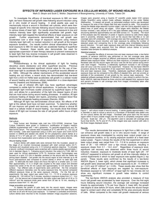
  • 1. EFFECTS OF INFRARED LASER EXPOSURE IN A CELLULAR MODEL OF WOUND HEALING Mark D. Skopin and Scott C. Molitor, Department of Bioengineering, University of Toledo, Toledo OH To investigate the efficacy of low-level exposure to 980 nm laser Images were acquired using a Quantix 57 scientific grade digital CCD camera light, we have measured cell growth rates following wound induction using (Roper Scientific) using custom made software designed to run under Matlab an in vitro model of wound healing. A small pipette was used to (Mathworks). To assess cell growth, an automated analysis routine was developed to measure the area of the imaged field that was covered by cells. This procedure mechanically induce a wound in fibroblast cell cultures, which were then utilized the Matlab edge detection function edge( ) with the Canny algorithm option to imaged at specific time intervals following wound induction and exposure detect lines of pixels along cell edges. Once edges were detected, the Matlab to various doses of laser light. Our results show that exposure to low and function imclose( ) was utilized to fill in the gaps between edge lines using circular medium intensity laser light significantly accelerate cell growth; high structuring elements approximately one cell width across (12 - 15 pixels). The result intensity laser light negated the beneficial effects of laser exposure on cell of this analysis was the detection of pixels in regions covered by cells where edges growth. Further experiments demonstrated that cell growth was were in proximity, whereas regions such as the wound that had little or no cell accelerated over a wide range of exposure durations using medium coverage were left undetected. Cell coverage area was then quantified at each time interval following wound induction and laser exposure by adding the number of intensity laser light with no marked reduction in cell growth at the longest detected pixels and comparing this to the coverage area immediately following exposure durations. Our results confirm clinical observations that low- wound induction. For each laser exposure dose and time interval following wound level exposure to 980 nm laser light can accelerate healing of superficial induction, images were acquired from five different culture dishes to provide wounds. However, these results also demonstrate the need for repeated data samples for statistical analysis. appropriate supervision of laser therapy sessions to prevent overexposure Statistical analysis to laser light that may reverse increases in cell growth rates observed in To separate the effects of recovery time and laser exposure dose, a two-factor response to lower levels of laser exposure. analysis of variance (ANOVA) was performed on the cell coverage area data obtained from images acquired at different post-exposure elapsed times and from Introduction different laser exposure doses. Without any laser exposure, a complete re-growth of fibroblast cells into the wound region will occur over the 48 hour period during which Photobiotherapy is the clinical application of light for healing cells were imaged. Therefore, a statistically significant increase in cell coverage decubitus ulcers (bedsores) and other superficial wounds. Previous area will occur over the course of the experiment. However, the two-factor ANOVA studies have demonstrated significant clinical value for the use of low- procedure allows for the separation of two experiment factors, in this case elapsed level laser light to accelerate the healing of superficial wounds (Mester et time and laser exposure dose. Therefore, statistically significant effects of laser al., 1985). Although the cellular mechanisms of this accelerated wound exposure dose can be compared to the effects of elapsed time, and can provide an healing are not known, a recent study has demonstrated that low-level estimate of the acceleration of cell growth by the various laser exposures. The light from a 633 nm HeNe laser accelerates cell growth in a cellular model statistical software package Minitab 14 (Minitab Inc.) was utilized to perform the two- factor ANOVA; data was entered as three columns with elapsed time, exposure dose of wound healing and improves cellular metabolism in a dose-dependent and percent increase in cell coverage area from images immediately taken after manner (Hawkins and Abrahamse, 2006). wound induction. The use of infrared (IR) light may have significant advantages compared to visible light for clinical applications. In particular, the longer wavelength light minimizes scatter produced by superficial layers of the skin, and allows for a penetration of the light into deeper layers of skin that are most active during wound healing processes. In addition, IR light produces heating of deeper skin layers, promoting increased blood flow and to further accelerate healing processes (Dierickx, 2006). Although IR light has demonstrated clinical value, the effects of IR light at the cellular level have not been examined. To determine whether IR can improve cell growth and recovery, we have utilized a clinical IR laser in a cellular model of wound healing. Our results show that limited doses of IR light can increase the rate of cell growth within hours of light Figure 1: cell culture model of wound healing. A sterile pipette approximately 1 mm in diameter is used to induce a wound in a monolayer of fibroblast cells plated onto exposure. 35 mm culture dishes (far left image). Fibroblast cells grow back into the wound region within 8 hours (middle image) and the wound is completely overgrown within Methods 24 hours. Scale bar: 250 μm. The algorithm used to calculate cell coverage area Cell culture found that 64.9%, 76.7% and 97.7% of the imaged area was covered with cells in Fetal human skin fibroblast cells (cell line CCD-1070SK, American Type the three images displayed above. Culture Collection) were grown in Dulbecco’s modification of Eagle’s medium (DMEM) supplemented with 1 mM L-glutamine, 1% penicillin-streptomycin and 3% Results fetal bovine serum. The cultures were incubated at 37 ºC with 95% O2 - 5% CO2 at Our results demonstrate that exposure to light from a 980 nm laser 85% humidity. Cells were trypsinized using a 0.25% (w/v) trypsin and 0.03% EDTA solution in DMEM and seeded into sterile 35 mm polystyrene culture dishes at a can enhance cell growth rates in an in vitro wound model. A range of density of 7.0 x 10⁴ cells per cm2. Cells were incubated overnight to allow the cells exposure doses was investigated by varying laser output power over a to recover from trypsinization and to adhere to the bottom of the culture dishes. fixed exposure duration, or by varying exposure duration at a fixed laser Wound healing model output power. Figure 2 shows the results of the first experiment in which Culture dishes with plated cells were removed from the incubator, placed on laser output power was varied from 1.5 - 7.5 W to produce an exposure 2 the stage of an inverted microscope equipped with relief contrast optics (IX-71, level of 26 - 120 mW/cm over a two minute exposure, resulting in 2 Olympus) and visualized using a 4x, 0.13 numerical aperture objective. To simulate exposure doses from 3.1 - 14.4 J/cm . Regardless of exposure level, a wound, confluent monolayers were scratched with a sterile pipette approximately 1 significant cell recovery was observed within three hours of wound mm in diameter (figure 1). Following wound induction, the output of a 7.5W, 980 nm induction; however, exposure to moderate levels of laser light (26 - 97 laser used for clinical applications (VTR 75, Avicenna Laser Technologies) was 2 mW/cm ) appeared to enhance cell growth at all time intervals relative to focused on a spot 12.5 mm in diameter centered on the wound with the visible red aiming beam disabled. Cell growth into the wound region at time intervals up to 48 control experiments in which no laser exposure was applied (figure 2, top hours post-exposure was compared to control dishes in which no laser light was panel). These results were confirmed by the results of a two-factor used. To attenuate the laser output and focus the light on a smaller spot, a 3 mm ANOVA (figure 2, lower right), which shows that significant increases in 2 fiber approximately 1.5 m in length was coupled to the laser output and directed cell growth were observed with two minute exposures to 26 - 73 mW/cm 2 toward the center of the 35 mm culture dish approximately 10 mm above the dish (p < 0.01) and 97 mW/cm (p < 0.05). These results also show that the surface. Two different sets of experiments were performed: the first compared beneficial effects of laser exposure are negated by over-exposure: different exposure intensities over the same exposure time, the second compared 2 fibroblasts exposed to 120 mW/cm of laser light for two minutes did not different exposure times at the same exposure intensity. For the first set of experiments, the laser output was varied from 1.5 - 7.5 W to produce exposure show any significant increase in growth rates relative to control densities 26 - 120 mW/cm2 for 2 minutes, resulting in exposure doses from 3.1 - 14.4 experiments. J/cm2. For the second set of experiments, the laser output was fixed at 4.5 W or 73 The two-factor ANOVA analysis also provided a measure of how mW/cm2 and the exposure times were varied from 20 sec to 15 min, resulting in much cell growth was accelerated by laser exposure. Over the first eight exposure doses from 1.5 - 66 J/cm2. hours following wound induction, the average cell coverage increased Image acquisition and analysis linearly by approximately 1.7% per hour (figure 2, lower left); the growth To assess the growth of cells back into the wound region, images were rate begins to slow before 24 hours, when cells across the wound margin acquired at hourly intervals up to 8 hours after wound induction and laser exposure, begin to contact each other and completely fill the area previously devoid and then at 24 and 48 hours post exposure. To maintain a controlled environment, of cells. When compared to the mean cell growth measured at various culture dishes were returned to the incubator between image acquisition sessions.
  • 2. time intervals following wound induction, the 4 - 6% increase in cell 2 growth produced by 49 - 73 mW/cm of laser exposure over a two minute period represented an acceleration of wound healing by approximately 2.5 - 3.5 hours within the first eight hours of healing. This represents a sizeable acceleration in cell growth considering that the wounds from our in vitro model were nearly completed healed within 24 hours following wound induction. Despite the significant increases in cell growth across various time intervals and exposure levels, the two-factor ANOVA analysis did not find any significant interaction between elapsed time and exposure level. In other words, the various exposure levels showed consistent effects across all time intervals following wound induction, and there were no exposure levels whose effects were only observed at a particular time interval or subset of time intervals following wound induction. Figure 3: Top panel: cell growth in wound model as a function of time elapsed from wound induction and laser exposure dose. Vertical bars show % change in cell coverage area averaged across five experiments in which cells were not exposed to laser light, or in which cells were exposed to 73 mW/cm2 of light during exposures that varied from 20 sec to 15 min to give exposure doses from 1.5 - 65.7 J/cm2. Error bars show S.E.M across five experiments. Bottom left: confidence intervals from the two-factor ANOVA analysis demonstrate a significant increase in cell coverage area observed as early as 3 hours after wound induction regardless of laser exposure (* p < 0.05 for 3 hours, ** p < 0.01 for 4 hours and beyond). Bottom right: confidence intervals from the two-factor ANOVA analysis demonstrate a significant increase in cell coverage area for moderate laser exposure doses when compared to no laser exposure (** p < 0.01 for 1.5 - 8.8 J/cm2). No significant increase in cell coverage was observed at the second highest dose (21.9 J/cm2), and a significant decrease in cell coverage was observed at the highest exposure dose (* p < 0.05 for 65.7 J/cm2). Figure 2: Top panel: cell growth in wound model as a function of time elapsed from wound induction and laser exposure intensity. Vertical bars show % change in cell Conclusions coverage area averaged across five experiments in which cells were not exposed to Our results confirm the clinical observation that low-level exposure to laser light, or in which cells were exposed to 26 - 120 mW/cm2 of light during a two 980 nm of laser light can accelerate cell growth in a wound healing model. minute exposure by varying the laser power from 1.5 - 7.5 W to give exposure doses Because our measurements were obtained from an in vitro cell culture from 3.1 - 14.4 J/cm2. Error bars show S.E.M across five experiments. Bottom left: model, these results also suggest that the mechanisms involved in the confidence intervals from the two-factor ANOVA analysis demonstrate a significant acceleration of cell growth following laser exposure are cellular or increase in cell coverage area observed as early as 3 hours after wound induction regardless of laser exposure (** p < 0.01). Horizontal bars show 95% confidence molecular in nature. The hypothesis that IR light accelerates healing intervals with midline at mean value; error bars show 99% confidence intervals. processes by heating skin and promoting increased blood flow (Dierickx, Bottom right: confidence intervals from the two-factor ANOVA analysis demonstrate 2006) could not explain the increased cell growth rates in an in vitro cell a significant increase in cell coverage area for low and moderate levels of laser culture model. Our measurements suggested that IR exposure produced exposure when compared to no laser exposure (** p < 0.01 for 26 - 73 mW/cm2; * p temperature increases less than 2 ºC, and the use of a controlled < 0.05 for 97 mW/cm2). No significant increase in cell coverage was observed at the incubation environment between image acquisition intervals further highest exposure level (120 mW/cm2). minimizes the temperature variability in our experiments. Previous researchers have suggested that light exposure increases ATP levels by Figure 3 shows the results of the second experiment in which altering the energetic state of light-sensitive cytochromes within the inner exposure durations were varied from 20 sec - 15 min at a constant laser mitochondrial membrane that participate in oxidative phosphorylation 2 output power of 4.5 W to produce an exposure level of 73 mW/cm , (Karu et al., 1995); we are conducting further experiments to examine 2 resulting in exposure doses from 1.5 - 66 J/cm . As with changes in ATP levels following low-level exposure to 980 nm laser light. exposure level, significant cell recovery was observed within three hours Our results also demonstrate the importance of appropriate of wound induction regardless of exposure duration, and a wide range of supervision of laser light exposure in a clinical setting. In particular, the exposure durations appeared to enhance cell growth at all time intervals average cell growth rates formed a non-monotonic function of laser relative to control experiments in which no laser exposure was applied exposure levels (figure 2, lower right) and exposure doses (figure 3, lower (figure 3, top panel). These results were confirmed by the results of a right); with peak growth rates at moderate exposures, and reduced benefit two-factor ANOVA (figure 3, lower right), which shows that significant at higher exposure intensities and doses. This suggests that excessive 2 increases in cell growth were observed with 73 mW/cm exposures having light exposure could have potentially damaging effects that negate any 2 durations of 20 sec - 2 min to produce exposure doses of 1.5 - 8.8 J/cm initial benefit of light exposure. Therefore the appropriate exposure levels 2 (p < 0.01). Note that a long exposure of 15 min (65.7 J/cm ) produced a and durations must be selected in order to maximize cell growth rates. significant decrease in cell growth (p < 0.05), suggesting that long exposure to laser light at a moderate level can reverse the benefits of References lower exposure does. A comparison of these results to the mean cell Dierickx CC (2006) The role of deep heating for noninvasive skin rejuvenation. growth measured at various time intervals following wound induction Lasers Surg Med 38:799-807. (figure 3, lower left) showed that the 6% increase in cell growth produced 2 Hawkins DH, Abrahamse H (2006) The role of laser fluence in cell viability, by 73 mW/cm of laser exposure over a 20 sec - 2 minute period proliferation, and membrane integrity of wounded human skin fibroblasts represented an acceleration of wound healing by approximately two hours following helium-neon laser irradiation. Lasers Surg Med 38:74-83. within the first eight hours of healing. Furthermore, the two-factor ANOVA Karu T, Pyatibrat L, Kalendo G (1995) Irradiation with He-Ne laser increases ATP analysis did not find any significant interaction between elapsed time and level in cells cultivated in vitro. J Photochem Photobiol B 27:219-223. exposure duration despite the significant increases in cell growth across Mester E, Mester AF, Mester A (1985) The biomedical effects of laser application. various time intervals and various exposure durations. Lasers Surg Med 5:31-39.