
Recommended
More Related Content
What's hot
What's hot (20)
Similar to Endoscopic ear surgery
Similar to Endoscopic ear surgery (20)
Recently uploaded
Recently uploaded (20)
Endoscopic ear surgery
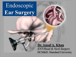
- 1. Endoscopic Ear Surgery Dr. Ausaf A. Khan ENT/Head & Neck Surgery HCM&D. Hamdard University
- 2. ENDOSCOPIC EAR SURGERY Prof. Dr. Ausaf Ahmed khan Professor and Head of Department Hamdard College of Medicine & Dentistry Hamdard University Hospital Karachi, Pakistan.
- 3. Introduction Otologic surgery has progressed rapidly over the past century Before1920’s; loupes/without microscope assistance 1950’s; refinement of the binocular microscopes Late 60’s; use of microscope to visualize middle ear was introduced 1990’s; endoscopes incorporated in middle ear surgery
- 4. Introduction to EES Endoscopic Ear Surgery (EES – the use of endoscopes to perform otologic surgery) has become, in experienced hands, a safe and reasonable approach for a variety of indications in ear surgery. The endoscope is slowly emerging as a valuable adjunct to the operative microscope & the sole means for visualization and performing many routine and complex otologic procedures for experienced surgeons in selected centers in US & elsewhere. The Endoscope is NOT meant to replace the microscope in all patients, but may serve a specialized purpose in select cases.
- 5. Introduction to EES Microscopic techniques, introduced in late 1950’s changed the character and outcome of ear surgery. Endoscope offers a same ‘game changing’ impact. By using endoscopes, the improved access to the tympanic cavity and proximal Eustachian tube has allowed us to have a better understanding of the primary disease process : impaired ventilation.
- 7. CONTENTS History of Endoscopic Ear Surgery Rationale for use of the endoscope in ear surgery Terminology How to get started Relevant instrumentation for EES Surgical ergonomics & OR setup Indications, C/I & safety considerations Endoscopic tympanoplasty steps Techniques to incorporate EES in practice Pearls and pitfalls/Technical tips/Promotional tips 10-commandments for the novice surgeons Summary
- 8. HISTORY OF OTOMICROSCOPY & ENDOSCOPIC EAR SURGERY
- 9. History of Otomicroscopy Otologic surgery has progressed rapidly over the past century Carl Olof Nylen 1921; The monocular microscope was first applied in ear surgery. Gunnar Holmgren 1922; developed the first binocular microscope for use in ear surgery. Otologists of that era in the first half of the 20th century mostly used loupes for visualization.
- 10. History of Otomicroscopy Otologic surgery has progressed rapidly over the past century 1953, Carl Zeiss in collaboration with physicist Hans Littman adapted and redesigned the ear microscope. 1950’s- The Zeiss OPMI-1 microscope became widely available and revolutionized otologic surgery.
- 11. HISTORY OF ENDOSCOPIC EAR SURGERY Paralleling the introduction of endoscopes for sinus surgery in the 1990s, otology is facing a similar paradigm shift. “Otoendoscopy” (The use of endoscopy to visualize the ear) was introduced in the late1960s. Poor image resolution at that time, in comparison to the operative microscope limited its application.
- 12. HISTORY OF ENDOSCOPIC EAR SURGERY Ear Endoscopy from the 1960s to the 1980s Mer and colleagues in 1967: examined cadaver’s ears & living animals’ ears through an iatrogenic myringotomy. Nomura 1982: The first myringotomy was published - used an angled rigid endoscope and called it the needle otoscope. Nomura’s focus was on middle ear photography. 1989, Kimura and colleagues in Japan: used an ultrathin fiberscope that was inserted in living patients under LA through the eustachian tube orifice in Nasoph.
- 13. HISTORY OF ENDOSCOPIC EAR SURGERY Ear Endoscopy from the 1960s to the 1980s (From Mer SB, Derbyshire AJ, Brushenko A, et al. Fiberoptic endotoscopes for examining the middle ear. Arch Otolaryngol 1967;85(4):387–93) Early endoscopic views of the middle ear, 1967.
- 14. HISTORY OF ENDOSCOPIC EAR SURGERY Endoscopy Ear Surgery in the 1990’s The true beginnings of EES took place in the 1990’s. Otologic surgeons started to use endoscopic approaches not only for inspection but also to guide intervention. McKennan in California – Second look Mastoidectomies (Transcutaneous Mastoidoscopy) – to avoid another postauricular incision during second-look surgery for cholesteatoma. Rosenberg and Silverstein – investigated this mastoidoscopy approach further by first examining the mastoid endoscopically via a postauricular keyhole approach then formally opening the mastoid via the postauricular approach.
- 15. One advantage of ear endoscopy over binocular otomicroscopy is the wide field of view
- 16. Microscopic and endoscopic views of the right middle ear. Daniel Lee, MD Massachusetts Eye and Ear Infirmary Harvard Medical School
- 17. HISTORY OF ENDOSCOPIC EAR SURGERY Endoscopy Ear Surgery in the 1990’s Muaaz Tarabichi – embraced the endoscope as a sole mode of visualization for ear surgery, and by the late 1990s published an important series on the endoscopic management of cholesteatoma.
- 19. Tarabichi M – Endoscopic management of Acquired Cholesteatoma. Am J Otol. 1997; 18: 5444-5449 38 adults with acquired cholesteatoma 36 underwent transcanal EES 29/30 disease free at 1 year 10/13 disease free at 2 years 4/6 disease free at 2 years (on surgical exploration) Transcanal Endoscopic resection of Cholesteatoma is safe and effective.
- 20. The main contribution of the endoscope in my experience has not been a technical one, but rather the different perspective of cholesteatoma and cholesteatoma surgery that it afforded me. Cholesteatoma is a manifestation of advanced retraction of the tympanic membrane, with the sac advancing into the tympanic cavity proper and then on to its extensions (ST,FR,HyT). Only in advanced cases, it proceeds further to mastoid cavity proper. The endoscope allowed a better understanding of cholesteatoma and the way it travels through the temporal bone Therefore, the most logical approach to cholesteatoma is Tarabichi M – Endoscopic management of Acquired Cholesteatoma. Am J Otol. 1997; 18: 5444-5449.
- 21. Right ear: Note the retraction and cholesteatoma. H: Handle of malleus
- 23. HISTORY OF ENDOSCOPIC EAR SURGERY Endoscopy Ear Surgery in the 2000’s During this decade, more investigators and otologic surgeons explored the potential benefits of endoscopic techniques. Number of publications in peer-reviewed journals dramatically increased. Otologic surgeons tried their hands at performing a variety of classic otologic procedures endoscopically and reported their experiences as well as technical tips and limitations. Video clips of various endoscopic ear surgeries could be found on different websites and on YouTube.
- 24. HISTORY OF ENDOSCOPIC EAR SURGERY Endoscopy Ear Surgery in the 2000’s The International Working Group on Endoscopic Ear Surgery (IWGEES) formed as a consortium of otologists interested in endoscopic ear surgery. The group promotes endoscopic ear surgery and provides educational materials and seminars.
- 25. Dr. Nirmal Patel
- 26. Daniel Lee, MD Massachusetts Eye and Ear Infirmary Harvard Medical School
- 27. João Flávio Nogueira Assistant Professor ENT Universidade Estadual do Ceará – UECE Director Sinus & Oto Centro Fortaleza, Brazil.
- 29. Rational for EES The operative microscope, pioneered in the 1950s & 1960s, is essential for otologic surgery as it provides 1. excellent illumination, 2. depth perception and magnification, 3. binocular vision, 4. ability to work with 2 hands, and 5. capacity to capture HD images and video. Despite these advantages, the microscope is limited when constrained by small surgical corridors: the External Auditory Canal.
- 30. Transcanal microscopic view is limited by size of speculum. Transcanal endoscopic view is wider than the microscope.
- 31. Rational for EES In cases with a small surgical corridor, additional soft tissue incisions (endaural or postauricular) or bone removal (canalplasty, atticotomy, removal of ossicles, and canal up or down mastoidectomy) are sometimes needed to access middle ear disease. This is especially true when the EAC is small, when there is a prominent anterior bony overhang & when the middle ear disease extends to the attic,
- 32. Rational for EES The endoscope allows for excellent visualization of the entire tympanic membrane, middle ear because A wide-angle lens & Illumination emerges from the distal tip. With the introduction of 3-CCD camera systems and wide-format digital displays, endoscopes now provide an immersive and high-fidelity visual experience for the surgeon that is also shared by observers in the operating room.
- 37. Main Advantages of EES 1. Using the ear canal as the natural conduit to the tympanic cavity 2. High quality resolution and magnification 3. Restoring normal middle ear & mastoid ventilation routes 4. Preserving as much normal anatomy as possible by minimizing unnecessary dissection of bone and soft tissue 5. Decreasing the need for drilling 6. Avoidance of postauricular approaches and minimizing damage to neurovascular structures
- 38. Philosophy (of the experts) in EES David D. Pothiar Toronto General Hospital
- 39. Drawbacks of EES include; Challenging one handed dissection without suction in other hand Lack of 3 dimensional view – reliance on motion parallax to assess depth perception Lack of exposure to these techniques during surgical training Limited instrumentation
- 40. Basic differences between endoscopic and microscopic ear surgery Endoscope Microscope Number of hands available for dissection One handed (optional 2-handed) Two handed Typical surgical approach Transcanal (can be postauricular for combined cases as well as via the antrum following CWU mastoidectomy Transcanal with speculum +- endaural incision or postaural Resolution High High Binocular vision No Yes Field of vision Wide Narrow Ability to look around corners Yes (0-70degrees) No
- 41. Terminology of EES OTOENDOSCOPY It involves the use of rigid (or flexible) endoscope for inspection of the outer ear, middle ear, mastoid, or lateral skull base. E.E.S It involves the use of the endoscope for simultaneous visualization and dissection of the outer ear, middle ear, and mastoid. This applies to transcanal, transmeatal (canal wall down cavity), trans-mastoid, and transcranial lateral skull base approaches. TRANSCANAL- E.E.S (TEES) It refers to EES techniques in which the EAC is used as the primary surgical portal to access the TM, middle ear, and in very specialized cases, the inner ear and lateral
- 42. HOW TO GET STARTED
- 43. EES Instruments If you have FESS sinuscopes and a middle ear instruments tray you are ready to start… Rigid sinus endoscopes A light source A HD 3-CCD Camera A HD video monitor Basic otological surgical instruments set Few specialized instruments
- 45. 4.0 mm 3.0 mm
- 46. A basic otology Instrument set for Middle ear surgery
- 47. Panetti Endoscopic Instrument set for Middle ear surgery
- 49. Surgical ergonomics & OT setup
- 51. Hand positioning and placement of the endoscope A standard otologic chair that has armrests is essential for EES. Both forearms and elbows should rest on the table, patient shoulder, or armrest to maintain wrist stability and minimize fatigue. The endoscope may be held in a similar fashion as during sinus surgery, with the hand placed partly along the shaft and camera head. The endoscope should be stabilized gently along the cartilaginous meatus.
- 52. Left vs. right ear cases For the right-handed surgeon, it is recommended to start with left-sided EES cases as dissection of routine and complex middle ear disease is much easier than the right ear. Use dominant hand for dissection in both left and right ear cases.
- 53. Indications for Endoscopic Ear Surgery External ear Exostosis Canalplasty Debridement & Bx. EAC cholesteatoma Middle ear Myringotomy Myringo/Tympanoplasty Ossiculoplasty Cholesteatoma Tumors (glomus) Stapedectomy Inner ear/Skullbase Intracochlear schwannoma Small symptomatic neoplasm of IAC fundus or facial N. Petrous apex cyst Perilymph fistula repair Middle cranial fossa SCC dehiscence repair Post. Fossa/CP angle Identification of residual schwannoma in IAC
- 54. Contraindications & potential complications No known absolute contraindications to EES. Any otologic case that may be performed via microscopic techniques may be assisted by the use of an endoscope. Potential complications of EES are identical to that of traditional microscopic ear surgery; Direct damage to ossicles Direct damage to facial nerve Heat damage to inner ear Heat damage to facial nerve There is no reason to believe that complications for EES are higher than microscope-based approaches. Contraindications & potential complications
- 55. Safety considerations specific to EES Potential of thermal injury from tip of endoscope: Power of light source no greater than 50% and A safe distance of >5 mm from inner ear structures Use of 0 scopes is encouraged until comfort is gained using highly angled scopes i.e. 30 and 45.
- 56. Before you begin EES Visit the IWGEES website www.iwgees.org Look at the video clips Visit the SEES website www.sydneyendoscopyear.com Read the SEES dissection guide, watch the videos Visit an IWGEES member Attend 1 (or two) Hands-on dissection course.
- 57. EES courses Harvard, USA Vanderbilt, USA St. Louis MI, USA Glasgow Toronto, Canada Sydney, Australia Bern, Switzerland Cape town, SA Fortaleza, Brazil Modena, Italy Nice, France Yamagata, Japan Dubai, UAE Alexandria, Egypt Jeddah, Saudi Arabia
- 58. A 3-step process to introduce EES into your surgical practice 1. Use the endoscope during chronic ear surgery after the microscope-based dissection to a) Look for hidden disease. b) Examine the retrotympanum, epitympanum, and hypotympanum with a 30° endoscope. c) Examine the antrum through the ear canal with a 30° endoscope. d) Assess the ossicular chain and round window.
- 59. 2. Perform an easy transcanal procedure, including a) Endoscopic examination under anesthesia of EAC and TM before microscope dissection to document abnormality. b) Cerumen removal. c) Myringotomy & PE tube placement. d) Myringoplasty. 3. Use the microscope to begin the tympanomeatal flap; then complete elevation with EES techniques a) Switch to a 0° endoscope before dissection of the A 3-step process to introduce EES into your surgical practice
- 67. PROMOTIONAL TIPS TECHNICAL TIPS MEEI 10 COMMANDMENTS
- 68. Promotional tips No soft tissue injury No head bandage/dressing No scar No removal of Sutures Day-case Minimum requirement of analgesia Good view, recording Everyone is engaged Good educational tools
- 69. TECHNICAL TIPS Success in EES comes form the accumulation of many tiny tips & pearls 1. Inject the EAC and surrounding tissues thoroughly 2. Place cottonoids with 1:1000 adrenaline in EAC during preparation of case 3. While you are waiting 1. Trim EAC hairs 2. Clean debris and cerumen 4. Placing the endoscope in the EAC is critical each time 1. Use instruments to push tragus forward 2. Place in canal under screen view
- 70. 5. Raising the tympanomeatal flap is often the most difficult part: Once you reach the MEar, everything settles down 6. Make the tympanomeatal flap more lateral than you might expect 7. Raise the flap with a cottonoid +/- suction elevator 8. Be liberal with the cottonoids 9. Irrigate 10. 5 minutes ‘by the clock’ will solve almost every bleed 11. Take your time TECHNICAL TIPS Success in EES comes form the accumulation of many tiny tips & pearls
- 71. The MEEI “10 Commandments” of EES for the novice surgeon 1. Participate in an EES course and practice EES in a temporal bone laboratory. 2. Essential EES surgery equipment: includes 0 and 30 endoscopes, 3-CCD HD camera, HD monitor, and standard otologic instrument set. 3. Discuss with OR team, anesthesiologist & ancillary staff, the setup for EES before beginning any case 4. The light source should be no greater than 50%.
- 72. 6. Trim ear canal hair before the start of EES cases. 7. Avoid using endoscope holders 8. Initial cases of EES; use the endoscope to look for hidden disease after using the microscope and then transitioning to “easy” procedures 9. For angled endoscopes; use two hands to introduce the endoscope into canal and middle ear: be aware of “blind spots”. 10. Finally, keep practicing and expect setbacks. The MEEI “10 Commandments” of EES for the novice surgeon
- 73. Time 3 hours 3 hours 2.3 hours 2.3 hours 2 hours Microscope Case Endoscope Case David D. Pothiar Toronto General Hospital
- 74. The LEARNING curve microscope endoscope David D. Pothiar Toronto General Hospital
- 75. The BENEFIT curve microscope endoscope David D. Pothiar Toronto General Hospital
- 77. Summary Advancing technique with many historical precedents Excellent tool for CSOM Advances in anatomy of relevant structures Expanding indications Early days Rapidly developing field Requires commitment and practice
- 78. A truth passes through three stages. First it is ridicule. Second it is violently opposed. Third it is accepted as being self evident - Arthur Schopenhauer
- 79. Gunner Holmgren (1875-1954) father of fenestration surgery. Raymond Carhart (1912-1975) first described Carhart notch. Julius Lempert (1890-1968) developed one-stage fenestration surgery. Samuel Rosen (in 1953) proposed stapes mobilization. John Shea Jr. (1924-2015 ) father of modern stapes surgery. John W. House President - House Ear Institute. We are all dwarfs seated on Giant’s shoulder. If we can see far this is not because we are tall, this is because we are seated on Giant’s shoulder. Iftikhar Salahuddin The Aga Khan University Hospital.
Editor's Notes
- Tympanomeatal flap has been elevated, middle ear has been entered, and the cholesteatoma sac has been exposed C; Chorda R: Round window S; Chol sac A; Annulus