2. carotid artery aneurysms with slight interval improvement in
focal stenosis of the right common carotid artery. It also
revealed suspected mild stenosis of the right subclavian
artery origin (►Fig. 2).
The patient continues to be totally asymptomatic on 20 mg
of prednisone with a plan of slow taper over 6 to 12 months’
period.
Discussion
Takayasu arteritis is an uncommonly rare, chronic large vessel
vasculitis with poorly understood pathogenesis and etiolo-
gy.1,2
Women are 8.5 times more likely to be affected than
men, with a typical onset between 10 and 40 years of age.2,3
Despite a worldwide distribution, the disease is most com-
mon in Asian populations; Japan has an estimated 150 new
cases per year, compared with 1 to 3 new cases per year per
million people in the United States and Europe.3,4
The disease
primarily affects the aorta and its branches, with initial
lesions often occurring in the left mid or proximal subclavian
artery.5,6
Arterial wall thickening is caused by inflammatory
processes and can result in narrowing, occlusion, or rarely
dilation in varying degrees, yielding a wide range of symp-
toms and presentations.7
Early systemic features of the
disease may include fever, night sweats, weight loss, mild
anemia, myalgia, malaise, or arthralgia.8
As the disease pro-
gresses, other manifestations may include headaches, caro-
tidynia, myocardial ischemia, or erythema nodosum.8
Neurological features comprise postural dizziness, seizures,
or amaurosis that can be secondary to hypertension or brain
ischemia.8
Physical examination reveals vascular bruits which are
heard in a majority of patients, in addition to diminished or
absent pulses accompanying right/left blood pressure dis-
crepancies and limb claudication.2
Hypertension is also com-
mon and reflects renal artery stenosis, seen in 28 to 75% of
patients.2,9
Diagnosis is based on history and presenting symptoms, in
conjunction with imaging of the arterial tree.10
MR, comput-
ed tomography (CT), or catheter-based angiography usually
shows smooth, tapered narrowing, or occlusion of the lumen
accompanied by increased arterial wall thickness.10
Although
catheter-based angiography provides information on vessel
lumen anatomy, CT or MR angiography allows clear
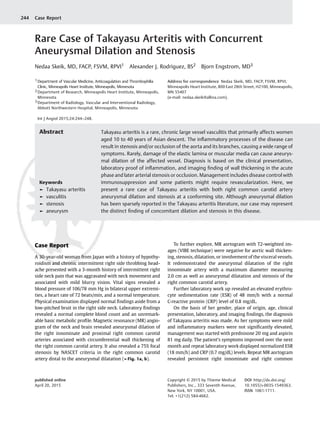
Fig. 1 (a) A three-dimensional time-of-flight images from the mag-
netic resonance angiogram. A coronal subtracted maximum intensity
projection demonstrates a right innominate artery aneurysm (long
bottom arrow), right proximal common carotid artery aneurysmal
dilation (short middle arrow), and right common carotid artery
stenosis more distally (top arrow). (b) An axial image demonstrating
severe wall thickening and aneurysmal dilatation in the right proximal
common carotid artery (long and short arrows).
Fig. 2 A three-dimensional time-of-flight coronal subtracted maxi-
mum intensity projection image from the follow-up magnetic reso-
nance angiogram neck demonstrates persistent right innominate and
right common carotid artery (RCCA) aneurysms with slight interval
improvement in focal stenosis of the RCCA (long arrow), and suspected
mild stenosis of the right subclavian artery origin (short arrow).
International Journal of Angiology Vol. 24 No. 3/2015
Takayasu Arteritis with Aneurysm and Stenosis Skeik et al. 245
3. visualization of wall thickening, in addition to dilation,
stenosis, or aneurysms.11,12
This method is preferred to
invasive angiography that unnecessarily exposes patients to
high-dose radiation and procedure-related risk.11
Laboratory
findings such as elevated ESR and CRP levels are mostly
nonspecific, but they can reflect the underlying inflammatory
process in Takayasu arteritis.3,11
Inflammation accompanies Takayasu arteritis and can
lead to damage of the elastic lamina and muscular media,
which can rarely cause aneurysmal dilation of the vessel.
Scarring has also been shown to progress from the adven-
titia and compromise the vascular lumen.12
In addition,
stenotic arterial lesions can be a result of intimal prolifera-
tion.12
A report by Seko et al found that infiltrating cells in
the aortic tissue largely comprised of killer cells, particu-
larly gamma delta T lymphocytes.13
Another study found
anti-endothelial antibody levels nearly 20 times greater
than normal in the serum of 18 of 19 patients with
Takayasu.14
Differential diagnoses include atherosclerosis,
giant cell arteritis, cerebral vasculitis, Behcet vasculitis,
fibromuscular dysplasia, Cogan syndrome, late sequela of
Kawasaki disease, and connective tissue disorders such as
Marfan syndrome and Ehlers–Danlos syndrome
(►Table 13,9–11,15–20
).
Table 1 Differential diagnoses that should be considered when evaluating a patient for Takayasu arteritis with signs/symptoms and
differentiating tests3,9–11,15–20
Disease/condition Differentiating signs/symptoms Differentiating tests
Giant cell arteritis Inflammatory medium and large vessel
arteritis predominantly affecting external carotid
artery in elders. Polymyalgia rheumatica
may be concurrent. Jaw claudicating is common.
Lower extremity involvement is less common.
Imaging with CT or MRA; GCA is more
likely to involve cranial artery than
lower extremity circulation.
Cerebral vasculitis Rare inflammatory medium and small cerebrovascular
vasculitis leading to variety of symptoms
including headache, difficulty with body part
coordination, confusion,
changes in sensation or perception and stroke.
Cerebral angiography or MRI of the
brain: endothelial thickening
leading to stenosis.
Brain biopsy.
Behcet disease A triad of oral and genital ulceration with uveitis.
Often accompanied by a peripheral
arthritis. May have arterial and/or venous thrombosis.
Angiography: reveals saccular
dilation of involved arteries or
thrombotic occlusion.
CSF examination supportive but not
diagnostic; increased inflammatory
cells and protein.
Cogan syndrome Inflammatory medium and large vessel vasculitis with
systemic involvement including
anterior eye and ear leading to blurred vision,
eye redness, pain, and photosensitivity
as well as hearing loss and dizziness.
Clinical diagnosis.
Slit-lamp examination.
Kawasaki disease Inflammatory medium vessel vasculitis with
high-grade fever, strawberry-tongue, marked
lymphadenopathy, red eyes with uveitis or
conjunctivitis, rash and peeling of the skin on the
palms and soles that typically affects children
younger than 5 years.
Clinical diagnosis using set criteria.
Angiography reveals saccular dilation of
affected arteries. Predominantly
affects the coronaries.
Marfan syndrome Connective tissue disease caused by the
misfolding of fibrillin. Typically tall patients with long
limbs. May have a family history of Marfan syndrome.
Susceptible to lens dislocation.
Clinical diagnosis.
Family history.
Genetic testing.
Ehlers–Danlos syndrome Inherited connective tissue disorder causing
hypermobile joints or paper-thin skin scars.
Angiography: may reveal saccular
dilation of involved arteries.
Genetic testing.
Atherosclerosis Typically patients are elder than 40 years.
Risk factors include hyperlipidemia, hypertension,
smoking, and diabetes.
Angiography: arterial stenosis. Lesions are
usually at the vessel
origin and or bifurcations.
Fibromuscular dysplasia Nonatherosclerotic, noninflammatory vascular
disease with abnormal vascular
wall growth that is more common in females.
Most commonly affects renal and carotid arteries.
Angiography: characteristic
beading of affected arteries.
Aorta usually not involved.
Abbreviations: CT, computed tomographic; CSF, cerebrospinal fluid; GCA, giant cell arteritis; MRA, magnetic resonance angiogram; MRI, magnetic
resonance imaging.
International Journal of Angiology Vol. 24 No. 3/2015
Takayasu Arteritis with Aneurysm and Stenosis Skeik et al.246
4. Treatment is aimed at suppressing the systemic inflam-
mation and controlling related symptoms. Glucocorticoids
have been shown to be mostly effective in halting inflamma-
tion and reversing elevation of acute phase reactants.9
In
patients with glucocorticoid-resistant active disease, metho-
trexate and/or azathioprine may be considered.21
There is
new evidence indicating that rituximab, a B cell targeting
medication might be helpful in patients with refractory
Takayasu arteritis.1,22
Our patient’s diagnosis of Takayasu arteritis was based on
the presentation of indicative clinical symptoms, along with
CT and MR angiography revealing stenosis, wall thickening,
and ultimately dilation of parts of the aortic branch. Success-
ful resolution of symptoms and normalization of ESR levels on
prednisone validated the presence of inflammation accom-
panying this disease. Although Takayasu arteritis is rare, the
presentation of the disease with associated aneurysmal dila-
tion of arch vessels is exceedingly rare.23
Furthermore, the
presentation of the disease with concomitant aneurysmal
dilation and stenosis or occlusion is even less common.24,25
In
a review of 106 patients with Takayasu arteritis over 50 years,
only 6 patients were found to have accompanying carotid
aneurysms.26
Of these six patients, three had associated
arterial occlusions: one with occlusion of the left radial artery,
one with occlusion of the celiac artery and stenosis of the
abdominal aorta, and one with coarctation of the aorta. Only
two patients displayed concomitant wall thickening along
with aneurysmal dilation.26
Interestingly, in all these six
cases, the stenoses or wall thickening was found in areas
discrete from the aneurysmal lesions. Only two cases have
been reported of aneurysmal dilation and stenosis at a
conforming site.24,25
Some authors believe that blood pressure, rather than
disease location seems more closely linked to dilation and
appears to be the major risk factor for Takayasu arteritis
morbidity.27,28
In our case, we believe the aneurysmal dila-
tion was a result of vascular wall inflammation and thicken-
ing, as the patient did not demonstrate elevated blood
pressure. Death in Takayasu arteritis is most commonly
caused by a cerebrovascular event or congestive heart failure.9
In rare cases, aneurysmal lesions can rupture or dissect,
necessitating the need for urgent surgical intervention.29,30
One must carefully weigh the decision to operate on patients
with Takayasu arteritis because of the inherent inflammatory
properties of the disease, with complications such as hemor-
rhage, valve and suture line detachment, pseudoaneurysm
formation, and paravalvular leakage.30
We did not consider
any surgical or endovascular intervention for our patient as
she is completely asymptomatic.
Conclusion
Takayasu arteritis with associated aneurysmal dilation of the
arch vessels has been sparsely reported in the litera-
ture.23,26,27,30
Occlusion or stenosis accompanying this dila-
tion has been an even more seldom condition.24–26
Some
patients may also have simultaneous wall thickening.25,27
Our article represents a unique case among scarce reports
in the Takayasu literature, with aneurysmal dilation and
stenosis occurring in the same arch vessel caused by wall
thickening. Only two other Takayasu cases we found in the
literature described the dilation and stenosis/occlusion at an
analogous site. We recommend taking Takayasu arteritis in
consideration when evaluating patients with aortic branch
stenosis and or dilation.
Funding
No financial support was needed or provided by any
source.
Note
This article is original and is not under consideration by
another journal, and has not been previously published.
Acknowledgment
We acknowledge Alexander Rodriguez for his hard work in
preparing this article.
References
1 Skeik N, Rumery KK, Udayakumar PD, Crandall BM, Warrington KJ,
Sullivan TM. Concurrent Takayasu arteritis with common variable
immunodeficiency and moyamoya disease. Ann Vasc Surg 2013;
27(2):240.e13–240.e18
2 Lupi-Herrera E, Sánchez-Torres G, Marcushamer J, Mispireta J,
Horwitz S, Vela JE. Takayasu’s arteritis. Clinical study of 107 cases.
Am Heart J 1977;93(1):94–103
3 Arend WP, Michel BA, Bloch DA, et al. The American College of
Rheumatology 1990 criteria for the classification of Takayasu
arteritis. Arthritis Rheum 1990;33(8):1129–1134
4 Koide K. Takayasu arteritis in Japan. Heart Vessels Suppl 1992;
7:48–54
5 Hata A, Noda M, Moriwaki R, Numano F. Angiographic findings of
Takayasu arteritis: new classification. Int J Cardiol 1996;54
(Suppl):S155–S163
6 Cid MC, Font C, Coll-Vinent B, Grau JM. Large vessel vasculitides.
Curr Opin Rheumatol 1998;10(1):18–28
7 Sharma BK, Jain S, Sagar S. Systemic manifestations of Takayasu
arteritis: the expanding spectrum. Int J Cardiol 1996;54(Suppl):
S149–S154
8 Johnston SL, Lock RJ, Gompels MM. Takayasu arteritis: a review.
J Clin Pathol 2002;55(7):481–486
9 Kerr GS. Takayasu’s arteritis. Rheum Dis Clin North Am 1995;
21(4):1041–1058
10 Kissin EY, Merkel PA. Diagnostic imaging inTakayasu arteritis. Curr
Opin Rheumatol 2004;16(1):31–37
11 Yamada I, Numano F, Suzuki S. Takayasu arteritis: evaluation with
MR imaging. Radiology 1993;188(1):89–94
12 Tso E, Flamm SD, White RD, Schvartzman PR, Mascha E, Hoffman
GS. Takayasu arteritis: utility and limitations of magnetic reso-
nance imaging in diagnosis and treatment. Arthritis Rheum 2002;
46(6):1634–1642
13 Seko Y, Minota S, Kawasaki A, et al. Perforin-secreting killer cell
infiltration and expression of a 65-kD heat-shock protein in aortic
tissue of patients with Takayasu’s arteritis. J Clin Invest 1994;
93(2):750–758
14 Eichhorn J, Sima D, Thiele B, et al. Anti-endothelial cell antibodies
in Takayasu arteritis. Circulation 1996;94(10):2396–2401
International Journal of Angiology Vol. 24 No. 3/2015
Takayasu Arteritis with Aneurysm and Stenosis Skeik et al. 247
5. 15 Epocrates® Online. Takayasu arteritis. Available at: https://online.
epocrates.com/u/29351064/Takayasuþarteritis. Accessed Janu-
ary 6, 2015.
16 Weyand CM, Goronzy JJ. Medium- and large-vessel vasculitis. N
Engl J Med 2003;349(2):160–169
17 Ishikawa K. Diagnostic approach and proposed criteria for the
clinical diagnosis of Takayasu’s arteriopathy. J Am Coll Cardiol
1988;12(4):964–972
18 Kerr GS, Hallahan CW, Giordano J, et al. Takayasu arteritis. Ann
Intern Med 1994;120(11):919–929
19 Berlit P. Diagnosis and treatment of cerebral vasculitis. Ther Adv
Neurol Disord 2010;3(1):29–42
20 Kessel A, Vadasz Z, Toubi E. Cogan syndrome—pathogenesis,
clinical variants and treatment approaches. Autoimmun Rev
2014;13(4-5):351–354
21 Hoffman GS, Leavitt RY, Kerr GS, Rottem M, Sneller MC, Fauci AS.
Treatment of glucocorticoid-resistant or relapsing Takayasu
arteritis with methotrexate. Arthritis Rheum 1994;37(4):
578–582
22 Hoyer BF, Mumtaz IM, Loddenkemper K, et al. Takayasu arteritis is
characterised by disturbances of B cell homeostasis and responds
to B cell depletion therapy with rituximab. Ann Rheum Dis 2012;
71(1):75–79
23 Perrotta S, Rådberg G, Perrotta A, Lentini S. Aneurysmatic disease
in patients with Takayasu disease: a case review. Herz 2012;37(3):
347–353
24 Caballero PE. Common carotid artery aneurysm revealing Takaya-
su’s arteritis. J Stroke Cerebrovasc Dis 2011;20(6):556–558
25 Renker M, Baumgartner I, Diehm N. Takayasu arteritis presenting
with extensive bilateral aneurysms of the common carotid arter-
ies. Eur Heart J 2012;33(4):435
26 Tabata M, Kitagawa T, Saito T, et al. Extracranial carotid aneurysm
in Takayasu’s arteritis. J Vasc Surg 2001;34(4):739–742
27 Sueyoshi E, Sakamoto I, Hayashi K. Aortic aneurysms in patients
with Takayasu’s arteritis: CT evaluation. AJR Am J Roentgenol
2000;175(6):1727–1733
28 Regina G, Fullone M, Testini M, et al. Aneurysms of the supra-aortic
trunks in Takayasu’s disease. Report of two cases. J Cardiovasc Surg
(Torino) 1998;39(6):757–760
29 Geraldes R, Batista P, Pedro LM, Fernandes A, Melo TP. Takayasu
arteritis presenting with internal carotid artery dissection. Cere-
brovasc Dis 2012;33(4):408–409
30 Song MH, Nakayama T, Hattori K, Tokuda Y, Mabuchi Y, Ueda Y.
Aortic root aneurysm in Takayasu arteritis syndrome: exploration
in active phase and repair in inactive phase. J Thorac Cardiovasc
Surg 2008;136(4):1084–1085
International Journal of Angiology Vol. 24 No. 3/2015
Takayasu Arteritis with Aneurysm and Stenosis Skeik et al.248