1. Protocol for Uterine Tissue
Homogenization in the Bullet Blender™
The protocol described in this document is for the use of the Bullet Blender™ for the
homogenization of uterine tissue (from a variety of animals). Note that the time and
speed settings may differ due to the variation in consistency/texture of different tissue
from species to species. This protocol does not specify a particular buffer - you may
choose which is most appropriate for your downstream application (nucleic acid isolation,
protein extraction, etc.).
Materials Required: uterine tissue, saline, aspirator, Bullet Blender™, homogenization
buffer, pipettor, microcentrifuge tubes, 0.9 to 2.0 mm stainless
steel bead blend (part number SSB14B) or 1.6 mm stainless steel
beads (part number SSB16).
Instructions
1. Cut uterine tissue into appropriately sized pieces for analysis (50mg-150mg, if your
tissue is not already smaller) and place into microcentrifuge tubes.
2. OPTIONAL: Wash tissue 3x with ~1mL PBS. Aspirate. NOTE: This step removes
some external contaminants (blood, etc.).
3. Add a mass of the stainless steel beads equal to 3X the mass of the sample. One
scoop of stainless steel blend ≈ 220mg. One scoop of 1.6 mm stainless steel beads
≈ 186mg.
4. Add 2 volumes of buffer for every mass of tissue, minimum of 25μL.
5. Close the microcentrifuge tubes.
6. Place tubes into the Bullet Blender™.
7. Set controls for SPEED 8 and TIME 5 minutes. Press Start.
8. After the run, remove tubes from the instrument.
9. Visually inspect samples. If homogenization is unsatisfactory, run for another three
minutes at the SPEED 10.
10. Remove sample tubes from the Bullet Blender™, add the appropriate buffer and
proceed with your downstream application.
SAFETY NOTE!!!
When using a centrifuge to separate your homogenate from the
debris and beads, make sure your tubes are balanced.
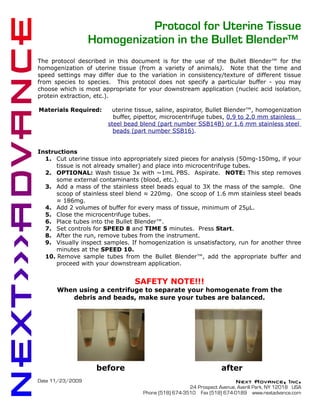
before after
Date 11/23/2009 Next Advance, Inc.
24 Prospect Avenue, Averill Park, NY 12018 USA
Phone (518) 674-3510 Fax (518) 674-0189 www.nextadvance.com