Acromioclavicular instability, rotator cuff : dynamic ultrasound

https://osteopathie-adhesiolyse.com In 1/3 of acromioclavicular instabilities, Wagner found a dynamic instability (Fig. 4, 5), which has never been described before, and which seems to potentiate the risk of conflict. Indeed, we find this dysfunction 1 in 2 times in the case of a complete tear of the cuff. This instability is linked with a hyper-laxity, which adds to the arthrosis of the joint. During the abduction, it is as if the clavicle were pulled downwards at the same time as the acromion follows the motion of the humerus. This phenomenon appears to be cased by a retraction of the anterior fascia and especially by a contraction of the subclavicle muscle, which is painful when put under pressure. Manual de-contracting manipulation can be proposed.

Recommended
More Related Content
More from Adhésiolyse Manuelle Dynamique-Osteopathie-Ho Pun Cheung
More from Adhésiolyse Manuelle Dynamique-Osteopathie-Ho Pun Cheung (10)
Recently uploaded
Recently uploaded (20)
Acromioclavicular instability, rotator cuff : dynamic ultrasound
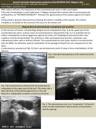
- 1. With rotator cuff pain, the importance of the acromioclavicular joint is often overlooked. If the role of osteophytes is well understood, P. Wagner showed that in almost half of acromioclavicular arthropathies, an “INFERIOR INSTABILITY” increases the agressiveness of the clavicle against the rotator cuff. He described a dynamic ultrasound test showing this inferior instability of the clavicle. This inferior instability is correlated to the severity of the injury to the rotator cuff. In the two tiers of rotator cuff pathology, Wagner found osteophytosis (Fig. 1) at the upper part of the acromioclavicular joints. In these cases of acromioclavicular osteoarthritis (Fig. 2), it is probable that an inferior osteophytosis could be aggressive against the rotary cuff. Radiological assessment with Lamy incidence must be demonstrated. This arthrosis is often accompanied by synovitis, sometimes very inflammatory either with or without effusion. The acromioclavicular joint space may be in communication with the BSAD. An infiltration permits visualization of the passage of liquid from one compartment to the other. A subcutaneous synovial cyst (Fig. 3), from a acromioclavicular point of view, is also a manifestation of this effusion. Fig 1. Rounded osteophystosis of the distal end of the clavicle Fig. 2 Acromioclavicular arthrosis with the production of osteophytes at the upper part of the joint. The lower side is likely identical, with potential aggression on the cuff. Requires a radiographic incidence centered on the joint. Fig. 3. The subcutaneous cyst is an “evagination” of the joint cavity. Careful examination indeed shows communication with the spacing. Acromio-clavicular degenerative syndrome of the ROTATOR CUFF: Wagner’s test Thierry Ho-Pun-Cheung, sport doctor, osteopath, Montpellier June 2014 Pierre Wagner, physician, Nimes Degenerative acromioclavicular osteophytes and synovites
- 2. Fig. 6a 6b. Same anomaly. Patient who has had a resection of the distal end of the clavicle. - supraspinatus at neutral rotation - infra-spinous at forced medial rotation - sub-scapular at forced lateral rotation In 1/3 of acromioclavicular instabilities, Wagner found a dynamic instability (Fig. 4, 5), which has never been described before, and which seems to potentiate the risk of conflict. Indeed, we find this dysfunction 1 in 2 times in the case of a complete tear of the cuff. This instability is linked with a hyper-laxity, which adds to the arthrosis of the joint. During the abduction, it is as if the clavicle were pulled downwards at the same time as the acromion follows the motion of the humerus. This phenomenon appears to be cased by a retraction of the anterior fascia and especially by a contraction of the subclavicle muscle, which is painful when put under pressure. Manual de-contracting manipulation can be proposed. Acromioclavicular instability Fig. 4. Inferior instability of the clavicle. Due to the subclavicle muscle, sometimes associated with scapular dyskinesia, the clavicle does not follow the rocking motion of the scapula, creating an ultrasound impression of the lower discrepancy. Fig. 5. Acromioclavicular in adduction: the upper surface of the clavicle (C) is higher than the acromion (A). Joint disjunction allows observation of bursitis through the acromioclavicular space. The synovial under the upper ligament is a little too visible (arrow). During the anterolateral elevation of the arm, the clavicle will be lower and closer to the acromion, driving the synovial below the superior ligament. Surgical resection (Fig. 6) of the distal end of the clavicle is often ineffective, as it aggravates the instability (Fig. 7, 8). Fig. 7. Evolution of the resection: changes with scar fibrosis and inflammation. Fig. 8. This patient continues to suffer three years after the intervention; the articular space is very modified and remains inflammatory, in spite of several cortisone infiltrations. We understand that lower osteophytosis attacks the cuff, particularly: Acromioclavicular atypia (Fig. 9, 10) Fig. 9. Abnormalappearanceand bilateralof an acromioclaviclar,without history. The two sides are separated by 6.5 mm. The space is filledupon ultrasound:remainsof meniscus or fibrosis? Fig. 10. An incidental discovery, without history, the clavicle is dislocated in the upper position.