20140802 crc trainging image for ra & as 講義版
•Download as PPTX, PDF•
4 likes•921 views

Clinical Research Coordinator training program

Recommended
More Related Content
Similar to 20140802 crc trainging image for ra & as 講義版
Similar to 20140802 crc trainging image for ra & as 講義版 (20)
More from Kailen Tsai
More from Kailen Tsai (10)
20140802 crc trainging image for ra & as 講義版
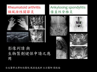
- 1. Rheumatoid arthritis 類風溼性關節炎 Ankylosing spondylitis 僵直性脊椎炎 台北醫學大學附設醫院 風濕免疫科 主治醫師 蔡凱倫 影像判讀 與 生物製劑健保申請之應 用
- 2. The rapid expansion of the therapeutic bioagents has the potential to dramatically improve RA patient care. NEJM 2001 NEJM, 2006. NEJM 2012 Others: 小分子標靶藥物 Tofacitinib(Xeljanz Or Anti-IL17…..
- 6. X-ray
- 11. RA clinical coarse The role of image? Radiography, Ultrasound, CT MRI
- 12. 類風溼性關節炎 臨床進程 時間 健康 滑膜發炎 臨床症狀, 關節痛,腫 無症狀期 早期類風濕性 關節炎 晚期類風濕性 關節炎 基因與環境因素 自體抗體產生 RF, anti-CCP 關節破壞 關節變形 X 光片 超音波 核磁共振
- 14. MRI and Miniarthroscopy of MCP Joints in RA . Arthritis Rheum 2001; 44:2492–2502. TI + contrast
- 15. Placebo arms
- 16. 類風溼性關節炎在核磁共振下可觀 察到的病理變化 Synovitis 滑膜炎 Tenosynovitis 肌腱滑膜炎 Bone Erosions 骨侵蝕 Bone Marrow Edema 骨質發炎水腫 Bursitis 滑囊炎
- 17. contrast-enhanced MRI depicted more abnormalities within the osseous structures of the rheumatoid wrist than corresponding fat-suppressed T2-weighted fast spin-echo imaging. Contrast-enhanced T1-weighted images are considered more sensitive and specific in the assessment of acute synovitis AJR:187, August 2006
- 18. Tenosynovitis AJR:189, December 2007 T1FS + C Effusion: dark(T1) ; bone: dark(T1 white -> T1FS :no fat signals, dark) Synovitis, contrast median: water
- 19. Contrast-enhanced axial T1- weighted fat-saturated MR image
- 22. 骨骼肌肉超音波
- 23. 價格 敏感度 安排所 需等待 時間 是否有助急性發 炎診斷 骨折診斷 放射性 傳統X光片 便宜 差 快 否 是 是 核磁共振 昂貴 高 須排程 是,但視情況需 顯影劑 是 是 骨骼肌肉超 音波 便宜 高 快 是 否 無 類風濕性關節炎的影像診斷工具
- 24. Clinical applications in RA 髖關節 膝關節 腳踝 趾關節 肩關節 手肘 手腕 手指關節
- 26. 類風溼性關節炎 三大臨床特色 Synovitis Joint effusion(GS) Synovial hypertrophy(GS) Synovial vascularization(CD/PD) Bone erosion Tendon pathology A B C DE F
- 28. 滑膜發炎 滑膜增生 手腕關節
- 29. 0: no erosions 1: very small ( <1 mm) 2: small (1 - 2 mm) 3: moderate ( 2 - 4 mm) 4: large ( > 4 mm) 骨骼侵蝕 第二指關節 longitudinal Lateral views transverse
- 30. Power Doppler signals 能量都普勒 代表 發炎訊號
- 32. Power Doppler signals代表active inflammation,訊號越強,代表發炎 越厲害。 有證據顯示之前Power Doppler signals 發現發炎越厲害,之後骨面磨損侵蝕 越嚴重,關節破壞越嚴重。 消滅 Power Doppler signals為什麼那 麼重要?
- 36. Right 2nd MCP
- 38. 56歲女性,診斷類風溼性關節炎已十多年,服用DMARD及生物製劑,關 節腫痛起起伏伏,但今年初生物製劑開始被減藥。 ESR,CRP 並不高,但病患近日仍主訴手指疼痛益增 5/13 12/4 經過半年,雖然病患關節外觀早已 變形,但是從超音波可見其骨頭仍 持續破壞 Right hand 2nd MCP
- 39. 達標治療 Treat to target 除了傳統的我們知道的 DAS28 超音波也可以當作一個治 療追蹤指標 MSU treat to target 發炎訊號的消失 關節積水變少 滑膜增生變薄
- 40. Objective. To investigate the construct validity and reliability of US DAS compared with 28-joint DAS(DAS-28) in assessing joint inflammation and in prediction of structural damage in patients with RA. Rheumatology (Oxford). 2012 Jan;51(1):120-8. 超音波比DAS28在評估關節發炎程度和預測 結構破壞上更準確性和可靠性
- 41. 是否可以當作退場機制申覆上的輔助工具? ARTHRITIS & RHEUMATISM Vol. 64, No. 1, January 2012, pp 67– 76 Conclusion. For RA patients whose disease is in remission or who have low levels of disease activity, PD signals on ultrasonography could predict relapse or radiographic progression and identify those whose disease is adequately controlled, which is especially helpful when considering treatment tapering or interruption.. 能量嘟普勒訊號可以預測傳統X光上結構破 壞,和正確評估病患是否有適當的疾病控制
- 44. 正常脊椎 早期僵直性脊椎炎 晚期僵直性脊椎炎 發炎 沾黏融合 病情嚴重及控制不良者: 脊椎沾黏: 竹子狀,變形,駝背 脊椎沾黏嚴重: 喪失柔軟度,提高骨折風險,甚至有壓迫 神經導致下肢麻木問題。
- 45. [image tool: MRI] modified by Medscape Rheumatology education, Expanding the spectrum for TNF antagonist: Safty & Efficacy in the Spondyloarthropathies: Ankylosing Spondylitis Image tool: MRI, Ultrasound
- 46. Sacroiliitis 0 Normal 1 Some blurring of the joint margins - suspicious 2 Minimal sclerosis with some erosions 3 Severe erosions with widening of joint space +/- some ankylosis (Pseudo-widening of the joint space: Subchondral bone resorption— blurring ; Erosion sclerosis ;Calcification leading to ankylosis) 4 complete ankylosis Grade 0 Grade 1 Grade 3 Grade 4
- 54. anterior corner erosions at vertebral bodies (vertebral enthesitis) shiny corner sign (Romanus lesion) Vertebral body squaring Vertebral fusion
- 56. AS, Andersson lesion, discovertebral destruction, spine (severe back pain!)
- 57. 2~5年 Modified New York criteria 1984 1. 下背痛僵硬 休息無法減輕 三個月以上 2. 腰椎活動範圍受限 3. 擴胸範圍受限 4. X光有薦腸關節炎雙側2級或單側3級以上 4+ 1 or 2 or3
- 58. Spine
- 59. T1 white, STIR dark Bone marrow edema, STIR white, T1 dark Romanus lesion Active lesion Chronic lesion
- 60. SPARCC MRI spinal score (0–108) Arthritis Rheum. 2005 Aug 15;53(4):502-9. T2 SPARCC 2 SPARCC 13 Spondyloarthritis Research Consortium of Canada magnetic resonance imaging index
- 61. Arthritis Rheum. 2003 Apr;48(4):1126-36
- 62. STIR Frequency of active inflammation in the spine (vertebral units) Ann Rheum Dis 2013;72:967–973
- 63. Ann Rheum Dis 2013;0:1–7.
- 64. Enthesitis T1 T1FS + C C7~ T3 supraspinal ligament RadioGraphics 2005; 25:559 –570
- 67. Synovitis, SpA Synovitis, Tendonitis/ tenosynovitis Enthesopathy Dactylitis
- 68. Elemental lesions Ossification/ cortical bone changes Achilles Tendon
- 69. Achilles Tendon Cortical bone irregularities: erosion, doppler
- 71. Taipei Medical University Hospital HIP joint
- 73. 傳統X光片仍是評估關節炎很重要的工具 核磁共振與骨骼肌肉超音波對於關節炎軟組織部份 的評估較傳統X光片敏感許多。 類風濕性關節的重要病理病變為滑膜發炎,僵直性 脊椎炎的重要病理變化為著骨點發炎。 類風溼性關節炎的發炎評估以手腕關節與2nd MCP掌 指關節最為重要。 能量督普勒代表的是發炎訊號,超音波的達標治療 目標以消滅能量嘟普勒訊號為最重要。 Take home massage