2. Nucleic Acids Research, 1997, Vol. 25, No. 81560
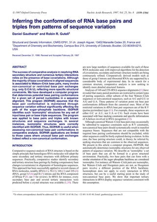
Figure 1. Construction of planar single H bond base pairs. (a) Stage 1.
Superimposition of H bond donors and acceptors onto H bonds of initial base
pairs. (b) Stage 2. Superimposition of glycosyl bonds onto glycosyl bonds of
initial base pairs. (c) Stage 3. Systematic construction of N-H..N and N-H..O
bonds, using the bond angles shown.
MATERIALS AND METHODS
The ISOPAIR program takes as input a set of base pair or triple
sequences (e.g. {G:C, A:G, U:A} or {C:G:A, G:C:U}), typically
obtained from covariation analysis. There is no limit to the
number of input sequences. Isopair first generates internally a list
of possible pairing conformations for each sequence and then
seeks sets of isomorphic conformations that can be formed with
every input sequence. Conformation sets are returned in the form
of PostScript or Brookhaven ‘pdb’ files. The program is written
in C. Unix executables are available through anonymous ftp at
igs-server.cnrs-mrs.fr, in directory /pub/ISOPAIR, or through
written request to gauthere@igs.cnrs-mrs.fr.
Initial generation of base pair conformations
An initial set of 28 double H-bond conformations available in the
literature (13) was constructed using interactive molecular
graphics. Single H-bond pairings are computer-generated. Their
construction is limited to planar structures and proceeds as follows.
(i) A first collection of single H-bond pairs is obtained by
superimposing each of the four bases onto the 28 double H-bond
pairs built previously. ISOPAIR performs superimpositions in
two ways. First, H-bond donors and acceptors are superimposed
onto the H-bonds of the 28 initial pairs (Fig. 1a). New pairing
structures that do not produce steric conflicts are stored. In the
example shown, a new C:U pair is generated.
(ii)Inasecondstage,glycosylbondofeachfournucleotidesare
superimposed onto glycosyl bonds of the 28 initial base pairs
(Fig. 1b). New structures that contain at least one H-bond and do
Figure 2. Base pair superimpositions used in the measure of isomorphism.
Glycosyl bond atoms N1 (or N9) and C1′ from the two base pairs to be
compared are superimposed and rms deviations are measured between these
two sets of four atoms. Angles α1 and α2 are measured as well.
not produce steric conflicts are stored. In the example shown, a
new A:A pair is created. This stage ensures that no single H-bond
conformation that is rigorously isomorphic to a known double
H-bond conformation is omitted.
(iii) Finally, additional single H-bond base pairs are sought
through a systematic connection of H-bond donors and acceptors
in all four bases, using the H-bond angles shown in Figure1c.
This procedure generates a total of 351 different pairing
structures. Base triple structures are generated by combining
Watson–Crick or wobble pairs with every non-canonical pair.
Defining the distance between two conformations
We define base pair isomorphism, I, as the ability to form while
retaining similar sugar–phosphate backbone conformations. We
must therefore quantify how well backbones from two different
base pairs can be superimposed. As rotations about the glycosyl
bond are possible, we can only compare the position of glycosyl
bonds and the angle they form in the pairs. Glycosyl bond atoms
(N1 or N9 and C1′) from the two pairs to be compared are thus
superimposed and their root mean square (rms) deviation is
measured, as well as the angles α1 and α2 represented in Figure 2.
Thehyperbolicfunctionsf1(α)orf2(rms)representedinFigure3are
then applied to convert rms and angle values into a distance value
comprised between 0 and 1. These functions increase quasi-
exponentially near zero, which quickly penalizes measures
departing from ideal values but, unlike exponential functions,
they are upper-bounded, which permits comparisons of independent
measures. The plateau reached with high α or rms values is not
a problem here since values in this range correspond to
uninteresting non-isomorphic conformations. The final distance
D is f1(α) × f2(rms). Base triple comparisons are performed
similarly, using three glycosyl bonds instead of two.
Selection of isomorphic sets of conformations
Given the input base pair or triple sequences {s1, s2, ..., sn} where
each sequence si has m possible conformations Ci = {ci,1, ci,2, ...,
ci,m}, we compute all the pairwise distances D(ci, cj) where ci
∈ Ci and cj ∈ Cj. Using a conventional branch and bound
3. 1561
Nucleic Acids Research, 1994, Vol. 22, No. 1Nucleic Acids Research, 1997, Vol. 25, No. 8 1561
Figure 3. Functions used in the measure of isomorphism. α1, α2 and rms
deviation are defined in here and text. The inflexion points off1 and f2 for α = π
and rms = 0.4 were chosen empirically. functions f1, f2 and D have the same
shape.
algorithm, we then construct all the conformation sets of the form
{c1, c2, ..., cn}, c1 ∈ C1, c2 ∈ C2, ..., cn ∈ Cn, satisfying:
I +
Si+1..n,j+1..nD(ci, cj)
n2
t t
This selects conformation sets for which the average pairwise
distance I (or isomorphism) is lower than a fixed thresholdt. Note
that the lower I, the higher the isomorphism. After visual
inspection of isomorphic structures, we empirically set the value
of t at 3 × 10⁻³. A set of conformations for which I is lower than
this value is said ‘isomorphic’.
Constraints
When the input set of base pair sequences yield several
isomorphic solutions, further criteria are needed to distinguish the
most interesting ones. ISOPAIR may optionally require that
solutions contain at least one double H-bond pair. This is referred
to as the ‘double H-bond’ constraint. Users can also prohibit
conformations involving variable glycosyl bond orientations (syn
andanti) in thesame isomorphicset. Thiscan beused for instance
to avoid certain solutions containing syn pyrimidines.
ISOPAIR can also exclude pairings that can be formed by
sequences that are not in the input set. For instance, if an
isomorphic structure is found for the covariation {A:A, G:G} and
this structure can also be formed with {C:G}, we may consider
this structure as ‘wrong’, as it provides no strict rationale for the
sequence observation. This constraint, which we term ‘uniqueness’,
is useful when it can be reasonably argued that unobserved
sequences are indeed counter selected. Uniqueness (U) of a
isomorphic set is defined asthe shortest average distance between
the structures in that isomorphic set and any structure that can be
formed with other base pair sequences. An isomorphic set with
isomorphism I and uniqueness U is considered as unique if
U > 3 × 10⁻³ or U > 2 × I (empirical thresholds). Due to the high
number of base triple conformations, the current version of
ISOPAIR cannot test uniqueness for base triples. Base triples, can
optionally be constrained to occur in the major groove.
Base pair sequences
Base pair exchanges in tRNA were obtained from a sequence
alignment adapted from that of Sprinzl (14), containing 895 type
I nuclear tRNA and tRNA gene sequences. Type I and type II
tRNAs differ in the size of their variable loop (4 or 5 nt in type I
tRNAs, 10–24 nt in type II tRNAs) and in the position of their
tertiary interactions.
RESULTS
A first simple question that can be addressed with ISOPAIR is the
number of base pairs that may potentially adopt a common
structure. There are 96 possible sets of two different base pairs
({A:A,A:C}, {A:A, A:G}, {A:A,A:U}, etc.) after removal of
equivalent sets such as {A:A,A:U} and {A:A,U:A}. We ran
ISOPAIR for each set and counted the number of solutions. For
each set, ISOPAIR finds at least one isomorphic solution with an
I value below 3 × 10⁻³, that is with only mild differences in
glycosyl bond angles and positions (data not shown). This result
is not surprising if one considers the large variety of single
H-bond conformations. Most of these common structures,
however, are not unique to the input pairing set. For instance, the
Watson–Crick conformation is a solution for the input set {U:A,
A:U}, but this conformation may also be achieved with G:C or
C:G. When studying sequence variations issued from comparative
sequence analysis, this type of non-specific solution is questionable
as it cannot explain why only certain base pair sequences are
observed. This is why we introduced the ‘uniqueness’ constraint
(see Materials and Methods), that discards solutions that can also
be obtained with sequences not in the input set. Now of the same
96 sequence sets analyzed earlier, only 33 have a unique solution
(Fig. 4). The uniqueness constraint thus considerably reduces the
number of structures to be considered.
To test ISOPAIR’s ability to reproduce known pairing geometries
from typical covariations, the program was given canonical
combinations of Watson–Crick and G:U sequences. As expected,
the input set {A:U, U:A, G:C, C:G} produces the Watson–Crick
conformation as the most isomorphic solution, with anI value of
5.3 × 10⁻⁶. This result is obtained whether or not ‘uniqueness’ is
imposed on solutions, confirming what we already knew about
the conformation of this set of pairings. When G:U or U:G are
added to the four Watson–Crick sequences, the most isomorphic
solution has an I value of 7.3 × 10⁻⁵ and, surprisingly, does not
contain Watson–Crick nor wobble conformations. This solution,
shown in Figure 5a, only contains single H-bond base pairs. The
expected Watson–Crick/wobble solution (Fig. 5b) ranks fourth
but is the best solution involving double H-bond pairs. The
number of H-bonds in base pairs could thus be important in
ranking solutions. A correct prediction can be achieved here by
choosing the solution that involves the highest number of
H-bonds. The I value for this solution (8.2 × 10⁻⁵) is an order of
magnitude higher than for Watson–Crick sequences alone, due to
the significant difference in glycosyl bond positions between
Watson–Crick and wobble pairs.
Applying the uniqueness constraint to the {A:U, U:A, G:C,
C:G, G:U} input set eliminates the Watson–Crick/wobble
solution. This was expected, since a wobble structure can also
form with an A:C pair. The input set {A:U, U:A, G:C, C:G, G:U,
4. Nucleic Acids Research, 1997, Vol. 25, No. 81562
Figure 4. Unique isomorphic structures for combinations of two base pairing sequences. Asterisks indicate the presence of several isomorphic conformations for the
input sequences. In this case, the solution shown was selected based on: (i) the presence of double H bond pairings; (ii) the lowestI value. The presence of both syn
and anti conformations in the same solution was purposely not checked in these ISOPAIR runs. Numbers below each base pair refer to the internal numbering of base
pair structures in ISOPAIR.
U:G} does not produce the Watson–Crick/wobble solution,
whetherornottheuniquenessconstraintisused.Thisisconsistent
with the large deviation observed when superimposing wobble pairs
G:U and U:G.
tRNA
Transfer RNA sequences provide a number of covariations that
can be related to known pairing structures. An important unusual
covariation is that found at position 15:48, known as the Levitt
pair (10). The vast majority of tRNAs contain either G:C or A:U
at this position, and Klug et al. (15) have suggested that this
sequence constraint was consistent with the parallel reverse
Watson–Crick pair present at this position, since bases other than
G:C or A:U would induce a significant backbone displacement if
paired similarly. ISOPAIR finds more than 10 different isomorphic
structures for A:U and G:C, one of which is the reversed
Watson–Crick pairing found in tRNA crystal structures. However,
the only ‘unique’ solution containing a double H-bond pair is
indeed the parallel reverse Watson–Crick pairing observed in
tRNA crystal structures (Fig. 6a).
Certain cysteine tRNA do not have the usual G:C or A:U Levitt
pair, but have instead a G:G pair (16) that cannot adopt a
reverse-Hoogsteen conformation. This can be regarded as a threat
Figure 5. ISOPAIR results for input set {A:U, U:A, G:C, C:G, G:U}. (a) Highest
ranking solution. (b) Fourth ranking solution. Numbers below each base pair
refer to the internal numbering of base pair structures in ISOPAIR.
to our initial assumption that sequences not compatible with a
required base pairing would be excluded by selection. It has been
shown, however, that this G:G Levitt pair is a determinant for the
aminoacylation of tRNACys (17). In this case, the absence of
isomorphism is thus related to a variation in the structure and
5. 1563
Nucleic Acids Research, 1994, Vol. 22, No. 1Nucleic Acids Research, 1997, Vol. 25, No. 8 1563
Figure 6.Sequence variations in tRNA alignments and corresponding base pair
or base triple structures. (a–d) The structures shown are isomorphic sets
produced by ISOPAIR using the input sequence in the left column. The last
column indicates the rank of this isomorphic solution in terms ofI value, in the
presence or absence of ‘double H bond’ constraint. (e) tRNAPhe sequences for
base triple 10:25:45 and the structure observed in yeast tRNAPhe. ISOPAIR
does not identify this structure.
function of the RNA molecule, consistent with our initial assump-
tion.
Other unusual covariation patterns in tRNA are observed at
base triple positions. Since the structure of these base triples
varies considerably (18), they have been studied independently
for each tRNA species (Asp, Phe, etc.). In yeast tRNAPhe, base
triples occur at positions 12:23:9, 13:22:46 and 10:25:45. At
position 12:23:9, the two most predominant sequences are U:A:A
and G:C:G in all 895 tRNA sequences in the database, as well as
in each of the tRNA species for alanine, phenylalanine, asparagine
and tryptophan. We sought isomorphic structures for these two
triple sequences. When using the ‘double H-bond’ constraint (see
Materials and Methods), the highest ranking solution (Fig.6b) is
that observed in the tRNAPhe crystal structure (19). This solution
ranks sixth without the ‘double H-bond’ constraint.
At positions 13:22:46, tRNAPhe sequences are either C:G:G or
U:G:G. The most isomorphic solution for these sequences is in
agreement with the crystal structure, provided that the ‘double
H-bond’ constraint is used (Fig. 6c). In tRNAAsp species, the
predominant sequences for this triple are U:G:A and C:G:G. The
yeast tRNAAsp crystal structure (20) is shown in Figure 6d
(U:G:A sequence). ISOPAIR predicts this structure fourth in
terms ofI value for the sequence {U:G:A, C:G:G}, even when the
‘double H-bond’ constraint is used.
The predominant sequences at positions 10:25:45 in tRNAPhe
speciesareG:C:GandG:C:U.TheyeasttRNAPhe structure(Fig.6e)
cannot be identified by ISOPAIR using these sequences, whatever
constraint is used. This result could be expected since (i) the
peculiar single H-bond G:G interaction in this base triple does not
follow ISOPAIR’s rules for base pair construction, and (ii) the
structure observed for G:C:G cannot form with sequence G:C:U,
implying an absence of isomorphism at this position.
These test runs for tRNA triples (Fig.6b–e) were all performed
without using the ‘major groove’ constraint. This constraint
imposes that solutions contain only major groove base triples
(which is the case in all tRNA base triples). In the absence of the
‘double H-bond’ constraint, discarding minor groove triples
slightly improves the ranking of the correct solutions in Figure6b–d
(data not shown).
DISCUSSION
We have presented a computed program (ISOPAIR) capable of
seeking base pair conformations that are common to a given set
of sequences. This program is intended for use in comparative
sequence analysis when unusual base covariations are observed
at specific RNA positions. Our underlying assumption was that
any base covariation inferred from comparative analysis was
amenable to a set of isomorphic base pair structures that could all
form with a similar orientation of the sugar–phosphate backbone.
This led to a definition of isomorphism based on comparisons of
glycosyl bond positions and orientations.
Results obtained with tRNA base pairs and triples indicate that
ISOPAIR may indeed be useful as an investigative tool for base
pair conformations. Actual conformations often lie within the
highest ranking solutions, although selecting the right solution
cannot be guaranteed by the program. Most ISOPAIR runs
generate multiple solutions (Fig.4) and whether or not preference
should be given to solutions with double H-bond pairs, pairs with
syn or anti glycosyl bonds or pairs with a unique conformation
remains an expert’s task. Our tests with tRNA sequences suggest
that correct solutions most often involve at least one double
H-bond pair. However, parameters such as the number and
variability of available sequences and prior knowledge of structural
constraints in the vicinity of the base pair are essential in reaching
a correct conclusion.
Another important factor to consider when interpreting sequence
covariations is the nature of the constraints underlying an
interaction. Our model explicitly seeks base pairs that can form
with a similar orientation of the sugar–phosphate backbone. In
many cases however, this constraint is not predominant or is
combined with others. These involve purine:pyrimidine constraints,
that account for covariations such as {A:U, G:C} or {A:U, G:C,
G:U} observed in 16S and 23S rRNA (11), or the exposure of
specific atoms to tertiary interactions or binding of external
factors. Unusual covariations may also result from particular
tertiary environments. For instance, certain pairing sequences
could be excluded because they would result in unwanted tertiary
interactions with surrounding residues. The ISOPAIR program
can also be useful in identifying this variety of constraints, as it
generates sets of similar structures that can be displayed or saved
as three-dimensional coordinates for a detailed search of common
structural properties.
6. Nucleic Acids Research, 1997, Vol. 25, No. 81564
ACKNOWLEDGEMENTS
R.R.G thanks the W. M. Keck Foundation for its support of RNA
science on the Boulder campus. This work was supported in part
by grants from the NIH (GM48207) and the Colorado RNA
center to R.R.G.
REFERENCES
1 Holley,R., Agpar,J., Everett,G., Madison,J., Marquisee,M., Merrill,S.,
Penswick,J. and Zamir,A. (1965) Science, 147, 1462–1465.
2 Fox,G. and Woese,C. (1975) Nature, 256, 505–507.
3 Noller,H. and Woese,C. (1981) Science, 212, 403–411.
4 Glotz,C., Zwieb,C. and Brimacombe,R. (1981) Nucleic Acids Res., 9,
3287–3306.
5 Michel,F., Jacquier,A. and Dujon,B. (1982) Biochimie, 64, 867–881.
6 Davies,R., Waring,R., Ray,J., Brown,T. and Scazzocchio,C. (1982)Nature,
300, 719–724.
7 Michel,F., Umesono,K. and Ozeki,H. (1989) Gene, 82, 5–30.
8 James,B., Olsen,G., Liu,J. and Pace,N. (1988) Cell, 52, 19–26.
9 Gutell,R.R. (1993) Curr. Biol., 3, 313–322.
10 Levitt,M. (1969) Nature, 224, 759–763.
11 Gutell,R.R. (1996) Comparative sequence analysis and the structure of 16S
and 23S RNA in Zimmerman,R.A. and Dahlberg,A.E. (eds) Ribosomal
RNA. Structure, Evolution, Processing and Function in Protein
Biosynthesis. CRC Press, pp. 111–128.
12 Bubienko,E., Cruz,P., Thomason,J.F. and Borer,P.N. (1983) Prog. Nucleic
Acids Res. Mol. Biol., 30, 41–90.
13 Saenger,W. (1984) Principles of Nucleic Acid Structure. Springer Verlag,
New York, p. 120.
14 Sprinzl,M., Dank,N., Nock,S. and Schon,A. (1991) Nucleic Acids Res., 19,
2127–2171.
15 Klug,A., Ladner,J. and Robertus,J. (1974) J. Mol. Biol., 89, 511–516.
16 Mazzara,G. and McClain,W. (1977) J. Mol. Biol., 117, 1061–1079.
17 Hou,Y.M., Westhof,E. and Giegé,R. (1993) Proc. Natl. Acad. Sci. USA, 90,
6776–6780.
18 Gautheret,D., Damberger,S.H. and Gutell,R.R. (1995)J. Mol. Biol., 248,
27–43.
19 Quigley,G. and Rich,A. (1976) Science, 194, 796–806.
20 Westhof,E., Dumas,P. and Moras,D. (1985) J. Mol. Biol., 184, 119–145.