SlideShare a Scribd company logo
1 of 16
Download to read offline
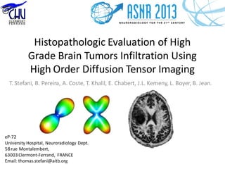
Histopathologic Evaluation of High
Grade Brain Tumors Infiltration Using
High Order Diffusion Tensor Imaging
T. Stefani, B. Pereira, A. Coste, T. Khalil, E. Chabert, J.L. Kemeny, L. Boyer, B. Jean.
eP-72
University Hospital, Neuroradiology Dept.
58 rue Montalembert,
63003 Clermont-Ferrand, FRANCE
Email: thomas.stefani@aitb.org
Summary
• 1 Purpose
• 2 Materials and Methods
• 3 Results
• 4 Discussion
• 5 Conclusion
• 6 Bibliography
The authors of this study have no conflict of interest to disclose.
1 Purpose
• Delineation of high grade brain tumors is critical for treatment
planning, follow up and prognosis. Diffusion Weighted Imaging is
widely used in neuro oncology for this purpose [1-3].
• High grade brain tumors are heterogeneous and infiltrative lesions.
Tumoral cell dissemination is known to be found beyond from the
tumor visible margins therefore analysis of the Brain Adjacent to
Tumor (BAT)remains challenging with conventional MRI sequences [4].
• Diffusion Tensor Imaging (DTI) is sensitive to changes in white matter
fiber bundles microstructure [8].
• The aim of this study was to evaluate the peripheral white matter
infiltration of high grade brain tumors using high order Diffusion
Tensor Imaging.
2 Materials and Methods
• Monocentric prospective study, from April 2012 to April 2013.
• Local IRB approval and informed consent from all patients were obtained.
• All patients from 18 to 85 years of age having a suspected cerebral high grade tumor
(WHO III-IV) on CT scans were screened to be included the protocol.
• Exclusion criteria: MRI contraindication, severe renal failure, cerebral biopsy
contraindication, pregnancy.
TotalTotal
17 patients screened
IncludedIncluded
7 patients
Biopsy RefusalBiopsy Refusal
2 patients
Post-CTSurgicalPost-CTSurgical
Treatment
3 patients
Not WHO III-IVNot WHO III-IV
5 patients
(1 WHO II, 2 metastasis,
1 lymphoma,
1 atypical meningioma)
BiopsyBiopsy
12 patients
DiffusionTensor MRIDiffusionTensor MRI
14 patients
excluded
excluded
excluded
2 Materials and Methods
• Seven patients were prospectively included and underwent 3 T clinical
MRI scans (Signa MR750, GE Medical Systems, Milwaukee, Wis).
– Usual diagnostic MRI protocol included :
• 3D-T1GE (Repetition Time (TR) 8.8 ms, Echo Time (TE) 3.54 ms, 1x1x1.2mm3)
beforeand after contrast media injection
• T2-SE (TR 4923 ms, TE 100.23 ms, 1x1x4mm3)
• Diffusion(TR 8500 ms, TE minimum, 1x1x2mm3, b0-b500-b1000 s/mm2)
• T2 FLAIR (TR 11000 ms, TE 142.97 ms, Inversion Time (TI) 2350 ms, 1x1x4mm3)
• Perfusion Weighted (Echo Planar Imagine (EPI), TR 2000 ms, TE 14.4 ms,
2x2x4mm3)
• Multivoxel Spectroscopy (Point Resolved Spectroscopy (PRESS), TE 144 ms and
TE 35ms, 2x2x2cm3)
– DiffusionTensor Sequence :
• Diffusion Tensor Imaging 20 directions (EPI, TR 8000, TE minimum,
1x1x2mm3, b0-b1000 s/mm2, sequence duration: 8 minutes).
• Diffusion Tensor Imaging 60 directions (EPI, TR 8000, TE minimum,
1x1x2mm3, b0-b3000 s/mm2 sequence duration: 16 minutes).
• The 20 directions sequence was done prior to the 60 directions sequence to decrease the probability of bad
sequences associated with motion artifacts, or premature termination of examination.
2 Materials and Methods
• Patients then underwent surgical CT guided stereotactic tumor biopsy
targeted with Leksell frame (Iplan Stereotaxy 3.0.2,Brainlab), according to
standard procedure, maximum 1 week after MRI procedure (mean delay
= 4 days).
• An extra biopsy (10mm length, 1mm diameter) was performed in normal-appearing
white matter adjacent to the tumor along the biopsy pathway 3 to 8 mm
awayfrom the enhancementarea.
• The coordinates of the center of the biopsy window were noted.
Fig. 1. CT guided stereotactic biopsy planning. Target is located along
the biopsy pathway, 3 to 4 mm away from the enhancement area.
2 Materials and Methods
• Biopsies were fixed with formaldehyde.
• Histopathologicanalysis :
– WHO classification of the tumor.
• Using immunohistochemical markers :
– MAP2 (microtubule associated protein 2), Ki67.
• Semi quantitative estimation of tumor infiltration percentage(IP):
– Number of atypical cells / Total number of cells in the field of view.
– (Field1mm x 2mm, Magnification x10).
Fig. 2. Brain adjacent to tumor biopsy sample, field
1x2mm, magnification 10. Ki67 immunologic marker
showing moderate proliferative rate.
2 Materials and Methods
• Analysis of DT-MRI datasets was done using in-house software using MATLAB scripts (R2012a.
Natick, Massachusetts: The MathWorksInc) courtesy of Angelos Barmpoutis, PhD [5].
– Fourth order tensor resolution :
• 9 x 9 Matrix diagonalisation :
 6 Eigenvalues i
 6 EigenvectorsUi
Fig. 3. Fourth order tensor and its eigenvalues/vectors orientation distribution
fonction (ODF).
2 Materials and Methods
– Computation of the tensor scalars [6], at the biopsy locus (MRI and biopsy CT images
coregistration, 1x1x1mm3 precision, EPI spatial distortions correction using Iplan
Stereotaxy 3.0.2,Brainlab)
• GA (Generalized Anisotropy)
• GV (Variance)
• MD (Mean Diffusion)
with
Fig. 4. Fourth order tensor scalar computation at the biopsy
locus (blue dot).
2 Materials and Methods
• Statistical analysis (Spearman’s rank correlation) was done using STATA
10.0 (Stata Corporation, College Station, TX, USA).
– 30 subjects needed (risk  = 5%, statistical power  = 90%).
• Primary analysis:
– Correlation coefficient was evaluated between the 4th order diffusion Generalized
Anisotropy (GA) distribution and the tumor infiltration percentage (TI).
• Secondary analysis:
– Correlation coefficient was evaluated between the 4th order diffusion tensor metrics
distributions (GV, MD) and the tumor infiltration percentage.
3 Results
• 7 prospective patients(3M, 4F).
• Age ranging from 52 to 86 year old (mean age value = 63).
• Homogeneous population of glioblastoma multiforme (GBM).
• Tumor InfiltrationPercentage(TI) values :
– Mean TI value= 0.16 [0;1].
• Tensor scalars mean values:
– GA (biopsy) = 0.36 [0.18;0.68].
– GV (biopsy)= 0.0028 [0.0009;0.0086].
– MD (biopsy)= 0.00065 [0.00035;0.00075].
3 Results
• We observed a significant correlationbetween :
– GA and tumor infiltration percentage (TI) (r=-0.98 p=0.006).
– GV and tumor infiltration percentage (TI) (r=-0.98 p=0.006).
• MD showed no significant correlation(r=0.11 p=0.76) with TI.
Fig. 5. GA maps.
Left: (patient number: 03) Right frontoparietal
glioblastoma showing low GAsignal in a
peripheral ROI (red arrow) and 0% TI on
histology.
Right: (patient number: 07) Left parietal
glioblastoma showing high GAsignal in a
peripheral ROI (yellow arrow) and an 10% TI
on histology.
4 Discussion
• In this study we introduced 4th order diffusion tensor which offers an
alternative model to conventionnal 2nd order tensor (3 x 3 matrix).
– Jayachandra et al [7], showed that anisotropy could be estimated more accurately for
complex microstructure with 4th order DTI compared to 2nd order DTI.
– Simpler modelisation compared to other techniques (Qball, High Angular Resolution DTI).
• Provenzale et al [8] showed that there is a significant difference in Fractional
Anisotropy (FA) decrease for peritumoral NAWM between gliomas and meningiomas
but not for peritumoral regions (hyperintense areas on T2-weighted MR images),
showing that DTI may be sensitive to subtle microstructure changes.
• Stadlbauer et al [9] showed FA is better than MD for assessment and delineation of
different degrees of pathologic changes (ie, TI) in glioma but their retrospective
population washeterogeneous(WHO Grade II and III gliomas).
• Ellingson et al [10] showed limitations of conventional 2nd order DTI compared to 4th
order DTI when evaluating glioblastomas but needed careful tissue validation as they
didn’tcorrelate their results to a biopsy with cell quantification.
4 Discussion
• In our study we showed that GA and GV were significatively correlated
to Tumor Infiltration Percentage in glioblastoma.
• Butthis study has some limitations:
– Few subjects : only 7 patients (this study is still recruiting).
– Non fixed distance from tumor to biopsy due to surgeon target choice.
– Semi quantitative estimation of percentage infiltration.
– Coregistration accuracy: 1mm accuracy (3mm diameter ROI and correction of EPI
spatial distortions)
– 60 directions GA maps have a lower signal to noise ratio (SNR) compared to 20
directions sequence.
5 Conclusion
•These preliminary results despite the little number of inclusions, show that
GA and GV measurements derived from high order diffusion tensor estimation
are correlated (p=0.006, p=0.006) to tumor infiltration percentage of the local
environment of glioblastoma.
 GA derived from fourth order diffusion tensor could be used for
assessment and delineation of different degrees of pathologic changes (ie,
Tumor Infiltration Percentage) in glioblastoma.
•These results need to be confirmed at the end of the inclusion plan (30
patients completed with DTI and biopsy) in order to validate a clinical non
invasive method to delineate real infiltration margins of GBM without the use
of extra biopsy in the vicinity of high grade gliomas.
• 1. Chenevert, T.L., and al., Diffusion magnetic resonance imaging: an early surrogate
marker of therapeutic efficacy in brain tumors. J Natl Cancer Inst, 2000. 92(24): p. 2029-36.
• 2. Khayal, I.S., and al., Evaluation of diffusion parameters as early biomarkers of disease
progression in glioblastoma multiforme. Neuro Oncol, 2010. 12(9): p. 908-16.
• 3. Gerstner, E.R., and al., Infiltrative patterns of glioblastoma spread detected via
diffusion MRI after treatment with cediranib. Neuro Oncol, 2010. 12(5): p. 466-72.
• 4. Hayat, M.A., Growth of Malignant Gliomas In Vivo: High-Resolution Diffusion Tensor
Magnetic Resonance Imaging in Tumors of the Central Nervous System, Volume 4. 2012,
Springer Science.
• 5. Barmpoutis A., and al., Symmetric positive 4th order tensors & their estimation from
diffusion weighted MRI. Inf Process Med Imaging 2007; 20:308–319.
• 6. Ozarslan, E., B.C. Vemuri, and T.H. Mareci, Generalized scalar measures for diffusion
MRI using trace, variance, and entropy. Magn Reson Med, 2005. 53(4): p. 866-76.
• 7. Jayachandra, M.R., et al., Fiber tracking of human brain using fourth-order tensor and
high angular resolution diffusion imaging. Magn Reson Med, 2008. 60(5): p. 1207-17.
• 8. Provenzale, J.M., et al., Peritumoral brain regions in gliomas and meningiomas:
investigation with isotropic diffusion-weighted MR imaging and diffusion-tensor MR imaging.
Radiology, 2004. 232(2): p. 451-60.
• 9. Stadlbauer, A., et al., Gliomas: histopathologic evaluation of changes in directionality
and magnitude of water diffusion at diffusion-tensor MR imaging. Radiology, 2006. 240(3): p.
803-10.
• 10. Ellingson, B.M., et al., High order diffusion tensor imaging in human glioblastoma.
Acad Radiol, 2011. 18(8): p. 947-54.
6 Bibliography

More Related Content

What's hot

INDUCTION CHEMOTHERAPY WITH TPF IN HEAD & NECK CANCERS
INDUCTION CHEMOTHERAPY WITH TPF IN HEAD & NECK CANCERS INDUCTION CHEMOTHERAPY WITH TPF IN HEAD & NECK CANCERS
INDUCTION CHEMOTHERAPY WITH TPF IN HEAD & NECK CANCERS Paul George
 
The Influence of Transurethral Resection of Bladder Tumor on Staging of Bladd...
The Influence of Transurethral Resection of Bladder Tumor on Staging of Bladd...The Influence of Transurethral Resection of Bladder Tumor on Staging of Bladd...
The Influence of Transurethral Resection of Bladder Tumor on Staging of Bladd...asclepiuspdfs
 
FRACTAL PARAMETERS OF TUMOUR MICROSCOPIC IMAGES AS PROGNOSTIC INDICATORS OF C...
FRACTAL PARAMETERS OF TUMOUR MICROSCOPIC IMAGES AS PROGNOSTIC INDICATORS OF C...FRACTAL PARAMETERS OF TUMOUR MICROSCOPIC IMAGES AS PROGNOSTIC INDICATORS OF C...
FRACTAL PARAMETERS OF TUMOUR MICROSCOPIC IMAGES AS PROGNOSTIC INDICATORS OF C...csandit
 
HOLISTIC APPROACH IN WHOLE BRAIN RADIATION IN BRAIN METS
HOLISTIC APPROACH IN WHOLE BRAIN RADIATION IN BRAIN METSHOLISTIC APPROACH IN WHOLE BRAIN RADIATION IN BRAIN METS
HOLISTIC APPROACH IN WHOLE BRAIN RADIATION IN BRAIN METSKanhu Charan
 
Step by Step Stereotactic Planning of Meningioma: A Guide to Radiation Oncolo...
Step by Step Stereotactic Planning of Meningioma: A Guide to Radiation Oncolo...Step by Step Stereotactic Planning of Meningioma: A Guide to Radiation Oncolo...
Step by Step Stereotactic Planning of Meningioma: A Guide to Radiation Oncolo...Kanhu Charan
 
HNSS Poster Draft v6
HNSS Poster Draft v6HNSS Poster Draft v6
HNSS Poster Draft v6Matthew Crowson
 
Secondary Malignancy after Treatment of Prostate Cancer. Radical Prostatectom...
Secondary Malignancy after Treatment of Prostate Cancer. Radical Prostatectom...Secondary Malignancy after Treatment of Prostate Cancer. Radical Prostatectom...
Secondary Malignancy after Treatment of Prostate Cancer. Radical Prostatectom...asclepiuspdfs
 
Topic of the month.... Radiotherapy of intracranial meningiomas
Topic of the month.... Radiotherapy of intracranial meningiomasTopic of the month.... Radiotherapy of intracranial meningiomas
Topic of the month.... Radiotherapy of intracranial meningiomasProfessor Yasser Metwally
 
craniospinal irradiation
craniospinal irradiation craniospinal irradiation
craniospinal irradiation Mohammad Ashour
 
APRIL 2021 ONCOLOGY CARTOONS
 APRIL 2021 ONCOLOGY CARTOONS APRIL 2021 ONCOLOGY CARTOONS
APRIL 2021 ONCOLOGY CARTOONSKanhu Charan
 
JUNE 2021 ONCOLOGY CARTOONS
JUNE 2021 ONCOLOGY CARTOONSJUNE 2021 ONCOLOGY CARTOONS
JUNE 2021 ONCOLOGY CARTOONSKanhu Charan
 
Step-by-Step Stereotactic Radiotherapy Planning of Vestibular Schwannoma: A G...
Step-by-Step Stereotactic Radiotherapy Planning of Vestibular Schwannoma: A G...Step-by-Step Stereotactic Radiotherapy Planning of Vestibular Schwannoma: A G...
Step-by-Step Stereotactic Radiotherapy Planning of Vestibular Schwannoma: A G...Kanhu Charan
 
Scca maxillary sinus bioline journal
Scca maxillary sinus bioline journalScca maxillary sinus bioline journal
Scca maxillary sinus bioline journalJanel Enriquez
 
Anti-lymphangiogenic properties of mTOR inhibitors in head and neck squamous ...
Anti-lymphangiogenic properties of mTOR inhibitors in head and neck squamous ...Anti-lymphangiogenic properties of mTOR inhibitors in head and neck squamous ...
Anti-lymphangiogenic properties of mTOR inhibitors in head and neck squamous ...Enrique Moreno Gonzalez
 
Perceived benefits and barriers to exercise for recently treated patients wit...
Perceived benefits and barriers to exercise for recently treated patients wit...Perceived benefits and barriers to exercise for recently treated patients wit...
Perceived benefits and barriers to exercise for recently treated patients wit...Enrique Moreno Gonzalez
 
Personalised medicine in rt dr. ashutosh
Personalised medicine in rt   dr. ashutoshPersonalised medicine in rt   dr. ashutosh
Personalised medicine in rt dr. ashutoshAshutosh Mukherji
 
Hormone naive prostate cancer
Hormone naive prostate cancerHormone naive prostate cancer
Hormone naive prostate cancerkamali purushothaman
 

What's hot (20)

INDUCTION CHEMOTHERAPY WITH TPF IN HEAD & NECK CANCERS
INDUCTION CHEMOTHERAPY WITH TPF IN HEAD & NECK CANCERS INDUCTION CHEMOTHERAPY WITH TPF IN HEAD & NECK CANCERS
INDUCTION CHEMOTHERAPY WITH TPF IN HEAD & NECK CANCERS
 
The Influence of Transurethral Resection of Bladder Tumor on Staging of Bladd...
The Influence of Transurethral Resection of Bladder Tumor on Staging of Bladd...The Influence of Transurethral Resection of Bladder Tumor on Staging of Bladd...
The Influence of Transurethral Resection of Bladder Tumor on Staging of Bladd...
 
FRACTAL PARAMETERS OF TUMOUR MICROSCOPIC IMAGES AS PROGNOSTIC INDICATORS OF C...
FRACTAL PARAMETERS OF TUMOUR MICROSCOPIC IMAGES AS PROGNOSTIC INDICATORS OF C...FRACTAL PARAMETERS OF TUMOUR MICROSCOPIC IMAGES AS PROGNOSTIC INDICATORS OF C...
FRACTAL PARAMETERS OF TUMOUR MICROSCOPIC IMAGES AS PROGNOSTIC INDICATORS OF C...
 
HOLISTIC APPROACH IN WHOLE BRAIN RADIATION IN BRAIN METS
HOLISTIC APPROACH IN WHOLE BRAIN RADIATION IN BRAIN METSHOLISTIC APPROACH IN WHOLE BRAIN RADIATION IN BRAIN METS
HOLISTIC APPROACH IN WHOLE BRAIN RADIATION IN BRAIN METS
 
Step by Step Stereotactic Planning of Meningioma: A Guide to Radiation Oncolo...
Step by Step Stereotactic Planning of Meningioma: A Guide to Radiation Oncolo...Step by Step Stereotactic Planning of Meningioma: A Guide to Radiation Oncolo...
Step by Step Stereotactic Planning of Meningioma: A Guide to Radiation Oncolo...
 
HNSS Poster Draft v6
HNSS Poster Draft v6HNSS Poster Draft v6
HNSS Poster Draft v6
 
Secondary Malignancy after Treatment of Prostate Cancer. Radical Prostatectom...
Secondary Malignancy after Treatment of Prostate Cancer. Radical Prostatectom...Secondary Malignancy after Treatment of Prostate Cancer. Radical Prostatectom...
Secondary Malignancy after Treatment of Prostate Cancer. Radical Prostatectom...
 
Topic of the month.... Radiotherapy of intracranial meningiomas
Topic of the month.... Radiotherapy of intracranial meningiomasTopic of the month.... Radiotherapy of intracranial meningiomas
Topic of the month.... Radiotherapy of intracranial meningiomas
 
craniospinal irradiation
craniospinal irradiation craniospinal irradiation
craniospinal irradiation
 
Oligo final
Oligo finalOligo final
Oligo final
 
APRIL 2021 ONCOLOGY CARTOONS
 APRIL 2021 ONCOLOGY CARTOONS APRIL 2021 ONCOLOGY CARTOONS
APRIL 2021 ONCOLOGY CARTOONS
 
Ca prostata
Ca prostataCa prostata
Ca prostata
 
JUNE 2021 ONCOLOGY CARTOONS
JUNE 2021 ONCOLOGY CARTOONSJUNE 2021 ONCOLOGY CARTOONS
JUNE 2021 ONCOLOGY CARTOONS
 
Step-by-Step Stereotactic Radiotherapy Planning of Vestibular Schwannoma: A G...
Step-by-Step Stereotactic Radiotherapy Planning of Vestibular Schwannoma: A G...Step-by-Step Stereotactic Radiotherapy Planning of Vestibular Schwannoma: A G...
Step-by-Step Stereotactic Radiotherapy Planning of Vestibular Schwannoma: A G...
 
Scca maxillary sinus bioline journal
Scca maxillary sinus bioline journalScca maxillary sinus bioline journal
Scca maxillary sinus bioline journal
 
Anti-lymphangiogenic properties of mTOR inhibitors in head and neck squamous ...
Anti-lymphangiogenic properties of mTOR inhibitors in head and neck squamous ...Anti-lymphangiogenic properties of mTOR inhibitors in head and neck squamous ...
Anti-lymphangiogenic properties of mTOR inhibitors in head and neck squamous ...
 
Perceived benefits and barriers to exercise for recently treated patients wit...
Perceived benefits and barriers to exercise for recently treated patients wit...Perceived benefits and barriers to exercise for recently treated patients wit...
Perceived benefits and barriers to exercise for recently treated patients wit...
 
Personalised medicine in rt dr. ashutosh
Personalised medicine in rt   dr. ashutoshPersonalised medicine in rt   dr. ashutosh
Personalised medicine in rt dr. ashutosh
 
Recurrent Nasopharyngeal Carcinoma
Recurrent Nasopharyngeal CarcinomaRecurrent Nasopharyngeal Carcinoma
Recurrent Nasopharyngeal Carcinoma
 
Hormone naive prostate cancer
Hormone naive prostate cancerHormone naive prostate cancer
Hormone naive prostate cancer
 

Viewers also liked

Imaging of brain tumours
Imaging of brain tumoursImaging of brain tumours
Imaging of brain tumoursYerragunta Tirumal
 
Advanced MRI Imaging Combined with Intraoperative MRI for Brain Tumors
Advanced MRI Imaging Combined with Intraoperative MRI for Brain TumorsAdvanced MRI Imaging Combined with Intraoperative MRI for Brain Tumors
Advanced MRI Imaging Combined with Intraoperative MRI for Brain TumorsAllina Health
 
Presentation1.pptx, radiological anatomy of the brain and pituitary gland
Presentation1.pptx, radiological anatomy of the brain and pituitary glandPresentation1.pptx, radiological anatomy of the brain and pituitary gland
Presentation1.pptx, radiological anatomy of the brain and pituitary glandAbdellah Nazeer
 
Brain CT Anatomy and Basic Interpretation Part I
Brain CT Anatomy and Basic Interpretation Part IBrain CT Anatomy and Basic Interpretation Part I
Brain CT Anatomy and Basic Interpretation Part ISakher Alkhaderi
 
How to read a Head CT, CT Brain
How to read a Head CT, CT BrainHow to read a Head CT, CT Brain
How to read a Head CT, CT BrainGauhar Azeem
 
Brain tumor neuroimaging 4 17th may 02
Brain tumor neuroimaging   4 17th may 02Brain tumor neuroimaging   4 17th may 02
Brain tumor neuroimaging 4 17th may 02PS Deb
 
Imaging in pediatric brain tumors
Imaging in pediatric brain tumorsImaging in pediatric brain tumors
Imaging in pediatric brain tumorsDr.Suhas Basavaiah
 
Normal CT BRAIN
Normal CT BRAINNormal CT BRAIN
Normal CT BRAINNeurologyKota
 
Diagnostic Imaging of Brain Tumors
Diagnostic Imaging of Brain TumorsDiagnostic Imaging of Brain Tumors
Diagnostic Imaging of Brain TumorsMohamed M.A. Zaitoun
 
MRI imaging of brain tumors. A practical approach.
MRI imaging of brain tumors. A practical approach. MRI imaging of brain tumors. A practical approach.
MRI imaging of brain tumors. A practical approach. hazem youssef
 
MRI SECTIONAL ANATOMY OF BRAIN
MRI SECTIONAL ANATOMY OF BRAIN MRI SECTIONAL ANATOMY OF BRAIN
MRI SECTIONAL ANATOMY OF BRAIN Vipin Kumar
 
MRI brain; Basics and Radiological Anatomy
MRI brain; Basics and Radiological AnatomyMRI brain; Basics and Radiological Anatomy
MRI brain; Basics and Radiological AnatomyImran Rizvi
 
Normal mri brain
Normal mri brainNormal mri brain
Normal mri brainNeurologyKota
 
Mri brain anatomy Dr Muhammad Bin Zulfiqar
Mri brain anatomy Dr Muhammad Bin ZulfiqarMri brain anatomy Dr Muhammad Bin Zulfiqar
Mri brain anatomy Dr Muhammad Bin ZulfiqarDr. Muhammad Bin Zulfiqar
 

Viewers also liked (16)

Imaging of brain tumours
Imaging of brain tumoursImaging of brain tumours
Imaging of brain tumours
 
Advanced MRI Imaging Combined with Intraoperative MRI for Brain Tumors
Advanced MRI Imaging Combined with Intraoperative MRI for Brain TumorsAdvanced MRI Imaging Combined with Intraoperative MRI for Brain Tumors
Advanced MRI Imaging Combined with Intraoperative MRI for Brain Tumors
 
Presentation1.pptx, radiological anatomy of the brain and pituitary gland
Presentation1.pptx, radiological anatomy of the brain and pituitary glandPresentation1.pptx, radiological anatomy of the brain and pituitary gland
Presentation1.pptx, radiological anatomy of the brain and pituitary gland
 
Brain CT Anatomy and Basic Interpretation Part I
Brain CT Anatomy and Basic Interpretation Part IBrain CT Anatomy and Basic Interpretation Part I
Brain CT Anatomy and Basic Interpretation Part I
 
How to read a Head CT, CT Brain
How to read a Head CT, CT BrainHow to read a Head CT, CT Brain
How to read a Head CT, CT Brain
 
Brain tumor neuroimaging 4 17th may 02
Brain tumor neuroimaging   4 17th may 02Brain tumor neuroimaging   4 17th may 02
Brain tumor neuroimaging 4 17th may 02
 
Brain Tumors
Brain TumorsBrain Tumors
Brain Tumors
 
Imaging in pediatric brain tumors
Imaging in pediatric brain tumorsImaging in pediatric brain tumors
Imaging in pediatric brain tumors
 
Normal CT BRAIN
Normal CT BRAINNormal CT BRAIN
Normal CT BRAIN
 
Diagnostic Imaging of Brain Tumors
Diagnostic Imaging of Brain TumorsDiagnostic Imaging of Brain Tumors
Diagnostic Imaging of Brain Tumors
 
MRI imaging of brain tumors. A practical approach.
MRI imaging of brain tumors. A practical approach. MRI imaging of brain tumors. A practical approach.
MRI imaging of brain tumors. A practical approach.
 
Ct brain basics and anatomy
Ct brain basics and anatomyCt brain basics and anatomy
Ct brain basics and anatomy
 
MRI SECTIONAL ANATOMY OF BRAIN
MRI SECTIONAL ANATOMY OF BRAIN MRI SECTIONAL ANATOMY OF BRAIN
MRI SECTIONAL ANATOMY OF BRAIN
 
MRI brain; Basics and Radiological Anatomy
MRI brain; Basics and Radiological AnatomyMRI brain; Basics and Radiological Anatomy
MRI brain; Basics and Radiological Anatomy
 
Normal mri brain
Normal mri brainNormal mri brain
Normal mri brain
 
Mri brain anatomy Dr Muhammad Bin Zulfiqar
Mri brain anatomy Dr Muhammad Bin ZulfiqarMri brain anatomy Dr Muhammad Bin Zulfiqar
Mri brain anatomy Dr Muhammad Bin Zulfiqar
 

Similar to Asnr

Results of Stereotactic Body Radiotherapy (SBRT) for Management of Hepatic Tu...
Results of Stereotactic Body Radiotherapy (SBRT) for Management of Hepatic Tu...Results of Stereotactic Body Radiotherapy (SBRT) for Management of Hepatic Tu...
Results of Stereotactic Body Radiotherapy (SBRT) for Management of Hepatic Tu...Premier Publishers
 
Digital Pathology: Precision Medicine, Deep Learning and Computer Aided Inter...
Digital Pathology: Precision Medicine, Deep Learning and Computer Aided Inter...Digital Pathology: Precision Medicine, Deep Learning and Computer Aided Inter...
Digital Pathology: Precision Medicine, Deep Learning and Computer Aided Inter...Joel Saltz
 
MANAGEMENT OF LOW GARDE GLIOMA
MANAGEMENT OF LOW GARDE GLIOMAMANAGEMENT OF LOW GARDE GLIOMA
MANAGEMENT OF LOW GARDE GLIOMAKanhu Charan
 
Precision Radiotherapy: Tailoring Treatment for Individualised Cancer Care.pptx
Precision Radiotherapy: Tailoring Treatment for Individualised Cancer Care.pptxPrecision Radiotherapy: Tailoring Treatment for Individualised Cancer Care.pptx
Precision Radiotherapy: Tailoring Treatment for Individualised Cancer Care.pptxDr. Rituparna Biswas
 
Dr. Thomas Yankeelov: Integrating Advanced Imaging and Biophysical Models to...
Dr. Thomas Yankeelov:  Integrating Advanced Imaging and Biophysical Models to...Dr. Thomas Yankeelov:  Integrating Advanced Imaging and Biophysical Models to...
Dr. Thomas Yankeelov: Integrating Advanced Imaging and Biophysical Models to...Dawn Yankeelov
 
Scortotesticular radiation technique
Scortotesticular radiation techniqueScortotesticular radiation technique
Scortotesticular radiation techniqueKanhu Charan
 
Fractal Parameters of Tumour Microscopic Images as Prognostic Indicators of C...
Fractal Parameters of Tumour Microscopic Images as Prognostic Indicators of C...Fractal Parameters of Tumour Microscopic Images as Prognostic Indicators of C...
Fractal Parameters of Tumour Microscopic Images as Prognostic Indicators of C...cscpconf
 
Evolution of treatment strategies of brain tumors
Evolution of treatment strategies of brain tumorsEvolution of treatment strategies of brain tumors
Evolution of treatment strategies of brain tumorsAnil Gupta
 
NTCP MODELLING OF ACUTE TOXICITY IN CARCINOMA CERVIX TREATED WITH CONCURRENT ...
NTCP MODELLING OF ACUTE TOXICITY IN CARCINOMA CERVIX TREATED WITH CONCURRENT ...NTCP MODELLING OF ACUTE TOXICITY IN CARCINOMA CERVIX TREATED WITH CONCURRENT ...
NTCP MODELLING OF ACUTE TOXICITY IN CARCINOMA CERVIX TREATED WITH CONCURRENT ...Dr. Rituparna Biswas
 
Upper Rectal Cancer: Benefit After Preoperative Chemoradiation Versus Upfront...
Upper Rectal Cancer: Benefit After Preoperative Chemoradiation Versus Upfront...Upper Rectal Cancer: Benefit After Preoperative Chemoradiation Versus Upfront...
Upper Rectal Cancer: Benefit After Preoperative Chemoradiation Versus Upfront...daranisaha
 
Upper Rectal Cancer: Benefit After Preoperative Chemoradiation Versus Upfront...
Upper Rectal Cancer: Benefit After Preoperative Chemoradiation Versus Upfront...Upper Rectal Cancer: Benefit After Preoperative Chemoradiation Versus Upfront...
Upper Rectal Cancer: Benefit After Preoperative Chemoradiation Versus Upfront...JohnJulie1
 
Upper Rectal Cancer: Benefit After Preoperative Chemoradiation Versus Upfront...
Upper Rectal Cancer: Benefit After Preoperative Chemoradiation Versus Upfront...Upper Rectal Cancer: Benefit After Preoperative Chemoradiation Versus Upfront...
Upper Rectal Cancer: Benefit After Preoperative Chemoradiation Versus Upfront...eshaasini
 
Upper Rectal Cancer: Benefit After Preoperative Chemoradiation Versus Upfront...
Upper Rectal Cancer: Benefit After Preoperative Chemoradiation Versus Upfront...Upper Rectal Cancer: Benefit After Preoperative Chemoradiation Versus Upfront...
Upper Rectal Cancer: Benefit After Preoperative Chemoradiation Versus Upfront...semualkaira
 
Upper Rectal Cancer: Benefit After Preoperative Chemoradiation Versus Upfront...
Upper Rectal Cancer: Benefit After Preoperative Chemoradiation Versus Upfront...Upper Rectal Cancer: Benefit After Preoperative Chemoradiation Versus Upfront...
Upper Rectal Cancer: Benefit After Preoperative Chemoradiation Versus Upfront...NainaAnon
 
Clinics of Oncology | Oncology Journals | Open Access Journal
Clinics of Oncology | Oncology Journals | Open Access JournalClinics of Oncology | Oncology Journals | Open Access Journal
Clinics of Oncology | Oncology Journals | Open Access JournalEditorSara
 
Upper Rectal Cancer: Benefit After Preoperative Chemoradiation Versus Upfront...
Upper Rectal Cancer: Benefit After Preoperative Chemoradiation Versus Upfront...Upper Rectal Cancer: Benefit After Preoperative Chemoradiation Versus Upfront...
Upper Rectal Cancer: Benefit After Preoperative Chemoradiation Versus Upfront...semualkaira
 

Similar to Asnr (20)

Results of Stereotactic Body Radiotherapy (SBRT) for Management of Hepatic Tu...
Results of Stereotactic Body Radiotherapy (SBRT) for Management of Hepatic Tu...Results of Stereotactic Body Radiotherapy (SBRT) for Management of Hepatic Tu...
Results of Stereotactic Body Radiotherapy (SBRT) for Management of Hepatic Tu...
 
medulloblastoma
medulloblastomamedulloblastoma
medulloblastoma
 
Digital Pathology: Precision Medicine, Deep Learning and Computer Aided Inter...
Digital Pathology: Precision Medicine, Deep Learning and Computer Aided Inter...Digital Pathology: Precision Medicine, Deep Learning and Computer Aided Inter...
Digital Pathology: Precision Medicine, Deep Learning and Computer Aided Inter...
 
MANAGEMENT OF LOW GARDE GLIOMA
MANAGEMENT OF LOW GARDE GLIOMAMANAGEMENT OF LOW GARDE GLIOMA
MANAGEMENT OF LOW GARDE GLIOMA
 
The Breast Journal 2011 Diffusion weighted Imaging in Evaluating the Response...
The Breast Journal 2011 Diffusion weighted Imaging in Evaluating the Response...The Breast Journal 2011 Diffusion weighted Imaging in Evaluating the Response...
The Breast Journal 2011 Diffusion weighted Imaging in Evaluating the Response...
 
Precision Radiotherapy: Tailoring Treatment for Individualised Cancer Care.pptx
Precision Radiotherapy: Tailoring Treatment for Individualised Cancer Care.pptxPrecision Radiotherapy: Tailoring Treatment for Individualised Cancer Care.pptx
Precision Radiotherapy: Tailoring Treatment for Individualised Cancer Care.pptx
 
Dr. Thomas Yankeelov: Integrating Advanced Imaging and Biophysical Models to...
Dr. Thomas Yankeelov:  Integrating Advanced Imaging and Biophysical Models to...Dr. Thomas Yankeelov:  Integrating Advanced Imaging and Biophysical Models to...
Dr. Thomas Yankeelov: Integrating Advanced Imaging and Biophysical Models to...
 
Scortotesticular radiation technique
Scortotesticular radiation techniqueScortotesticular radiation technique
Scortotesticular radiation technique
 
Melanoma
MelanomaMelanoma
Melanoma
 
Using shave biopsies
Using shave biopsiesUsing shave biopsies
Using shave biopsies
 
Fractal Parameters of Tumour Microscopic Images as Prognostic Indicators of C...
Fractal Parameters of Tumour Microscopic Images as Prognostic Indicators of C...Fractal Parameters of Tumour Microscopic Images as Prognostic Indicators of C...
Fractal Parameters of Tumour Microscopic Images as Prognostic Indicators of C...
 
Evolution of treatment strategies of brain tumors
Evolution of treatment strategies of brain tumorsEvolution of treatment strategies of brain tumors
Evolution of treatment strategies of brain tumors
 
NTCP MODELLING OF ACUTE TOXICITY IN CARCINOMA CERVIX TREATED WITH CONCURRENT ...
NTCP MODELLING OF ACUTE TOXICITY IN CARCINOMA CERVIX TREATED WITH CONCURRENT ...NTCP MODELLING OF ACUTE TOXICITY IN CARCINOMA CERVIX TREATED WITH CONCURRENT ...
NTCP MODELLING OF ACUTE TOXICITY IN CARCINOMA CERVIX TREATED WITH CONCURRENT ...
 
Upper Rectal Cancer: Benefit After Preoperative Chemoradiation Versus Upfront...
Upper Rectal Cancer: Benefit After Preoperative Chemoradiation Versus Upfront...Upper Rectal Cancer: Benefit After Preoperative Chemoradiation Versus Upfront...
Upper Rectal Cancer: Benefit After Preoperative Chemoradiation Versus Upfront...
 
Upper Rectal Cancer: Benefit After Preoperative Chemoradiation Versus Upfront...
Upper Rectal Cancer: Benefit After Preoperative Chemoradiation Versus Upfront...Upper Rectal Cancer: Benefit After Preoperative Chemoradiation Versus Upfront...
Upper Rectal Cancer: Benefit After Preoperative Chemoradiation Versus Upfront...
 
Clinics of Oncology | Oncology Journals | Open Access Journal
Clinics of Oncology | Oncology Journals | Open Access JournalClinics of Oncology | Oncology Journals | Open Access Journal
Clinics of Oncology | Oncology Journals | Open Access Journal
 
Upper Rectal Cancer: Benefit After Preoperative Chemoradiation Versus Upfront...
Upper Rectal Cancer: Benefit After Preoperative Chemoradiation Versus Upfront...Upper Rectal Cancer: Benefit After Preoperative Chemoradiation Versus Upfront...
Upper Rectal Cancer: Benefit After Preoperative Chemoradiation Versus Upfront...
 

Recently uploaded

97111 47426 Call Girls In Delhi MUNIRKAA
97111 47426 Call Girls In Delhi MUNIRKAA97111 47426 Call Girls In Delhi MUNIRKAA
97111 47426 Call Girls In Delhi MUNIRKAAjennyeacort
 
Hematology and Immunology - Leukocytes Functions
Hematology and Immunology - Leukocytes FunctionsHematology and Immunology - Leukocytes Functions
Hematology and Immunology - Leukocytes FunctionsMedicoseAcademics
 
Call Girls Thane Just Call 9910780858 Get High Class Call Girls Service
Call Girls Thane Just Call 9910780858 Get High Class Call Girls ServiceCall Girls Thane Just Call 9910780858 Get High Class Call Girls Service
Call Girls Thane Just Call 9910780858 Get High Class Call Girls Servicesonalikaur4
 
call girls in Connaught Place DELHI 🔝 >༒9540349809 🔝 genuine Escort Service ...
call girls in Connaught Place  DELHI 🔝 >༒9540349809 🔝 genuine Escort Service ...call girls in Connaught Place  DELHI 🔝 >༒9540349809 🔝 genuine Escort Service ...
call girls in Connaught Place DELHI 🔝 >༒9540349809 🔝 genuine Escort Service ...saminamagar
 
Kolkata Call Girls Services 9907093804 @24x7 High Class Babes Here Call Now
Kolkata Call Girls Services 9907093804 @24x7 High Class Babes Here Call NowKolkata Call Girls Services 9907093804 @24x7 High Class Babes Here Call Now
Kolkata Call Girls Services 9907093804 @24x7 High Class Babes Here Call NowNehru place Escorts
 
Russian Call Girls Gunjur Mugalur Road : 7001305949 High Profile Model Escort...
Russian Call Girls Gunjur Mugalur Road : 7001305949 High Profile Model Escort...Russian Call Girls Gunjur Mugalur Road : 7001305949 High Profile Model Escort...
Russian Call Girls Gunjur Mugalur Road : 7001305949 High Profile Model Escort...narwatsonia7
 
Low Rate Call Girls Mumbai Suman 9910780858 Independent Escort Service Mumbai
Low Rate Call Girls Mumbai Suman 9910780858 Independent Escort Service MumbaiLow Rate Call Girls Mumbai Suman 9910780858 Independent Escort Service Mumbai
Low Rate Call Girls Mumbai Suman 9910780858 Independent Escort Service Mumbaisonalikaur4
 
Housewife Call Girls Hsr Layout - Call 7001305949 Rs-3500 with A/C Room Cash ...
Housewife Call Girls Hsr Layout - Call 7001305949 Rs-3500 with A/C Room Cash ...Housewife Call Girls Hsr Layout - Call 7001305949 Rs-3500 with A/C Room Cash ...
Housewife Call Girls Hsr Layout - Call 7001305949 Rs-3500 with A/C Room Cash ...narwatsonia7
 
Call Girls In Andheri East Call 9920874524 Book Hot And Sexy Girls
Call Girls In Andheri East Call 9920874524 Book Hot And Sexy GirlsCall Girls In Andheri East Call 9920874524 Book Hot And Sexy Girls
Call Girls In Andheri East Call 9920874524 Book Hot And Sexy Girlsnehamumbai
 
Housewife Call Girls Bangalore - Call 7001305949 Rs-3500 with A/C Room Cash o...
Housewife Call Girls Bangalore - Call 7001305949 Rs-3500 with A/C Room Cash o...Housewife Call Girls Bangalore - Call 7001305949 Rs-3500 with A/C Room Cash o...
Housewife Call Girls Bangalore - Call 7001305949 Rs-3500 with A/C Room Cash o...narwatsonia7
 
Call Girl Koramangala | 7001305949 At Low Cost Cash Payment Booking
Call Girl Koramangala | 7001305949 At Low Cost Cash Payment BookingCall Girl Koramangala | 7001305949 At Low Cost Cash Payment Booking
Call Girl Koramangala | 7001305949 At Low Cost Cash Payment Bookingnarwatsonia7
 
Russian Call Girl Brookfield - 7001305949 Escorts Service 50% Off with Cash O...
Russian Call Girl Brookfield - 7001305949 Escorts Service 50% Off with Cash O...Russian Call Girl Brookfield - 7001305949 Escorts Service 50% Off with Cash O...
Russian Call Girl Brookfield - 7001305949 Escorts Service 50% Off with Cash O...narwatsonia7
 
Call Girls Jayanagar Just Call 7001305949 Top Class Call Girl Service Available
Call Girls Jayanagar Just Call 7001305949 Top Class Call Girl Service AvailableCall Girls Jayanagar Just Call 7001305949 Top Class Call Girl Service Available
Call Girls Jayanagar Just Call 7001305949 Top Class Call Girl Service Availablenarwatsonia7
 
Call Girls Service Noida Maya 9711199012 Independent Escort Service Noida
Call Girls Service Noida Maya 9711199012 Independent Escort Service NoidaCall Girls Service Noida Maya 9711199012 Independent Escort Service Noida
Call Girls Service Noida Maya 9711199012 Independent Escort Service NoidaPooja Gupta
 
See the 2,456 pharmacies on the National E-Pharmacy Platform
See the 2,456 pharmacies on the National E-Pharmacy PlatformSee the 2,456 pharmacies on the National E-Pharmacy Platform
See the 2,456 pharmacies on the National E-Pharmacy PlatformKweku Zurek
 
College Call Girls Vyasarpadi Whatsapp 7001305949 Independent Escort Service
College Call Girls Vyasarpadi Whatsapp 7001305949 Independent Escort ServiceCollege Call Girls Vyasarpadi Whatsapp 7001305949 Independent Escort Service
College Call Girls Vyasarpadi Whatsapp 7001305949 Independent Escort ServiceNehru place Escorts
 
Book Call Girls in Kasavanahalli - 7001305949 with real photos and phone numbers
Book Call Girls in Kasavanahalli - 7001305949 with real photos and phone numbersBook Call Girls in Kasavanahalli - 7001305949 with real photos and phone numbers
Book Call Girls in Kasavanahalli - 7001305949 with real photos and phone numbersnarwatsonia7
 
Call Girls Electronic City Just Call 7001305949 Top Class Call Girl Service A...
Call Girls Electronic City Just Call 7001305949 Top Class Call Girl Service A...Call Girls Electronic City Just Call 7001305949 Top Class Call Girl Service A...
Call Girls Electronic City Just Call 7001305949 Top Class Call Girl Service A...narwatsonia7
 
Call Girls Viman Nagar 7001305949 All Area Service COD available Any Time
Call Girls Viman Nagar 7001305949 All Area Service COD available Any TimeCall Girls Viman Nagar 7001305949 All Area Service COD available Any Time
Call Girls Viman Nagar 7001305949 All Area Service COD available Any Timevijaych2041
 
VIP Call Girls Lucknow Nandini 7001305949 Independent Escort Service Lucknow
VIP Call Girls Lucknow Nandini 7001305949 Independent Escort Service LucknowVIP Call Girls Lucknow Nandini 7001305949 Independent Escort Service Lucknow
VIP Call Girls Lucknow Nandini 7001305949 Independent Escort Service Lucknownarwatsonia7
 

Recently uploaded (20)

97111 47426 Call Girls In Delhi MUNIRKAA
97111 47426 Call Girls In Delhi MUNIRKAA97111 47426 Call Girls In Delhi MUNIRKAA
97111 47426 Call Girls In Delhi MUNIRKAA
 
Hematology and Immunology - Leukocytes Functions
Hematology and Immunology - Leukocytes FunctionsHematology and Immunology - Leukocytes Functions
Hematology and Immunology - Leukocytes Functions
 
Call Girls Thane Just Call 9910780858 Get High Class Call Girls Service
Call Girls Thane Just Call 9910780858 Get High Class Call Girls ServiceCall Girls Thane Just Call 9910780858 Get High Class Call Girls Service
Call Girls Thane Just Call 9910780858 Get High Class Call Girls Service
 
call girls in Connaught Place DELHI 🔝 >༒9540349809 🔝 genuine Escort Service ...
call girls in Connaught Place  DELHI 🔝 >༒9540349809 🔝 genuine Escort Service ...call girls in Connaught Place  DELHI 🔝 >༒9540349809 🔝 genuine Escort Service ...
call girls in Connaught Place DELHI 🔝 >༒9540349809 🔝 genuine Escort Service ...
 
Kolkata Call Girls Services 9907093804 @24x7 High Class Babes Here Call Now
Kolkata Call Girls Services 9907093804 @24x7 High Class Babes Here Call NowKolkata Call Girls Services 9907093804 @24x7 High Class Babes Here Call Now
Kolkata Call Girls Services 9907093804 @24x7 High Class Babes Here Call Now
 
Russian Call Girls Gunjur Mugalur Road : 7001305949 High Profile Model Escort...
Russian Call Girls Gunjur Mugalur Road : 7001305949 High Profile Model Escort...Russian Call Girls Gunjur Mugalur Road : 7001305949 High Profile Model Escort...
Russian Call Girls Gunjur Mugalur Road : 7001305949 High Profile Model Escort...
 
Low Rate Call Girls Mumbai Suman 9910780858 Independent Escort Service Mumbai
Low Rate Call Girls Mumbai Suman 9910780858 Independent Escort Service MumbaiLow Rate Call Girls Mumbai Suman 9910780858 Independent Escort Service Mumbai
Low Rate Call Girls Mumbai Suman 9910780858 Independent Escort Service Mumbai
 
Housewife Call Girls Hsr Layout - Call 7001305949 Rs-3500 with A/C Room Cash ...
Housewife Call Girls Hsr Layout - Call 7001305949 Rs-3500 with A/C Room Cash ...Housewife Call Girls Hsr Layout - Call 7001305949 Rs-3500 with A/C Room Cash ...
Housewife Call Girls Hsr Layout - Call 7001305949 Rs-3500 with A/C Room Cash ...
 
Call Girls In Andheri East Call 9920874524 Book Hot And Sexy Girls
Call Girls In Andheri East Call 9920874524 Book Hot And Sexy GirlsCall Girls In Andheri East Call 9920874524 Book Hot And Sexy Girls
Call Girls In Andheri East Call 9920874524 Book Hot And Sexy Girls
 
Housewife Call Girls Bangalore - Call 7001305949 Rs-3500 with A/C Room Cash o...
Housewife Call Girls Bangalore - Call 7001305949 Rs-3500 with A/C Room Cash o...Housewife Call Girls Bangalore - Call 7001305949 Rs-3500 with A/C Room Cash o...
Housewife Call Girls Bangalore - Call 7001305949 Rs-3500 with A/C Room Cash o...
 
Call Girl Koramangala | 7001305949 At Low Cost Cash Payment Booking
Call Girl Koramangala | 7001305949 At Low Cost Cash Payment BookingCall Girl Koramangala | 7001305949 At Low Cost Cash Payment Booking
Call Girl Koramangala | 7001305949 At Low Cost Cash Payment Booking
 
Russian Call Girl Brookfield - 7001305949 Escorts Service 50% Off with Cash O...
Russian Call Girl Brookfield - 7001305949 Escorts Service 50% Off with Cash O...Russian Call Girl Brookfield - 7001305949 Escorts Service 50% Off with Cash O...
Russian Call Girl Brookfield - 7001305949 Escorts Service 50% Off with Cash O...
 
Call Girls Jayanagar Just Call 7001305949 Top Class Call Girl Service Available
Call Girls Jayanagar Just Call 7001305949 Top Class Call Girl Service AvailableCall Girls Jayanagar Just Call 7001305949 Top Class Call Girl Service Available
Call Girls Jayanagar Just Call 7001305949 Top Class Call Girl Service Available
 
Call Girls Service Noida Maya 9711199012 Independent Escort Service Noida
Call Girls Service Noida Maya 9711199012 Independent Escort Service NoidaCall Girls Service Noida Maya 9711199012 Independent Escort Service Noida
Call Girls Service Noida Maya 9711199012 Independent Escort Service Noida
 
See the 2,456 pharmacies on the National E-Pharmacy Platform
See the 2,456 pharmacies on the National E-Pharmacy PlatformSee the 2,456 pharmacies on the National E-Pharmacy Platform
See the 2,456 pharmacies on the National E-Pharmacy Platform
 
College Call Girls Vyasarpadi Whatsapp 7001305949 Independent Escort Service
College Call Girls Vyasarpadi Whatsapp 7001305949 Independent Escort ServiceCollege Call Girls Vyasarpadi Whatsapp 7001305949 Independent Escort Service
College Call Girls Vyasarpadi Whatsapp 7001305949 Independent Escort Service
 
Book Call Girls in Kasavanahalli - 7001305949 with real photos and phone numbers
Book Call Girls in Kasavanahalli - 7001305949 with real photos and phone numbersBook Call Girls in Kasavanahalli - 7001305949 with real photos and phone numbers
Book Call Girls in Kasavanahalli - 7001305949 with real photos and phone numbers
 
Call Girls Electronic City Just Call 7001305949 Top Class Call Girl Service A...
Call Girls Electronic City Just Call 7001305949 Top Class Call Girl Service A...Call Girls Electronic City Just Call 7001305949 Top Class Call Girl Service A...
Call Girls Electronic City Just Call 7001305949 Top Class Call Girl Service A...
 
Call Girls Viman Nagar 7001305949 All Area Service COD available Any Time
Call Girls Viman Nagar 7001305949 All Area Service COD available Any TimeCall Girls Viman Nagar 7001305949 All Area Service COD available Any Time
Call Girls Viman Nagar 7001305949 All Area Service COD available Any Time
 
VIP Call Girls Lucknow Nandini 7001305949 Independent Escort Service Lucknow
VIP Call Girls Lucknow Nandini 7001305949 Independent Escort Service LucknowVIP Call Girls Lucknow Nandini 7001305949 Independent Escort Service Lucknow
VIP Call Girls Lucknow Nandini 7001305949 Independent Escort Service Lucknow
 

Asnr

  • 1. Histopathologic Evaluation of High Grade Brain Tumors Infiltration Using High Order Diffusion Tensor Imaging T. Stefani, B. Pereira, A. Coste, T. Khalil, E. Chabert, J.L. Kemeny, L. Boyer, B. Jean. eP-72 University Hospital, Neuroradiology Dept. 58 rue Montalembert, 63003 Clermont-Ferrand, FRANCE Email: thomas.stefani@aitb.org
  • 2. Summary • 1 Purpose • 2 Materials and Methods • 3 Results • 4 Discussion • 5 Conclusion • 6 Bibliography The authors of this study have no conflict of interest to disclose.
  • 3. 1 Purpose • Delineation of high grade brain tumors is critical for treatment planning, follow up and prognosis. Diffusion Weighted Imaging is widely used in neuro oncology for this purpose [1-3]. • High grade brain tumors are heterogeneous and infiltrative lesions. Tumoral cell dissemination is known to be found beyond from the tumor visible margins therefore analysis of the Brain Adjacent to Tumor (BAT)remains challenging with conventional MRI sequences [4]. • Diffusion Tensor Imaging (DTI) is sensitive to changes in white matter fiber bundles microstructure [8]. • The aim of this study was to evaluate the peripheral white matter infiltration of high grade brain tumors using high order Diffusion Tensor Imaging.
  • 4. 2 Materials and Methods • Monocentric prospective study, from April 2012 to April 2013. • Local IRB approval and informed consent from all patients were obtained. • All patients from 18 to 85 years of age having a suspected cerebral high grade tumor (WHO III-IV) on CT scans were screened to be included the protocol. • Exclusion criteria: MRI contraindication, severe renal failure, cerebral biopsy contraindication, pregnancy. TotalTotal 17 patients screened IncludedIncluded 7 patients Biopsy RefusalBiopsy Refusal 2 patients Post-CTSurgicalPost-CTSurgical Treatment 3 patients Not WHO III-IVNot WHO III-IV 5 patients (1 WHO II, 2 metastasis, 1 lymphoma, 1 atypical meningioma) BiopsyBiopsy 12 patients DiffusionTensor MRIDiffusionTensor MRI 14 patients excluded excluded excluded
  • 5. 2 Materials and Methods • Seven patients were prospectively included and underwent 3 T clinical MRI scans (Signa MR750, GE Medical Systems, Milwaukee, Wis). – Usual diagnostic MRI protocol included : • 3D-T1GE (Repetition Time (TR) 8.8 ms, Echo Time (TE) 3.54 ms, 1x1x1.2mm3) beforeand after contrast media injection • T2-SE (TR 4923 ms, TE 100.23 ms, 1x1x4mm3) • Diffusion(TR 8500 ms, TE minimum, 1x1x2mm3, b0-b500-b1000 s/mm2) • T2 FLAIR (TR 11000 ms, TE 142.97 ms, Inversion Time (TI) 2350 ms, 1x1x4mm3) • Perfusion Weighted (Echo Planar Imagine (EPI), TR 2000 ms, TE 14.4 ms, 2x2x4mm3) • Multivoxel Spectroscopy (Point Resolved Spectroscopy (PRESS), TE 144 ms and TE 35ms, 2x2x2cm3) – DiffusionTensor Sequence : • Diffusion Tensor Imaging 20 directions (EPI, TR 8000, TE minimum, 1x1x2mm3, b0-b1000 s/mm2, sequence duration: 8 minutes). • Diffusion Tensor Imaging 60 directions (EPI, TR 8000, TE minimum, 1x1x2mm3, b0-b3000 s/mm2 sequence duration: 16 minutes). • The 20 directions sequence was done prior to the 60 directions sequence to decrease the probability of bad sequences associated with motion artifacts, or premature termination of examination.
  • 6. 2 Materials and Methods • Patients then underwent surgical CT guided stereotactic tumor biopsy targeted with Leksell frame (Iplan Stereotaxy 3.0.2,Brainlab), according to standard procedure, maximum 1 week after MRI procedure (mean delay = 4 days). • An extra biopsy (10mm length, 1mm diameter) was performed in normal-appearing white matter adjacent to the tumor along the biopsy pathway 3 to 8 mm awayfrom the enhancementarea. • The coordinates of the center of the biopsy window were noted. Fig. 1. CT guided stereotactic biopsy planning. Target is located along the biopsy pathway, 3 to 4 mm away from the enhancement area.
  • 7. 2 Materials and Methods • Biopsies were fixed with formaldehyde. • Histopathologicanalysis : – WHO classification of the tumor. • Using immunohistochemical markers : – MAP2 (microtubule associated protein 2), Ki67. • Semi quantitative estimation of tumor infiltration percentage(IP): – Number of atypical cells / Total number of cells in the field of view. – (Field1mm x 2mm, Magnification x10). Fig. 2. Brain adjacent to tumor biopsy sample, field 1x2mm, magnification 10. Ki67 immunologic marker showing moderate proliferative rate.
  • 8. 2 Materials and Methods • Analysis of DT-MRI datasets was done using in-house software using MATLAB scripts (R2012a. Natick, Massachusetts: The MathWorksInc) courtesy of Angelos Barmpoutis, PhD [5]. – Fourth order tensor resolution : • 9 x 9 Matrix diagonalisation :  6 Eigenvalues i  6 EigenvectorsUi Fig. 3. Fourth order tensor and its eigenvalues/vectors orientation distribution fonction (ODF).
  • 9. 2 Materials and Methods – Computation of the tensor scalars [6], at the biopsy locus (MRI and biopsy CT images coregistration, 1x1x1mm3 precision, EPI spatial distortions correction using Iplan Stereotaxy 3.0.2,Brainlab) • GA (Generalized Anisotropy) • GV (Variance) • MD (Mean Diffusion) with Fig. 4. Fourth order tensor scalar computation at the biopsy locus (blue dot).
  • 10. 2 Materials and Methods • Statistical analysis (Spearman’s rank correlation) was done using STATA 10.0 (Stata Corporation, College Station, TX, USA). – 30 subjects needed (risk  = 5%, statistical power  = 90%). • Primary analysis: – Correlation coefficient was evaluated between the 4th order diffusion Generalized Anisotropy (GA) distribution and the tumor infiltration percentage (TI). • Secondary analysis: – Correlation coefficient was evaluated between the 4th order diffusion tensor metrics distributions (GV, MD) and the tumor infiltration percentage.
  • 11. 3 Results • 7 prospective patients(3M, 4F). • Age ranging from 52 to 86 year old (mean age value = 63). • Homogeneous population of glioblastoma multiforme (GBM). • Tumor InfiltrationPercentage(TI) values : – Mean TI value= 0.16 [0;1]. • Tensor scalars mean values: – GA (biopsy) = 0.36 [0.18;0.68]. – GV (biopsy)= 0.0028 [0.0009;0.0086]. – MD (biopsy)= 0.00065 [0.00035;0.00075].
  • 12. 3 Results • We observed a significant correlationbetween : – GA and tumor infiltration percentage (TI) (r=-0.98 p=0.006). – GV and tumor infiltration percentage (TI) (r=-0.98 p=0.006). • MD showed no significant correlation(r=0.11 p=0.76) with TI. Fig. 5. GA maps. Left: (patient number: 03) Right frontoparietal glioblastoma showing low GAsignal in a peripheral ROI (red arrow) and 0% TI on histology. Right: (patient number: 07) Left parietal glioblastoma showing high GAsignal in a peripheral ROI (yellow arrow) and an 10% TI on histology.
  • 13. 4 Discussion • In this study we introduced 4th order diffusion tensor which offers an alternative model to conventionnal 2nd order tensor (3 x 3 matrix). – Jayachandra et al [7], showed that anisotropy could be estimated more accurately for complex microstructure with 4th order DTI compared to 2nd order DTI. – Simpler modelisation compared to other techniques (Qball, High Angular Resolution DTI). • Provenzale et al [8] showed that there is a significant difference in Fractional Anisotropy (FA) decrease for peritumoral NAWM between gliomas and meningiomas but not for peritumoral regions (hyperintense areas on T2-weighted MR images), showing that DTI may be sensitive to subtle microstructure changes. • Stadlbauer et al [9] showed FA is better than MD for assessment and delineation of different degrees of pathologic changes (ie, TI) in glioma but their retrospective population washeterogeneous(WHO Grade II and III gliomas). • Ellingson et al [10] showed limitations of conventional 2nd order DTI compared to 4th order DTI when evaluating glioblastomas but needed careful tissue validation as they didn’tcorrelate their results to a biopsy with cell quantification.
  • 14. 4 Discussion • In our study we showed that GA and GV were significatively correlated to Tumor Infiltration Percentage in glioblastoma. • Butthis study has some limitations: – Few subjects : only 7 patients (this study is still recruiting). – Non fixed distance from tumor to biopsy due to surgeon target choice. – Semi quantitative estimation of percentage infiltration. – Coregistration accuracy: 1mm accuracy (3mm diameter ROI and correction of EPI spatial distortions) – 60 directions GA maps have a lower signal to noise ratio (SNR) compared to 20 directions sequence.
  • 15. 5 Conclusion •These preliminary results despite the little number of inclusions, show that GA and GV measurements derived from high order diffusion tensor estimation are correlated (p=0.006, p=0.006) to tumor infiltration percentage of the local environment of glioblastoma.  GA derived from fourth order diffusion tensor could be used for assessment and delineation of different degrees of pathologic changes (ie, Tumor Infiltration Percentage) in glioblastoma. •These results need to be confirmed at the end of the inclusion plan (30 patients completed with DTI and biopsy) in order to validate a clinical non invasive method to delineate real infiltration margins of GBM without the use of extra biopsy in the vicinity of high grade gliomas.
  • 16. • 1. Chenevert, T.L., and al., Diffusion magnetic resonance imaging: an early surrogate marker of therapeutic efficacy in brain tumors. J Natl Cancer Inst, 2000. 92(24): p. 2029-36. • 2. Khayal, I.S., and al., Evaluation of diffusion parameters as early biomarkers of disease progression in glioblastoma multiforme. Neuro Oncol, 2010. 12(9): p. 908-16. • 3. Gerstner, E.R., and al., Infiltrative patterns of glioblastoma spread detected via diffusion MRI after treatment with cediranib. Neuro Oncol, 2010. 12(5): p. 466-72. • 4. Hayat, M.A., Growth of Malignant Gliomas In Vivo: High-Resolution Diffusion Tensor Magnetic Resonance Imaging in Tumors of the Central Nervous System, Volume 4. 2012, Springer Science. • 5. Barmpoutis A., and al., Symmetric positive 4th order tensors & their estimation from diffusion weighted MRI. Inf Process Med Imaging 2007; 20:308–319. • 6. Ozarslan, E., B.C. Vemuri, and T.H. Mareci, Generalized scalar measures for diffusion MRI using trace, variance, and entropy. Magn Reson Med, 2005. 53(4): p. 866-76. • 7. Jayachandra, M.R., et al., Fiber tracking of human brain using fourth-order tensor and high angular resolution diffusion imaging. Magn Reson Med, 2008. 60(5): p. 1207-17. • 8. Provenzale, J.M., et al., Peritumoral brain regions in gliomas and meningiomas: investigation with isotropic diffusion-weighted MR imaging and diffusion-tensor MR imaging. Radiology, 2004. 232(2): p. 451-60. • 9. Stadlbauer, A., et al., Gliomas: histopathologic evaluation of changes in directionality and magnitude of water diffusion at diffusion-tensor MR imaging. Radiology, 2006. 240(3): p. 803-10. • 10. Ellingson, B.M., et al., High order diffusion tensor imaging in human glioblastoma. Acad Radiol, 2011. 18(8): p. 947-54. 6 Bibliography