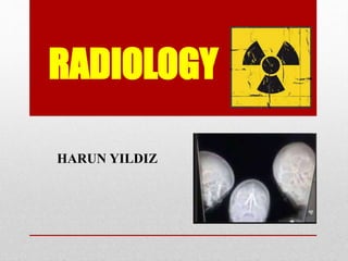
3. 1. Radiology
Radiology is a medical specialty. It uses imaging to
diagnose and heal disease. To diagnose or treat diseases,
radiographers use a variety of imaging techniques
such as X-ray radiography, fluoroscopy, computed
tomography (CT), nuclear medicine, ultrasound (US)
and magnetic resonance (MR).
3/16
4. 1. Radiology
Medical imaging is usually carried out by the radiographer.
Then, radiologist interprets or "reads" the images and
produces a report of their findings and diagnosis.
This report is then transmitted to the physician who ordered
the imaging. Specialist physicians often look at images
themselves although are less expert than radiologists.
4/16
6. Projection Radiography
• Wilhelm Conrad Röntgen discovered X-rays in 1895
and received the first Nobel Prize in Physics for their
discovery in 1901. Radiographs (or roentgenographs) are
produced by transmitting X-rays through a patient. In
film-screen radiography, an X-ray tube generates a
beam of X-rays. The X-rays strike a plate of sensors that
converts the signals generated into digital information,
which is transmitted and converted into an image
displayed on a computer screen.
• Due to its availability, speed, and lower costs,
radiography is often the first-line test of choice in
radiologic diagnosis. It is used various types of bone
diseases, bone tumors (especially benign bone tumors),
fractures, congenital skeletal anomalies, etc.
6/16
7. Computed Tomography
• Tomography was the first invented by the French
physician Boccage in 1915. The first
commercially useful CT scanner was invented
by Sir Godfrey Hounsfield in 1972. Hounsfield
and Allan McLeod Cormack shared the Nobel
Prize for Medicine in 1979.
• It is a medical imaging device. It is capable of
images of cross-section between 1 mm and 10
mm. It provides three-dimensional (3D) images.
Radiation (X-ray) in Tomography is available.
According to projection radiology, radiological
patient is exposed to relatively more X-rays.
7/16
8. Magnetic Resonance
MR uses strong magnetic fields to make them parallel
hydrogen protons within body tissues. It uses a radio signal.
An advantage of MR is its ability to produce multi-
directional images. It provides three-dimensional (3D)
images. MR gives the best soft tissue. MR has great benefit
in imaging the brain, spine, and musculoskeletal system.
No radiation is involved. There are no side effects.
8/16
9. Ultrasound
Medical ultrasonography uses ultrasound (high-frequency
sound waves) to visualize soft tissue structures in the body in real
time. No radiation is involved. It is used to view internal organs
in the abdomen. There are no side effects. The first ultrasound
images were static and two-dimensional (2D), but with modern
ultrasonography, it can be observed as 3D and 4D.
9/16
10. Fluoroscopy
Fluoroscopy and angiography are special applications of X-
ray imaging. This allows real-time imaging of structures
with a radiocontrast agent. Radiocontrast agents (such as
barium sulfate) are usually administered by swallowing. It
was used for evaluation of the gastrointestinal system.
10/16
12. (1) Hair: Losing of hair.
(2) Brain: Since brain cells do not reproduce, radiation kills
nerve cells and small blood vessels.
(3) Thyroid: The thyroid gland is susceptible to radioactive
iodine. It can destroy all or part of the thyroid.
(4) Blood System: It reduces blood's white cell and then the
victim more susceptible to infection.
12/16
13. (5) Heart: damaging to small blood vessels and probably
causing heart failure.
(6) Gastrointestinal System: Radiation damages to the
intestinal system lining such as bloody vomiting and
diarrhea.
(7) Reproductive System: It causes sterile (no children)
13/16
15. • Heal: to treat, to cure
• Intervene: to become involved in a situation in
order to try to stop a disease
• Benign: not likely to kill you
• Fracture: to break something in the body
• Congenital: people have from when they are
born
• Diarrhea: your solid waste is more liquid than
usual
15/16
16. • Air travel round trip (London-New York): 40
microSv
• Chest X-ray: 100 microSv
Sv (sievert): a measure of the health effect of low levels of radiation on the human body
16/16