Basic Spine Introduction for Understanding Back Problems
•
26 likes•5,099 views

Evaluation of Lumbar Spine Disease starts with understanding the clinical back grounds. It starts with good history and physical examination. This is a teaching lecture given twice by Prof. Dr. Mohamed Mohi Eldin, professor of neurosurgery, in the Basic Spine Course, Egyptian Medical Syndicate, Cairo, March 2009 and in 2010.

Recommended
Recommended
More Related Content
What's hot
What's hot (20)
Viewers also liked
Viewers also liked (19)
Similar to Basic Spine Introduction for Understanding Back Problems
Similar to Basic Spine Introduction for Understanding Back Problems (20)
More from Prof. Dr. Mohamed Mohi Eldin
More from Prof. Dr. Mohamed Mohi Eldin (20)
Recently uploaded
Recently uploaded (20)
Basic Spine Introduction for Understanding Back Problems
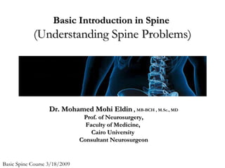
- 1. BBaassiicc IInnttrroodduuccttiioonn iinn SSppiinnee ((UUnnddeerrssttaannddiinngg SSppiinnee PPrroobblleemmss)) DDrr.. MMoohhaammeedd MMoohhii EEllddiinn ,, MB-BCH , M.Sc., MD PPrrooff.. ooff NNeeuurroossuurrggeerryy,, FFaaccuullttyy ooff MMeeddiicciinnee,, CCaaiirroo UUnniivveerrssiittyy CCoonnssuullttaanntt NNeeuurroossuurrggeeoonn Basic Spine Course 3/18/2009
- 5. Low back pain 15% 85% no specific specific pathology pathology NSLBP (mechanical)
- 6. “Mechanical” NSLBP • pain is worsened with movement • pain is improved with rest
- 7. Triage • Is the LBP due to serious pathology? • Duration of the LBP? • What treatment is indicated for the LBP?
- 8. Hypotheses generated about: • diagnosis • physical examination • treatment • prevention • contra-indications/precautions
- 9. Need knowledge about: • causes of LBP • pathology • tests (odds ratio, sensitivity and specificity) • treatment effects and efficacy
- 10. Serious spinal pathology • Cancer • Infection eg osteomyelitis • Cauda equina syndrome • Cord compression • Fracture (osteoporotic) • Inflammatory diseases/arthritides • Abdominal or cardio-thoracic pathology
- 11. Eliminate serious pathology (red flags) • unexplained weight loss • night pain • poor general health/systemic symptoms • fever • previous history of cancer • failure to improve with bed rest & therapy • history of trauma • steroid use (osteoporosis) • very severe pain/muscle spasm • Pain that worsens in supine • bowel/bladder frequency (cauda equina syndrome) • widespread neurological symptoms • non-mechanical behaviour of symptoms • Age > 50 years • Constant progressive non-mechanical pain • Persisting severe restriction of lumbar flexion
- 13. Typical Non-typical Presentation Presentation Kathryn Refshauge 13
- 14. Decisions • If suspect pathology, refer patient to appropriate health professional • If NSLBP, use knowledge (evidence-based practice) seriousnesss probability
- 15. Yellow flags • Previous history of LBP • Radiating leg pain, NR involvement • Poor fitness • Poor extensor endurance • Poor general health • Psychological distress (fear avoidance behaviour, depressed) • Much time lost from work • Disproportionate illness behaviour • Low job satisfaction • Personal problems (alcohol, marital, financial) • Adversarial medico-legal proceedings
- 16. Clinical Course acute sub-acute chronic 6 weeks 3 months Time acute sub-acute chronic most recover without intervention some recover very few recover psychosocial domain fear of activity acute sub-acute chronic Rx: spinal manual therapy McKenzie exercises spinal manual therapy exercises exercise cognitive behavioural therapy
- 17. Clinical Examination · observation · active movements · tension tests *** · palpation As applicable: · stress active movements · neurological examination · muscle performance · passive tests
- 19. multifidus
- 20. LLuummbbaarr SSppiinnee DDiisseeaassee • Low back pain is second to upper respiratory problems as a reason for visits to a physician • In the U.S., back pain is the most common cause of activity limitation in people younger than 45 years • Cost of low back pain to industry estimated $35- 75 billion
- 21. EEvvaalluuaattiioonn ooff LLuummbbaarr SSppiinnee DDiisseeaassee • Where to start? • What do we know? • What to do? • Who to consult? • What will they do?
- 22. What we know v. wwhhaatt wwee tthhiinnkk wwee kknnooww** % answering % very Topic ?s correctly confident SI joint pain 4.4 32.2 Lumbar stenosis 12.6 28.6 Leg length differences 42.0 27.0 Fibromyalgia 57.1 35.5 Myofascial pain (piri) 68.7 8.5 *J Am Geriatr Soc 54:1772-1777; 2006
- 23. EEvvaalluuaattiioonn ooff LLuummbbaarr SSppiinnee DDiisseeaassee From the top • Patient History: – Location of pain – Duration of pain – Character/quality of pain – Weakness – Numbness
- 24. Evaluation ooff LLuummbbaarr SSppiinnee DDiisseeaassee • Bowel, bladder or sexual dysfunction • Prior or current treatments including medication • Smoking (smokers complain of more severe symptoms and have less improvement postsurgically) • Obesity (obese patients more likely to suffer radicular pain or neurologic symptoms and carry more comorbidities) • Diabetes (may need neurophysiology testing) • Psychological factors: anxiety, depression, somatization symptoms, stressful responsibilities, job dissatisfaction, mental stress at work, negative body image, weakness in ego functioning (prospective predictors of developing back pain) • Activities that affect pain (e.g. leaning forward in spinal stenosis, sitting down, coughing, sneezing, Valsalva for herniated discs)
- 25. Evaluation ooff LLuummbbaarr SSppiinnee DDiisseeaassee • Physical Exam: – Strength – Sensation – Reflexes – Range of Motion – Palpation
- 26. Evaluation ooff LLuummbbaarr SSppiinnee DDiisseeaassee • Strength exam: – L3 - iliopsoas muscle (hip flexion), adductor longus (hip adduction) – L4 - quadriceps femoris (knee extension), tibialis anterior (dorsiflexion and inversion) • L5 - gluteus medius/minimus (thigh abduction and medial rotation), extensor hallicus longus (big toe extension), peroneus longus and brevis (plantar flexion and eversion) • S1 - gluteus maximus (thigh abduction), biceps femoris (hip extension), gastrocnemius (plantar flexion)
- 27. Evaluation ooff LLuummbbaarr SSppiinnee DDiisseeaassee LLoowweerr eexxttrreemmiittyy sseennssaattiioonn
- 28. EEvvaalluuaattiioonn ooff LLuummbbaarr SSppiinnee DDiisseeaassee • Reflexes: – L3 - iliopsoas reflex (meaningful?) – L4 - knee jerk – L5 - extensor hallicus reflex (meaningful?) – S1 - ankle jerk – Babinski - in adults, UMN lesion from motor strip to lower spinal cord
- 29. EEvvaalluuaattiioonn ooff LLuummbbaarr SSppiinnee DDiisseeaassee • Range of Motion: – Straight leg raise - most sensitive for sciatic pain syndromes – Pain in contralateral leg with straight leg raise is most specific for sciatic pain syndromes – Lumbar flexion/extension (lumbar stenosis worse with extension, better with flexion)
- 30. EEvvaalluuaattiioonn ooff LLuummbbaarr SSppiinnee DDiisseeaassee • ROM to rule out other causes of back/leg pain: internal and external hip rotation • Palpation over spine, SI joint, pelvis and hip
- 31. Evaluation ooff LLuummbbaarr SSppiinnee DDiisseeaassee • Clinical impression: – lumbar disc herniation: pain, paresthesias, weakness, depressed DTRs in an anatomic distribution (i.e. down lower extremity) – lumbar stenosis: diagnosis made mainly by history; low back/leg pain with walking or standing improved by sitting or lying down (not just standing still); no severe cramping in calf; no trophic changes in skin; a.k.a. neurogenic claudication
- 32. Evaluation ooff LLuummbbaarr SSppiinnee DDiisseeaassee • Clinical impression: – lumbar instability: pain with motion; improved with lying down; >5 mm motion on flexion/extension x-rays indicates unstable motion segment; look for defects in neural arch (lamina, pedicle, pars interarticularis); 30% of patients with degenerative spondylolisthesis (subluxation) will have progressive slippage – compression fracture: acute to subacute onset of pain, pain to palpation; +/- history of trauma/cancer – musculoskeletal: pain with active but not passive motion; point tenderness over joint; +/- history of trauma
- 33. Evaluation ooff LLuummbbaarr SSppiinnee DDiisseeaassee • If clinical suspicion high for ‘soft tissue’ (i.e. muscle, tendon, joint, ligament) source of symptoms then: NSAIDS, narcotics, antidepressant, cox-2 inhibitor, PT (exercise), +/- muscle relaxants, +/- chiropractor referral, +/- acupuncture, +/- behavioral therapy, ? corsets, ?massage, ?traction, ?TENS, ?epidural/facet injections, BUT…continue ordinary activities in the acute period* AND in the post-acute period begin conditioning activities to strengthen back, legs, abdomen to prevent recurrence^ • +/- = some evidence; ? = unknown • *NEJM 332:351-5, 1995 • ^JAMA 272:1286-91, 1994
- 34. Evaluation ooff LLuummbbaarr SSppiinnee DDiisseeaassee • Options when clinical suspicion low or diagnosis unclear: – 1. Observe (80-90% will resolve in <6 weeks) • most common diagnosis of acute (i.e. <6 weeks) back pain = “lumbar strain” • pathobiology ( any pain sensitive structure): muscle, tendon, ligaments, disc, facet joints, periosteum, meninges, blood vessels, or ‘degenerative changes*’ • NSAIDS, narcotics, antidepressant, cox-2 inhibitor, PT while you are observing – 2. Imaging • BUT ASK THE PATIENT: are you willing to have surgery or other invasive procedure if we do this work up? • For back pain pts: 4% will have compression fx, 1% will have a tumor, 3% will have a herniated disc
- 35. Imaging ooff LLuummbbaarr SSppiinnee DDiisseeaassee • If clinical suspicion high for intraspinal source of symptoms – i.e. radiculopathy, neurogenic claudication, lumbar instability, compression fx then: – 1) MRI, MRI, MRI unless there is a contraindication (see next slide) • Add contrast only if patient has had prior surgery or a history of cancer; perhaps with a demyelinating process like multiple sclerosis • If not sure; order without contrast and radiology will pick up the ones that do need it
- 36. Imaging ooff LLuummbbaarr SSppiinnee DDiisseeaassee – 2) If there is a contraindication to MRI then CT myelogram (contraindications to MRI = heart stent < 2 weeks old, defibrillator, pacemaker, pain pump, spinal cord or deep brain stimulator, prior lumbar spine instrumentation, programmable shunt) • Questions?: call radiology or specialist involved in placing device or hardware • If patient is too large for closed MRI then order open MRI • CT is WAY OVERUTILIZED as a spine diagnostic test and delivers A LOT of radiation to the patient
- 37. Imaging ooff LLuummbbaarr SSppiinnee DDiisseeaassee – 3) if signs of spondylolisthesis then flexion/extension x-rays (lateral) – 4) pain medications (NSAIDS, narcotics, +/- oral steroid taper, +/- muscle relaxant) – 5) Consultation after MRI or CT myelogram results show something other than ‘degenerative changes’*
- 38. Consultation aatt tthhee SSppiinnee CCeenntteerr • ‘Who’ should I send ‘what’ to? • General recommendations: – Acute pain problems – surgeons & pain management • Surgeons – usually after imaging – Active smokers will be strongly encouraged to stop – Poorly controlled diabetics (Hgb A1C > 7) will result in re-evaluation request with primary care prior to surgery • Pain management – does not require imaging – Chronic pain problems – physiatry & neurology • Does not require imaging
- 39. SSPPOORRTT SSttuuddyy** • Conservative therapy isn’t the worst idea for patients with a herniated disc and mild to moderate symptoms • 2-year prospective randomized trial of patients with radicular symptoms > 6 weeks and imaging evidence of a herniated disc – Randomized to surgery or PT, exercise, NSAIDS • LOTS of patients cross-over to opposite group if symptoms are too mild or too severe • BUT at 2-year follow-up, both surgery and conservative management was effective • *JAMA 2006;296:2451-2459
- 40. SShhoouulldd II rreeiimmaaggee?? • Have symptoms or signs changed significantly? • Has there been a recent intervention (e.g. surgery) or trauma? • Look at patient’s chart – has it been >1 year since last imaging? • If the answer to these 3 questions is “no” then reimaging is not indicated
- 41. SSuummmmaarryy • Start with good history and physical • Is this emergent, urgent or routine? • Is the cause most likely disc, stenosis, instability, compression fracture or soft tissue? • Typically start conservative and escalate as necessary
- 42. CClliinniiccaall HHiissttoorryy and Physical Examination on Spine Injury (Part I)
- 43. The Importance of History and Physical Examination • The most valuable service the correct diagnosis the magnitude of the problem the appropriate treatment
- 44. Image studies • Image studies Vs. Time-consuming process of history taking and P.E. • High false positive rate for spinal disease • No information about the source of pain • To confirm the diagnosis • To help guide any surgical procedure
- 45. History • Structural spine: vertebrae, joints Symptoms: axial • Neurologic spine: cord, cauda equina, nerve root Symptoms: peripheral, radicular
- 46. History • Backache In the lumbosacral junction? In the thoracolumbar junction? In the buttock and thigh? • Sciatica?
- 47. History • The ratio of back pain to leg pain symptoms • The pain intensity on a scale 1 to 10 • Functional impairment: Stable Deterioration • Psychosocial issues
- 48. Axial Symptoms: Back and Neck Pain • To characterize the nature of the pain Location Onset Duration Character Periodicity The precipitating factors The aggravating factors The relieving factors
- 49. Location • Local or diffuse, midline or paraspinous • Midline pain: spondylolisthesis or bony pathology • Paraspinous pain: muscular and spasm • Focal, highly localized pain: fracture, tumor, infection or single-level arthrosis or instability • Diffuse symptoms: DDD • Chronic, diffuse symptoms are seldom likely to warrant surgical treatment
- 50. Onset • Acute onset: acute injury • Insidious onset: repetitive trauma, degenerative disease or a progressive disorder • Insidiously but progressive rapidly: more serious underlying causes Pathological fracture: tumor, infection, or osteoporosis, Visceral disease: pancreatitis, AAA
- 51. Duration • Sprain/strain causing backache usually improves within 6 to 8 weeks of onset • Degenerative disease pain waxes and wanes over a period of years or decades • New pains, or pains that are new to longstanding backache
- 52. Character • Most backache: fairly focal pain intensified by activity and fatigue, improved by rest • Neoplasm/infection: boring, deep pain unrelieved by rest or recumbency • Instability: sharp, stabbing, incapacitating pain superimposed on a baseline ache a shift or “catch” with motion
- 53. Character • Discogenic pain: intensified by sitting and vibration exposure, flexion/extension, and axial loading • Discitis, osteomyelitis greater intensity, absolute intolerance of motion
- 54. Periodicity • Symptoms recur more frequently • To miss work several times in a given year Need further evaluation and more aggressive treatment
- 55. Factors that Precipitate, Aggravate, Relieve pain • Flexion: aggravate disc-related symptoms • Extension: irritate the facets • Motion: trigger instability, causing acute giving out or stabbing pain. • Whole body vibration: precipitate neuropeptide release that can sensitize nerve endings and directly irritate the disc
- 56. Factors that Precipitate, Aggravate, Relieve pain • Mechanical disorder: Pain caused by bending, lifting, twisting, or axial loading, and relieved by recumbancy • Discogenic pain: Pain aggravated by flexion, or by prolonged sitting or riding in a car Back pain caused by hypertension may be facet-related Leg pain in extension usually is a result of spinal stenosis
- 57. Factors that Precipitate, Aggravate, Relieve pain • Profound morning stiffness: requiring 30 mins to an hour to “loosen up” (inflammatory arthropathy) • Inquires about injury and the circumstances associated with the pain first appearing • Did pain come on immediately after an accident? • Was there an examination or radiography at that time?
- 58. Peripheral Symptoms: Arm and Leg Pain • Radiculopathy: Painful, hyperesthetic, numb, tingling, burning • HIVD Vs. Central spinal stenosis • Thoracic spine disorder: Belt-like radicular symptoms Herpes zoster; the pain is severe and predates the vesicles
- 59. Peripheral Symptoms: Arm and Leg Pain • Neurogenic Vs. Vascular claudication (The spine in flexion or extension) Walk Stand still Sit Uphill Downhill Pedaling a bicycle Lean forward over a shopping cart (on a counter)
- 60. Peripheral Symptoms: Arm and Leg Pain • Radicular pain reproduced by coughing, sneezing, or straining at stool (increasing intrathecal pressure) • Lhermitte’s sign • Resting the forearm over the head
- 61. Peripheral Symptoms: Arm and Leg Pain • Loss of bowel continence, urinary retention, and saddle anesthesia (accompanied by varying degrees of leg weakness) ---Cauda Equina Syndrome • Spasticity, and urinary incontinence (diffuse lower and upper extremity weakness) ---Spinal Cord Injury
- 62. Clinical History and Physical Examination on Spine Injury (Part II)
- 66. Low Back Pain (LBP) • 90% of all Americans • Minor insultsmajor injuries • Maintain normal lordotic and kyphotic curves to avoid injury
- 67. Clinical Anatomy • 5 vertebrae=lumbar spine • P.320, fig. 10-2 – Facets – Processes – Foramen – “Scotty Dog”
- 68. Evaluation • Primary role of ATC: – On-field evaluation: • Rule out (R/O) bony trauma which has, or may, damage to spinal cord – Clinical evaluation: • Evaluate specific cause of injury and devise a rehabilitation plan
- 69. History • Location of pain: – Localized or radiating? • Onset of pain: – Acute, chronic, insidious? • Consistency of pain: – Constant/intermittent? – Improves/Worsens with activity? • Mechanism: – Flex, ext, rotation, lat. Flex – Direct blow/trauma
- 70. History • PMH of injuries/surgery? • Smoker? • Bowel/bladder symptoms? – Incontinence or Ý frequency – Immediate referral • Referral history – Time in the medical system? – # of physicians seen?
- 71. Inspection/Observation • Sagittal curvature • Scoliosis • Frontal curvature • Normal curves • Standing posture • Shoulders • Head • Walking posture (gait)
- 72. Observation/ Inspection • Paravertebral muscles • Symmetry / spasm • PSIS level • Overall attitude
- 73. Palpation • Transverse processes • Spinous processes • PSIS • Paravertebral musculature – Symmetry – spasm
- 74. Functional testing • Gross ROM assessment only • Trunk Extension = 45º – Lordosis should increase • Trunk Flexion = 9045º – Lordosis should decrease • Rotation • Lateral flexion • Symmetry > Goniometry
- 75. Pathologies/Injuries • Muscle strains—p.353 • Facet joint syndrome-p. 353 • Disk lesion—p. 354 • Spondylopathies— p.292
- 76. Muscle Strains • Pain localized to paraspinal musculature & PSIS • Spasm probable • Limited flex. & ext. (pain) • No radiating pain • May not correlate to specific mechanism
- 77. Facet Joint Syndrome • Table 10-10,p.354 • ~40% of all LBP • Vague symptoms that mimic other pathologies • Common with repeated spine-loading activities • Localized pain • Often improves with activity • Nerve entrapment may result from compensatory posturing • Worsened by: – Repeated spine-loading activities (ext, side bending, rotation) – Poor LE flexibility – Poor Trunk strength
- 78. Disk lesion • Crack in annulus fibrosus herniation of nucleus pulposus • Pressure on nerve rootpain/burning sensation • “Bulge” ¹ pathology • Radiating pain into buttocks and down leg • MRI for best diagnosis • Altered standing posture • Symptoms Ý with activity • Bilateral or unilateral symptoms • Usually acute onset
- 79. Spondylopathies • Vertebral defect • May occur at any age/sports • Congenital? • Stress fx? • Common is sports with forced hyperextension • Generally occurs at L4- L5 or L5-S1 levels
- 80. Spondylolysis • Defect at pars interarticularis • Unilateral or bilateral • Signs/ Symptoms: – NL spinal alignment – LBP Ý during & after activity – Localized lumbar spine pain – NL flex; restricted ext. – (-) neuro. Test • X-rays show “collared” Scotty Dog
- 81. Spondylolysthesis • May occur with spondylolysis • Anterior displacement of proximal vertebrae on distal • Pain more intense/constant than spondylolysis • Neuro signs sometimes (+) if displacement worsens • Possible step-off deformity • X-rays show “decapitated” Scotty Dog • (+) Stork test
- 82. Straight leg raise test (SLR)— p.347, fig. Box 10-9 • Supine with knees extended • PROM hip flexion to point of discomfort or end of range • ß hip flexion and move into passive dorsiflexion • (+) = pain reproduced and recurs with reduced SLR • (-) =pain reproduced but does not return with reduced SLR • If pain does not recur: – Tight hamstrings
- 83. Well-leg SLR test p.348, Box 10-10 • Supine with knees extended • Passively raise one leg – Similar to SLR test – Raise leg with symptoms – Provocation test • (+)=Symptoms felt in the other leg (“well” leg)
- 84. Valsalva maneuver p. 344, Box 10-6 • Increasing intrathecal pressure to reproduce symptoms • (+)=Reproduced symptoms : Radiating pain or Numbness
- 85. Kernig’s Test—p. 346 • Box 10-8 • Provocation test to elongate the spinal cord • Active SLR until point of pain (knee straight) • Flex knee @ point of pain • (+)= pain in LB or radiating pain in LE • Brudzinski’s Test=Kernig with cervical flexion
- 86. Hoover test p.351, Box 10-13 • Tests compliance & effort • “Malingering” • Procedure: – Supine with knees extended – Active hip flexion – Pressure should be felt on opposite leg as SLR is attempted • (+)=No pressure=low effort
- 87. Babinski test p. 383, Box 11-3 • Tests presence of upper motor neuron pathology • Blunt device moved across plantar aspect of foot from calcaneus to 1st metatarsal head (great toe) – (-)=toe flexion – (+)=great toe extension with splaying of other toes • Normally (+) in newborns
- 88. Hamstring flexibility • Tripod sign • 90-90 position for testing • Tight hamstrings pelvic tilt Stretched extensors Pain/spasm
- 89. Strength tests • Isometric strength tests • Held for 60 sec. • Flexor strength testing • Extensor strength testing
- 90. Lifting Technique • Maintain natural curves – Sitting, standing, walking, lifting • 10:1 ratio • Use large LE muscles • Keep items close to body • Hip = axis (not LS) • Avoid rotating spine • Get help when needed
Editor's Notes
- I have here 2 diagrams of patients who presented to me with LB and leg pain. With every patient, we take a history that begins with getting a description of the symptoms: what symptoms the patient has, where the symptoms are located, a description of the type of symptoms and the intensity. The patient on the left has pain in the L/S and leg. The back pain is worse than the leg pain. This is a typical presentation of NSLBP. With our knowledge of pain patterns and anatomy, we know that the pain is likely to arise from either the L/S or the SIJ. Without any further information we already have hypotheses about the diagnosis, physical examination and treatment. We then get a lot more information from the history to check whether these hypotheses are correct. On the right, the patient has a more complex pattern of LBP and leg pain. The pain is worse in the leg, and both legs are involved. This is a very unusual pattern of pain, and is unlikely to arise from a single cause, unless it is a large tumour in the spinal canal, compressing the spinal cord. Our hypotheses for this patient, even at this stage in the history, would include serious pathology or some people with chronic pain have this kind of presentation. It is very unlikely that this patient has acute NSLBP.