SlideShare a Scribd company logo
1 of 24
Download to read offline
1 
ACUTE LYMPHOCYTIC LEUKEMIA BY DR. MAGDI AWAD SASI 2014 
ACUTE LYMPHOCYTIC LEUKEMIA Acute lymphoblastic leukemia (ALL) is a malignant (clonal) disease of the bone marrow in which early lymphoid precursors proliferate and replace the normal hematopoietic cells of the marrow. ALL is the most common type of cancer and leukemia in children in the United States. INCIDENCE: Acute leukaemia is an uncommon type of cancer. About 7,600 people are diagnosed with leukaemia each year in the UK. Of these, only about 650 people have acute lymphoblastic leukaemia. Acute lymphoblastic leukaemia((ALL)) is the most common type of cancer to affect children. Approximately 1 in every 2,000 children will develop it. About 85% of cases occur in children aged under 15, with the majority of cases developing in those between the ages of two and five years old. SYMPTOMS:  Acute lymphocytic leukemia (ALL) progress much faster and symptoms may worsen more quickly than chronic lymphocytic leukemia.  Patients with ALL present with either symptoms relating to direct infiltration of the marrow or other organs by leukemic cells, or symptoms relating to the decreased production of normal marrow elements.  Some leukemia symptoms, like night sweats, fever, fatigue and achiness, often resemble flu-like symptoms. If you have the flu, symptoms will likely subside as you get better. Make an appointment to see your doctor if the symptoms persist longer than expected.  Some general symptoms of leukemia include: 
Fever, chills ,Night sweats 
Fatigue, weakness 
Loss of appetite, weight loss 
Bone/joint pain, Body aches 
Abdominal discomfort 
Headaches 
Shortness of breath 
Frequent infections 
Easy bruising or bleeding 
Petechiae (small red spots under the skin) Fever is one of the most common symptoms of ALL, and patients with ALL often have fever without any other evidence of infection. However, in these patients, one must assume that all fevers are from infections until proved otherwise, because a failure to treat infections promptly and aggressively can be fatal. Infections are still the most common cause of death in patients undergoing treatment for ALL.
2 
ACUTE LYMPHOCYTIC LEUKEMIA BY DR. MAGDI AWAD SASI 2014 
Patients with ALL often have decreased neutrophil counts, regardless of whether their total white blood cell (WBC) count is low, normal, or elevated. As a result, these individuals are at an increased risk of infection. The prevalence and severity of infections are inversely correlated with the absolute neutrophil count (ANC), which is defined as the number of mature neutrophils plus bands per unit of volume. Infections are common when the absolute neutrophil count is less than 500/μL, and they are especially severe when it is less than 100/μL. 
Symptoms of anemia are common and include fatigue, dizziness, palpitations, and dyspnea upon even mild exertion. Other patients present with signs of bleeding. Bleeding can be the result of thrombocytopenia due to marrow replacement. Additionally, approximately 10% of patients with ALL have disseminated intravascular coagulation (DIC) at the time of diagnosis. These patients may present with hemorrhagic or thrombotic complications. 
Some patients present with palpable lymphadenopathy. Others, particularly those with T- cell ALL, present with symptoms related to a large mediastinal mass, such as shortness of breath. 
Infiltration of the marrow by massive numbers of leukemic cells frequently manifests as bone pain. This pain can be severe and is often atypical in distribution. 
About 10-20% of ALL patients may present with left upper quadrant fullness and early satiety due to splenomegaly. 
Although patients may present with symptoms of leukostasis (eg, respiratory distress, altered mental status) because of the presence of large numbers of lymphoblasts in the peripheral circulation, leukostasis is much less common in people with ALL than those with acute myelogenous leukemia (AML), and it occurs only in patients with the highest WBC counts (ie, several hundred thousand per μL). 
Patients with a high tumor burden, particularly those with severe hyperuricemia, can present in renal failure. Other potential signs and symptoms of ALL may include: 
Bleeding gums 
Frequent infections 
Nosebleeds 
Easy bruising 
Swollen lymph nodes around the neck, underarm, stomach or groin 
Shortness of breath 
Weight loss 
HEPATOMEGALLY AND SPLENOMEGALLY: 
 50% to 75% of patients with ALL & rare in AML. 
 The visceral involvement can produce symptoms of nausea , abdominal fullness and satiety. 
 Lymphoadenopathy – ALL ˃AML 80%// 50% 
 An anterior mediastinal mass ---T cell variant of ALL
3 
ACUTE LYMPHOCYTIC LEUKEMIA BY DR. MAGDI AWAD SASI 2014 
 Practical point—extramedullary leukemia can precede detectable involvement of B.M. 
EXTRAMEDULLARY TISSUES : 
Acute leukemia may infiltrate –skin, lung, eye, kidney ,nasopharynx 
Testicular involvement is common in male with ALL. 
CHLOROMAS----soft tissues masses of leukemic cells. 
SYMPTOMS RELATED TO THE EXPANDING MALIGNANT CELL MASS: 
Bony pain and sternal tenderness. 
This occur in half of patients. 
Osteolytic lesions are rare. 
Renal abnormalities due to: 
A. Leukemic infiltration 
B. Ureteral obstruction by urate nephropathy—enlarged LN, uric acid stone, infections ,haemorrhage GIT----bleeding, distention ,satiety ,constipation((due to organomegally or leukemic infiltrate)) . 
CNS SYMPTOMS: 
Cells infiltrate in subarachnoid space leads to leukemic meningitis or direct involvement of the brain or spinal cord parenchyma. 
Neurological symptoms are unusual at the time of diagnosis. 
CNS is a frequent site of relapse. ((particularly ALL )) 
Leukemic meningitis— 
1st symptoms---------headache and nausea 
With progression---- papillodema , cranial nerve palsies ,seizures ,altered mentation 
CSF----------------------- leukemic blast cell, increase protein, decrease glucose.
4 
ACUTE LYMPHOCYTIC LEUKEMIA BY DR. MAGDI AWAD SASI 2014 
METABOLIC DISTURBANCE : 
Hypokalemia and hyponatremia due to renal involvement . 
LDH—lactic dehydrogenase may be increased. Physical Examination:  Patients with ALL commonly have physical signs of anemia, including pallor and a cardiac flow murmur. Fever and other signs of infection, including lung findings of pneumonia, can also occur. Fever should be interpreted as evidence of infection, even in the absence of other signs.  Patients with thrombocytopenia usually demonstrate petechiae, particularly on the lower extremities. A large number of ecchymoses is usually an indicator of a coexistent coagulation disorder such as DIC.  Signs relating to organ infiltration with leukemic cells and, to a lesser degree, lymphadenopathy may be present.  Occasionally, patients have rashes that result from infiltration of the skin with leukemic cells. 
CLASSIFICATION OF ACUTE LEUKEMIA: 
1. FAB classification(French-American-British) :1970 
A classification of acute leukemia produced by a three-nation joint collaboration. 
A schema that divides acute leukemia into lymphoid–ALL or myeloid–AML cell lines. 
This is based on the microscopic features and cytochemistry of blast cells. 
Acute lymphoblastic leukemia is subdivided into three types(L1-L3). 
Of childhood ALL, 70% are predominantly L1, 27% are L2, and 3% or less are L3 or Burkitt cell type. 
In adults with ALL------ 30% are L1, 65% are L2, and 5% are L3. 
FAB classification, acute leukemia: Acute lymphocytic leukemia (ALL) 
L1 Small monotonous lymphocytes 
L2 Mixed L1- and L3-type lymphocytes 
L3 Large homogeneous blast cells 
ALL 
Disease of children 80% and young adults 
Boys more than the girl, whites more than non whites
5 
ACUTE LYMPHOCYTIC LEUKEMIA BY DR. MAGDI AWAD SASI 2014 
L1 
85% of children. 
Mild to moderate basophilia Cells are small , twice diameter of small lymphocytes 
Homogenous nuclear chromatin. 
Regular nuclear membrane. 
Small or no nucleolus. 
Scanty cytoplasm without vacuoles 
B cell.—pro B or pre B lineage 
Common ALL ((CALLA)) 
Tdt—postive----CD19 & CD10 
MPO is always negative 
L2 
15% adults. 
.Cells are larger, cleafted nuclei. .Variable nuclear chromatin. .An irregular nuclear shape. 
.Low nuclear cytoplasmic ratio. 
.More pleomorphic size and shape. 
.One or more prominent nucleoli. 
T cell/ B cell-pro B , pre B lineage Tdt positive , basophilia 
Anterior mediastinal mass 
High leukocyte count 
CD4 , CD5 ,CD1 ,CD2 , CD8 
L3 
Uncommon Large, homogeneous cells with fine stippled chromatin; regular nuclei; prominent nucleoli. 
˂5%, Large vasicular nuclei with abundant deeply basophilic cytoplasm The most distinguishing feature is prominent Cytoplasmic vacoulation. 
Have high mitotic index 
Leukemic form of burkitts lymphoma 
3 – 4 times diameter of lymphocytes 
Tdt negative 
Surface immunoglobulin positive,B CELL 
The vast majority of ALL ((80%)) have B antigen
6 
ACUTE LYMPHOCYTIC LEUKEMIA BY DR. MAGDI AWAD SASI 2014 
The leukemic blast (( B cell ALL )) express CD19 antigen. 
T lymphoblasts in patient with T cell ALL seem to be arrested at early intrathymic stages of maturation. 90% of cases have nuclear enzymes terminal deoxynucleotidyl transferase (Tdt). Tdt is absent in mature lymphocytes ,hairy cell leukemia , CLL . 
PROGNOSIS: 
Precursor B cell ALL 80% (( best )) 
T cell and pre B cell ALL (( intermediate )) 
B cell ( burkitts ) ((worst)) 
The major immunophenotypic classification of ALL is as follows: 
B cell lineage- 
1. Pro B ALL 
2. Pre B ALL 
3. B (surface Ig +) ALL (Burkitt's) 
T cell lineage*- 
1. T cell ALL 
2. Prothymic 
3. Early thymocyte 
4. Cortical (common) thymocyte 
5. Medullary (mature) thymocyte 
6. Mature T (peripheral) cells 
 * T cell ALL is usually not subclassified 
 **** 2YSMD do not need to know the different antigens. You should recognize CD3, CD5, and CD7 as pan T markers; CD19 as a pan B marker; CD4 as T-helper, and CD8 as T- suppressor.**** Risk Factors for Acute Lymphoblastic Leukemia For most people, the cause of ALL is unknown. For this reason, there is no known way to prevent it. However, there are a few known risk factors for this type of leukemia. But it is not yet known whether these risk factors are actual causes of the disease: 
Exposure to high levels of radiation to treat other types of cancer
7 
ACUTE LYMPHOCYTIC LEUKEMIA BY DR. MAGDI AWAD SASI 2014 
Exposure to certain chemicals such as benzene, a solvent used in oil refineries and other industries and present in cigarette smoke, certain cleaning products, detergents, and paint strippers 
Infection with human T-cell lymphoma/leukemia virus-1 (HTLV-1) in rarer cases outside the U.S. or Epstein-Barr virus (EBV), a related leukemia more commonly seen in Africa. 
Having an inherited genetic syndrome such as Down syndrome 
Being white 
Being male Laboratory tests for ALL include the following: 1. Complete blood count with differential 2. Peripheral blood smear 3. Coagulation studies 4. Chemistry profile, including lactic dehydrogenase, uric acid, liver function studies, and BUN/creatinine 5. Appropriate cultures (in particular, blood cultures, urine culture ,throat swab, stool culture ) in patients with fever or other signs of infection 6. Chest x-ray 7. Computed tomography 8. Multiple-gated acquisition scanning 9. Electrocardiography 10. Bone marrow aspiration and biopsy (definitive for confirming leukemia) 11. Immunohistochemistry 12. Flow cytometry 13. Cytogenetics 14. Polymerase chain reaction 15. Gene expression profiling 
National Comprehensive Cancer Network (NCCN) guidelines note that diagnosis of ALL generally requires the following :  Demonstration of ≥20% bone marrow lymphoblasts  Morphologic assessment of Wright/Giemsa–stained bone marrow aspirate smears  Hematoxylin and eosin (H&E)–stained bone marrow core biopsy and clot sections 
 Comprehensive flow cytometric immunophenotyping For optimal risk stratification and treatment planning in patients with ALL, the NCCN advises that bone marrow or peripheral blood lymphoblasts must be tested for specific recurrent genetic abnormalities, as follows : 1. Cytogenetics – Karyotyping of G-banded metaphase chromosomes 2. Interphase fluorescence in situ hybridization (FISH) 3. Reverse transcriptase polymerase chain reaction (RT-PCR) for fusion genes (eg, BCR-ABL) In addition, flow cytometric DNA index/ploidy testing can be done to assess for hyperdiploidy and hypodiploidy.
8 
ACUTE LYMPHOCYTIC LEUKEMIA BY DR. MAGDI AWAD SASI 2014 
CBC  A complete blood cell (CBC) count with differential demonstrates anemia and thrombocytopenia to varying degrees in individuals with (ALL). Patients with ALL can have a high, normal, or low white blood cell (WBC) count, but they usually exhibit neutropenia. The prevalence and severity of infections are inversely correlated with the absolute neutrophil count (ANC); infections are common when the absolute neutrophil count is less than 500/μL, and they are especially severe when it is less than 100/μL. Coagulation studies and chemistry profiles:  Abnormalities in the prothrombin time (PT) / activated partial thromboplastin time (aPTT) / fibrinogen / fibrin degradation products may suggest concomitant disseminated intravascular coagulation (DIC), which results in an elevated PT, decreased fibrinogen levels, and the presence of fibrin split products.  A review of the peripheral blood smear confirms the findings of the CBC count. Circulating blasts are usually seen. Schistocytes are sometimes seen if DIC is present.  A chemistry profile is recommended. Most patients with ALL have an elevated lactic dehydrogenase level (LDH), and they frequently have an elevated uric acid level. In addition, liver function tests and blood urea nitrogen (BUN)/creatinine determinations are necessary before the initiation of therapy. Cultures  Appropriate cultures, in particular blood cultures, should be obtained in patients with fever or with other signs of infection without fever. Imaging  Chest x-ray films may reveal signs of pneumonia and/or a prominent mediastinal mass in some cases of T-cell acute lymphoblastic leukemia (ALL).  Computed tomography (CT) scans can further define the degree of lymphadenopathy in some patients, including those with mediastinal masses.  Multiple-gated acquisition (MUGA) scans or electrocardiograms (ECGs) are needed when the diagnosis of acute lymphoblastic leukemia (ALL) is confirmed, because many chemotherapeutic agents used in the treatment of acute leukemia are cardiotoxic.  An ECG is recommended before the initiation of treatment. Bone Marrow Aspiration and Biopsy:  Bone marrow aspiration and biopsy are the definitive diagnostic tests to confirm the diagnosis of leukemia. Immunophenotyping helps to elucidate the subtype.  Aspiration slides should be stained for morphology with either Wright or Giemsa stain. The diagnosis of ALL is made when at least 30% lymphoblasts [FAB] classification) or 20% lymphoblasts ( [WHO] classification) are present in the bone marrow and/or peripheral blood.  In addition, slides should be stained with myeloperoxidase (MPO) (or Sudan black) and terminal deoxynucleotidyl transferase (TdT), unless another method is used, such as flow cytometry.  Bone marrow samples should also be sent for flow cytometry and cytogenetics. Approximately 15% of patients with ALL have a t(9;22) translocation (ie, Philadelphia [Ph] chromosome), but other chromosomal abnormalities may also occur, such as t(4;11), t(2;8), and t(8;14).
9 
ACUTE LYMPHOCYTIC LEUKEMIA BY DR. MAGDI AWAD SASI 2014 
Giemsa stain 
Trephine biopsy
10 
ACUTE LYMPHOCYTIC LEUKEMIA BY DR. MAGDI AWAD SASI 2014 
Histological Features: The older, traditional classification of (ALL) is the French-American-British (FAB) classification. This has now been replaced by the newer World Health Organization (WHO) classification but the FAB system is listed for historical purposes, as follows:  L1 – Small cells with homogeneous chromatin, regular nuclear shape, small or absent nucleolus, and scanty cytoplasm; subtype represents 25-30% of adult cases  L2 – Large and heterogeneous cells, heterogeneous chromatin, irregular nuclear shape, and nucleolus often large; subtype represents 70% of cases (most common)  L3 – Large and homogeneous cells with multiple nucleoli, moderate deep blue cytoplasm, and cytoplasmic vacuolization that often overlies the nucleus (most prominent feature); subtype represents 1-2% of adult cases WHO classification of ALL : B lymphoblastic leukemia/lymphoma: B lymphoblastic leukemia/lymphoma, NOS B lymphoblastic leukemia/lymphoma with recurrent genetic abnormalities  B lymphoblastic leukemia/lymphoma with t(9;22)(q34;q11.2), BCR-ABL1  B lymphoblastic leukemia/lymphoma with t(v;11q23); MLL rearranged  B lymphoblastic leukemia/lymphoma with t(12;21)(p13;q22) TEL-AML1 (ETV6-RUNX1)  B lymphoblastic leukemia/lymphoma with hyperdiploidy  B lymphoblastic leukemia/lymphoma with hypodiploidy  B lymphoblastic leukemia/lymphoma with t(5;14)(q31;q32) IL3-IGH  B lymphoblastic leukemia/lymphoma with t(1;19)(q23;p13.3) TCF3-PBX1 The WHO classifies the L1 and L2 subtypes of ALL as either precursor B lymphoblastic leukemia/lymphoblastic lymphoma or precursor T lymphoblastic leukemia/lymphoblastic lymphoma depending on the cell of origin. The L3 subtype of ALL is included in the group of mature B-cell neoplasms, as the subtype Burkitt lymphoma/leukemia. Immunohistochemistry A negative myeloperoxidase (MPO) stain and a positive terminal deoxynucleotidyl transferase (TdT) is the hallmark of the diagnosis of most cases of (ALL). However, positive confirmation of lymphoid (and not myeloid) lineage should be sought by flow cytometric demonstration of lymphoid antigens, such as CD3 (T-lineage ALL) or CD19 (B- lineage ALL), in order to avoid confusion with some types of myeloid leukemia (eg, M0), which also stain negative with myeloperoxidase . Polymerase Chain Reaction or Cytogenics Although more than 95% of cases of the L1 or L2 subtype of (ALL) are positive for Terminal deoxynucleotidyl transferase (TdT), TdT is not specific for ALL; TdT is absent in L3 (mature B-cell) ALL. However, TdT helps to distinguish ALL from malignancies of more mature lymphocytes (ie, non-Hodgkin lymphoma [NHL]). In cases of acute leukemia that are myeloperoxidase (MPO) negative and TdT positive, the distinction between (AML) and ALL is made on the basis of flow cytometry results. Patients with AML demonstrate myeloid markers such as CD33, whereas patients with ALL demonstrate lymphoid markers. Further confusion arises because some patients with ALL have aberrant expression of myeloid markers, such as CD13. However, if the cells are TdT positive, MPO negative, CD33 negative, and demonstrate lymphoid markers, the leukemia is considered ALL.
11 
ACUTE LYMPHOCYTIC LEUKEMIA BY DR. MAGDI AWAD SASI 2014 
Cytogenetic abnormalities occur in approximately 70% of cases of ALL in adults. These abnormalities include balanced translocations as occur in cases of AML. However, abnormalities of chromosome number (hypodiploidy, hyperdiploidy) are more common in ALL than in AML. Studies for bcr-abl analysis by (PCR) or cytogenetics may help distinguish patients with Philadelphia chromosome–positive (Ph+ ALL) from those with the lymphoid blastic phase of (CML). Most patients with Ph+ ALL have the p190 type of bcr-abl, whereas patients with lymphoid blastic CML have the p210 type of bcr-abl. 
Common Cytogenetic Abnormalities in ALL 
Abnormality Genes Involved 3-Year Event-Free Survival t(10;14)(q24;q11) HOX11/TCRA 75% 6q Unknown 47% 14q11 TCRA/TCRD 42% 11q23 MLL 18-26% 9p Unknown 22% 12 TEL 20% t(1;19)(q23;p13) PBX1/E2A 20% t(8;14)(q24;q32) t(2;8)(p12;q24) t(8;22)(q24;q11) c-myc/IGH IGK/c-myc c-myc/IGL 17% 80% t(9;22)(q34;q11) bcr-abl 5-10% 66%‡ t(4;11)(q21;q23) AF4-MLL 0-10%
12 
ACUTE LYMPHOCYTIC LEUKEMIA BY DR. MAGDI AWAD SASI 2014 
Eighty-five percent of cases of ALL are derived from B cells. The primary distinction is among the following (see below):  Early (pro-B) ALL, which is TDT positive, CD10 (CALLA) negative, surface immunoglobulin (Ig) negative  Precursor B ALL, which is TDT positive, CD10 (CALLA) positive, surface Ig negative  Mature B cell (Burkitt) ALL, which is TdT negative, surface Ig positive. Fifteen percent of these cases are derived from T cells. Immunophenotyping of ALL Cells – ALL of B-Cell Lineage (85% of cases of adult ALL) ALL Cells TdT CD19 CD10 CyIg SIg Early B-precursor ALL + + - - - Pre–B-cell ALL + + + + - B-cell ALL - + +/- +/- + ALL = acute lymphoblastic leukemia; Cylg = Cytoplasmic immunoglobulin; SIg =Surface immunoglobulin; TdT = terminal deoxynucleotidyl transferase. These cases are subclassified into different stages corresponding to the phases of normal thymocyte development. The early subtype is surface CD3 negative, cytoplasmic CD3 positive, and either double negative (CD4-, CD8-) or double positive (CD4+, CD8+). The latter subtype is surface CD3 positive, CD1a negative, and positive for either CD4 or CD8, but not both. Immunophenotyping of ALL Cells – ALL of T-Cell Lineage (15% of cases of adult ALL) 
ALL Cells 
TdT 
Surface CD3 
CD4/CD8 Early T-precursor ALL + 
- +/+ or -/- T-cell ALL + 
+ +/- or -/+ Treatment for acute leukemia: Patient need hospital with appropriate blood product support facilities, leukapheresis capabilities, or physicians and nurses familiar with the treatment of patients with leukemia . (ALL) is best treated by physicians who have significant experience in the treatment of patients with acute leukemia(( haematologist)). The four components of ALL treatment are induction, consolidation, maintenance, and central nervous system (CNS) prophylaxis. Patients with ALL require hospital admission for induction chemotherapy, and they require readmission for consolidation chemotherapy or for the treatment of toxic effects of chemotherapy. Surgical intervention may be required for the placement of a central venous catheter, such as a triple lumen, Broviac, or Hickman catheter.
13 
ACUTE LYMPHOCYTIC LEUKEMIA BY DR. MAGDI AWAD SASI 2014 
Supportive Care - Blood Products Patients with (ALL) have a deficiency in the ability to produce normal blood cells, and they need replacement therapy. This deficiency is temporarily worsened by the addition of chemotherapy. All blood products must be irradiated to prevent transfusion-related graft versus host disease, which is almost invariably fatal. Packed red blood cells are given to patients with a hemoglobin level ˂ 7-8 g/dL or at a higher level if the patient has significant cardiovascular or respiratory compromise. Platelets are transfused if the count is less than 10,000-20,000/μL. Patients with pulmonary or gastrointestinal hemorrhage receives platelet transfusions to maintain a value ˃ 50,000/μL. Patients with central nervous system CNS hemorrhage are transfused to achieve a platelet count of 100,000/μL. Fresh frozen plasma is given to patients with a significantly prolonged prothrombin time (PT). Cryoprecipitate is given if the fibrinogen level is less than 100 g/dL. 
Supportive Care - Therapy and Prophylaxis for Infection  Antibiotics are given to all febrile patients.  At a minimum, include a third-generation cephalosporin (or equivalent), usually with an amino glycoside.  In addition to this minimum, other antibiotic agents are added to treat specific documented or possible infections.  Patients with persistent fever after 3-5 days of antibacterial antibiotics should have an antifungal antibiotic (liposomal or lipid complex amphotericin, new generation azole or echinocandin) added to their regimen.  Patients with sinopulmonary complaints would receive anti-Aspergillus treatment. Particular care is warranted for patients receiving steroids as part of their treatment, because the signs and symptoms of infection may be subtle or even absent. 
The use of prophylactic antibiotics in neutropenic patients who are not febrile is controversial. However, most clinicians prescribe them for patients undergoing induction therapy. A commonly used regimen includes the following:  Ciprofloxacin (oral [PO] 500 mg twice daily [bid])  Fluconazole (200 mg PO daily), itraconazole (200 mg PO bid), or posaconazole (200 mg PO three times daily [tid])  Acyclovir (200 mg PO 5 times/d) or valacyclovir (500 mg PO daily) Once patients taking these antibiotics become febrile, they are switched to intravenous antibiotics. Most treatment plans for (ALL) have 3 steps. These are induction, consolidation, and maintenance.
14 
ACUTE LYMPHOCYTIC LEUKEMIA BY DR. MAGDI AWAD SASI 2014 
1. Induction therapy kills leukemia cells in the blood and bone marrow to induce remission. Treatments include chemotherapy and corticosteroids. Induction usually lasts 4 weeks and is done in a hospital. But ALL people have leukemia cells with a certain gene change. This gene is called the Philadelphia chromosome. These people will be treated with a tyrosine kinase inhibitor. 
Standard induction therapy typically involves either a four-drug regimen of vincristine, prednisone, anthracycline, and cyclophosphamide or L -asparaginase or a five-drug regimen of vincristine, prednisone, anthracycline, cyclophosphamide, and L -asparaginase given over the course of 4-6 weeks. Using this approach, complete remissions (CRs) are obtained in 65-85% of patients. 
The rapidity with which a patient's disease enters CR correlates with treatment outcome. 
In a large French study, patients with greater than 5% blasts in their bone marrow on day 15 had a lower response rate (34% vs 91%), worse disease-free survival, and worse overall survival than patients with low blast counts on day 15 2. Consolidation therapy kills any leukemia cells that may be present even though they don't show up in tests. If these cells regrow, they could cause a relapse. Treatments include more chemotherapy and may include stem cell transplant. This step may also include preventive treatment of the brain or spinal cord with radiation or chemotherapy. Consolidation usually takes several months but doesn't require staying overnight in the hospital.
15 
ACUTE LYMPHOCYTIC LEUKEMIA BY DR. MAGDI AWAD SASI 2014 
The use of consolidation chemotherapy in (ALL) is supported by several studies. 
Because most studies have showed a benefit to consolidation therapy, regimens using a standard 4- to 5-drug induction usually include consolidation therapy with Ara-C in combination with an anthracycline or epipodophyllotoxin. 3. Maintenance therapy also prevents any remaining leukemia cells from growing. This may be done using lower doses of chemotherapy than those used during induction or consolidation. Chemotherapy is given with pills and once-a-month intravenous (IV) treatment. Maintenance is often continued for up to 3 years, but during this time, most people are able to go back to being as active as they were before beginning treatment. When there are no signs of leukemia for 5 years, a person is usually considered cured. But if the leukemia doesn't go into remission, or if it comes back within the first few years, treatments may include more chemotherapy, a stem cell transplant, or joining a clinical trial for new treatments. The effectiveness of maintenance chemotherapy in adults with (ALL) has not been studied in a controlled clinical trial. However, several phase II studies without maintenance therapy have shown inferior results compared with historical controls. Although maintenance appears necessary, using a more intensive versus less intensive regimen does not appear to be beneficial. Intensification of maintenance therapy from a 12-month course of a four-drug regimen compared with a 14-month course of a seven-drug regimen did not show a difference in disease-free survival between the two groups. CNS prophylaxis: Patients with (ALL) frequently have meningeal leukemia at the time of relapse. A minority of patients have meningeal disease at the time of initial diagnosis. As a result, central nervous system (CNS) prophylaxis with intrathecal chemotherapy is essential. High-dose systemic chemotherapy reduces CNS relapse. However, early intrathecal chemotherapy is necessary to achieve the lowest risk of CNS relapse. CNS relapse rates were 3% for group 4 hyperfractionated cyclophosphamide, vincristine, doxorubicin, and dexamethasone (hyper-CVAD). All subjects received intrathecal chemotherapy starting in induction. High-risk subjects received 16 intrathecal treatments, and low-risk subjects received four intrathecal treatments. Newer Induction Approache: Standard induction regimens are modeled after pediatric programs and were originally developed when supportive care was significantly inferior to what is available today. Few antibiotics were available, and transfusion capabilities were minimal. Consequently, milder regimens were designed in an attempt to minimize early deaths during induction. With the addition of third-generation cephalosporins and sophisticated blood-banking techniques, the ability to support patients through a pancytopenic phase has increased dramatically. As a result, more intensive induction approaches are used by many physicians. Two notable examples are the Memorial Acute Lymphoblastic Leukemia – 2 (ALL-2) protocol and the hyper-CVAD (cyclophosphamide, vincristine, doxorubicin, and dexamethasone) protocol.
16 
ACUTE LYMPHOCYTIC LEUKEMIA BY DR. MAGDI AWAD SASI 2014 
A. ALL-2 protocol The ALL-2 protocol uses an intensive, high-dose, mitoxantrone-based, acute myelogenous leukemia (AML)-style induction regimen. Weiss et al reported treatment of 37 subjects with newly diagnosed ALL with this induction regimen followed by a first consolidation with vincristine, prednisone, L - asparaginase, and methotrexate; a second consolidation with Ara-C and etoposide; and then 2 years of maintenance therapy. Of these subjects, 84% achieved CR. The median remission duration was 17 months, and median survival was 20 months.- In a randomized phase III trial comparing the ALL-2 regimen with the L-20 regimen (vincristine, prednisone, cyclophosphamide and doxorubicin), the CR rate was 83% for patients receiving ALL-2 compared with 70% for patients receiving L- 20. Overall survival at 4 years was superior for patients receiving ALL-2 (40%) versus those receiving L-20 (22%). 
A. Hyper-CVAD regimen The hyper-CVAD regimen is based on the success achieved with short-term, dose- intensive chemotherapy regimens in children. It incorporates hyperfractionated cyclophosphamide and intensive doses of Ara-C and methotrexate in combination with dexamethasone and vincristine. Maintenance therapy with prednisone, vincristine (Oncovin), methotrexate, and mercaptopurine (Purinethol) (ie, POMP protocol) is given to patients with nonmature B-cell ALL. From 1992-2000, 288 patients received hyper-CVAD at MD Anderson Cancer Center, which 17% of patients had the Philadelphia (Ph) chromosome, and 13% had T-cell ALL. Overall, 92% of patients obtained a CR. The 5-year survival and percentage of patients in CR at 5 years were both 38%. Patients with Ph+ ALL had a 92% CR rate but only a 12% 5-year survival. Patients with T-cell ALL had a 75% CR rate and a 48% 5-year survival. Patients with Burkitt ALL had a 93% CR rate and a 67% 5-year survival. Newer modifications of the hyper-CVAD regimen include the addition of imatinib to patients whose leukemia is Philadelphia chromosome positive, and rituxan to patients whose leukemia is CD20 positive. Both of these approaches have resulted in improvements in disease-free survival 
More advanced regimen in treatment: Treatment of Mature B-Cell ALL Mature B-cell (ALL) is a special type (( 5% of adult ALL)) . The hallmark of mature B-cell ALL is the presence of surface immunoglobulin on the lymphoblasts. Using conventional regimens, only 30- 40% of patients enter complete remission (CR) and few patients survive long term. Newer short-term intensive therapies show improved results. A report of the hyper-CVAD regimen showed that disease in 93% of subjects entered CR, median survival was 16 months, and disease in 67% of subjects alive at 5 years.
17 
ACUTE LYMPHOCYTIC LEUKEMIA BY DR. MAGDI AWAD SASI 2014 
In a report by Hoelzer et al, with the use of regimens containing intensive cyclophosphamide and intermediate methotrexate or ifosfamide and high-dose methotrexate, CR rates were 63% (cyclophosphamide + intermediate methotrexate) and 74% (ifosfamide + high-dose methotrexate). Disease-free survival rates increased to 50% in the first group and 71% in the second group, and overall survival increased to 50% compared with 0% for historical controls. Although previously these patients were referred for transplantation in first remission, many physicians now defer transplantation for the time of relapse because of these improved results. Burkitt ALL cells are CD20 positive. This allows for the addition of targeted therapy with rituximab. Many studies are have demonstrated improved efficacy, including prolonged survival, when rituximab is added to chemotherapy in these patients. The combination of hyper-CVAD (hyperfractionated cyclophosphamide, vincristine, doxorubicin, and dexamethasone) plus rituxan resulted in an overall 3-year survival of 80% compared with 50% for historical controls treated without rituxan Treatment of Ph Chromosome–Positive ALL 
PH + In the past, Philadelphia chromosome–positive (Ph+) ALL was treated with the same regimens as other types of ALL, with poor results. However, the tyrosine kinase inhibitor imatinib inhibits the bcr-abl fusion protein of Ph+ ALL and thus allows targeted therapy of this disease. As a single agent, imatinib has limited activity. Imatinib The German Multicenter ALL (GMALL) trial conducted a randomized study of imatinib versus standard induction therapy for patients with Ph-positive ALL older than 55 years and reported the overall complete remission (CR) rate was 96.3% in patients randomly assigned to imatinib and 50% in patients allocated to standard chemotherapy. Severe adverse events were significantly more frequent during standard induction chemotherapy (90% vs 39%). The addition of imatinib to chemotherapy has resulted in significantly improved outcomes. The addition of imatinib to hyper-CVAD (hyperfractionated cyclophosphamide, vincristine, doxorubicin, and dexamethasone) resulted in a 3-year disease-free survival rate of 66% and overall survival of 55% compared with a 14% 3-year disease-free survival rate and 15% overall survival for patients treated with hyper-CVAD without imatinib. Newer tyrosine kinase inhibitors have been developed for patients with chronic myelogenous leukemia (CML) that has become resistant to imatinib. These agents are also being studied in Ph- positive ALL. Nilotinib and dasatinib Nilotinib is a tyrosine kinase inhibitor that has a higher binding affinity and selectivity for the ABL kinase than imatinib. Nilotinib has 20 to 50 times the inhibitory activity against imatinib-sensitive CML cell lines relative to imatinib. Dasatinib is a potent, orally active inhibitor of the BCR-ABL, c-KIT and the SRC family of kinases. Dasatinib is a more potent inhibitor of BCR-ABL and c-KIT than imatinib mesylate, and it is effective in patients with CML that is resistant to or intolerant of imatinib. The Gruppo Italiano Malattie Ematologiche dell'Adulto (GIMEMA) presented the interim results of a prospective study of dasatinib in patients with newly diagnosed Ph-positive ALL. Prednisone was started 7 days before the first dasatinib administration and continued until day 31. Dasatinib was
18 
ACUTE LYMPHOCYTIC LEUKEMIA BY DR. MAGDI AWAD SASI 2014 
administered for a total of 84 days. At the time of the report, all 23 patients treated showed a complete hematologic response by day 22. Although nilotinib and dasatinib are clearly active in Ph-positive ALL, it is likely that, similar to the results seen with imatinib, these responses will likely not be durable. Therefore each of these agents is currently being studied in combination with standard chemotherapy regimens. 
That said, these new tyrosine kinase inhibitors are not without their drawbacks and adverse events. Dasatinib has been associated with pleural effusions and pulmonary arterial hypertension while nilotinib has been linked to biochemical changes in liver function and QT-interval prolongation. Development of resistance may also occur with these agents. Ponatinib Ponatinib (Iclusig), a kinase inhibitor, was approved by the US Food and Drug Administration (FDA) in December 2012 for patients with Ph+ ALL that is resistant or intolerant to prior tyrosine kinase inhibitor therapy, including those with the T315I mutation. Because ponatinib has a high risk for thromboembolic events, its use is restricted to patients for whom no other tyrosine kinase inhibitor therapy is indicated. In October 2013, at the FDA’s request, ponatinib was temporarily removed from the market because of safety concerns. The FDA cited an increased risk for life-threatening blood clots and severe narrowing of blood vessels. Ponatinib was returned to the US market within 2 months, but with new safety measures to address the risk for serious cardiovascular and thrombotic events. The revised indications for patients with ALL are now limited to two groups: 1. Adults with T315I-positive Ph+ ALL. 2. Adults with Ph+ ALL for whom no other tyrosine kinase inhibitor therapy is indicated. The revised labeling also states that the optimal dose of ponatinib has not been determined. The recommended starting dose remains at 45 mg PO once daily with or without food, but additional information has been included regarding dose decreases and discontinuations. The boxed warning has been revised to include the risk for heart failure, including fatalities, and the incidence of vascular occlusion (at least 27%). 
Treatment of the Younger Adult Older children and younger adults with acute lymphoblastic leukemia (ALL) can be referred to either adult or pediatric hematologists. Usually, the patient will receive either an adult or pediatric regimen based on this referral pattern. However, several studies have suggested that younger patients are best treated on pediatric protocols. In a study by the Programme for the Study of Therapeutics for Haematological Malignancies (PETHEMA), adolescents and young adults were treated with a pediatric regimen (ALL-96), demonstrating a response to therapy that was similar to previously reported, although a slight increase in hematologic toxicity was observed in the adult patients.
19 
ACUTE LYMPHOCYTIC LEUKEMIA BY DR. MAGDI AWAD SASI 2014 
Transplantation: Most authorities agree that allogeneic transplantation should be offered to young patients with high-risk features whose (ALL) is in first remission. Young patients without adverse features should receive induction, consolidation, and maintenance therapy. In these patients, transplantation is reserved for relapse. Older patients whose disease is in complete remission (CR) may be considered for such investigational approaches as allogeneic transplantation with nonmyeloablative chemotherapy (ie, mini-transplants). Previously, patients with mature B-cell ALL would have been referred for transplantation when their disease was in first CR; however, with improving results from more intensive chemotherapy regimens, many clinicians are reserving transplantation for patients who have experienced relapse. Hematopoietic stem cell transplantation (HSCT) seems to be a valuable option for a subgroup of infants with mixed-lineage ALL carrying poor prognostic factors that include age younger than 6 months and either poor response to steroids at day 8 or leukocyte levels of 300 g/L or higher. However, when only high-risk patients were considered (ie, Philadelphia chromosome–positive (Ph+), null ALL; >35 y; white blood cell [WBC] count >30,000/μL; or time to CR > wk , allogeneic BMT proved superior to autologous BMT or chemotherapy with respect to overall survival rates (44% vs 20%) and disease-free survival rates (39% vs 14%). Other phase 2 studies have confirmed a benefit for high-risk patients who undergo allogeneic BMT, with as many as 50% achieving long- term remissions. Stem cell transplantation In the GOELAL02 study, patients with any high-risk feature (age >35 y, non–T-ALL, WBC >30,000, adverse cytogenetics: t[9;22], t[4;11], or t[1;19], or no CR after induction) received either allogeneic or autologous stem cell transplantation. For patients younger than 50 years, the 6-year overall survival rate was improved for patients receiving allogeneic transplantation (75%) compared with those receiving autologous transplantation (40%). The United Kingdom Medical Research demonstrated that matched related allogeneic transplantations for ALL in first complete CR provide the most potent antileukemic therapy and considerable survival benefit for standard-risk patients. However, the transplantation-related mortality for high-risk older patients was unacceptably high and abrogated the reduction in relapse risk. Allogeneic transplantation can also be effective therapy for patients who have experienced relapse after chemotherapy. Yet, although autologous transplantation is relatively safe, it is associated with a high relapse rate, making this modality of little use in patients with ALL. Unrelated donor transplantation For patients without a sibling donor, an alternative is an unrelated donor (URD) transplant. Weisdorf et al found that autologous BMT was associated with a lower transplant-related mortality rate, but URD transplantations had a lower risk of relapse. In patients whose disease was in second CR, URD transplantations resulted in a superior rate of disease-free survival. Although peripheral blood has come to be preferred to bone marrow as the source for stem cells from unrelated donors (about 75% of transplants), a randomized phase III trial found that peripheral-blood stem cells did not yield improved survival as compared with bone-marrow cells and were significantly associated with chronic graft-vs-host disease (GVHD); SOME suggested that peripheral-blood stem cells might be appropriate for patients at higher risk for graft failure and bone-marrow cells for all others
20 
ACUTE LYMPHOCYTIC LEUKEMIA BY DR. MAGDI AWAD SASI 2014 
Treatment of Relapsed ALL Patients with relapsed (ALL) have an extremely poor prognosis. Most patients are referred for investigational therapies. Young patients who have not previously undergone transplantation are referred for such therapy. Reinduction regimens include the hyper-CVAD (cyclophosphamide, vincristine, doxorubicin, dexamethasone) protocol and high-dose cytosine arabinoside (Ara-C)– based regimens. As noted earlier, the hyper-CVAD regimen is based on hyperfractionated cyclophosphamide and intermediate doses of Ara-C and methotrexate. The complete remission (CR) rate was 44% and median survival was 42 weeks. Arlin et al reported that 8 of 10 patients with relapsed ALL achieved CR with high-dose Ara-C and high-dose mitoxantrone . A similar regimen using a single high dose of idarubicin in combination with Ara-C (the Memorial ALL-3 protocol) resulted in CR rates of 58-78% in patients who experienced relapse. In August 2012, the US Food and Drug Administration (FDA) approved vincristine liposomal (Marqibo) for the treatment of Philadelphia chromosome negative (Ph-) ALL in adults. It is indicated for patients in second or greater relapse or whose disease has progressed following two or more anti-leukemia therapies. This product is a sphingomyelin/cholesterol liposome- encapsulated formulation of vincristine In December 2012, the FDA approved the kinase inhibitor ponatinib for Ph+ ALL that is resistant or intolerant to prior tyrosine kinase inhibitor therapy Novel and Experimental Drug Therapies A number of new drugs are in development for the treatment of ALL. Clofarabine is a novel nucleoside analogue that is approved for the treatment of pediatric patients with refractory or relapsed ALL. This agent inhibits DNA synthesis at both DNA polymerase I and at RNA reductase. Overall response rates average 25%. 506U78 (nelarabine [Arranon]) is a novel purine nucleoside that is a prodrug of guanine arabinoside (ara-G). This agent was approved as an orphan drug by the US Federal Drug Administration (FDA) in October 2005. Complete responses have been reported in 31% of patients and in 54% of patients with T-cell ALL. The dose-limiting toxicity of this drug is neurotoxicity Supportive Care - Growth Factors: The use of granulocyte colony-stimulating factor (G-CSF) during induction chemotherapy for (ALL) is supported by several studies. In a Groupe d'Etude et de Traitement de la Leucemie Aigue Lymphoblastique de l'Adulte (GET- LALA) study, in patients who received G-CSF, GM-CSF, or no growth factor during induction therapy, the median time for neutrophil recovery was 17 days for G-CSF, 18 days for GM-CSF, and 21 days for no growth factors Hyperuricemia and Tumor Lysis Syndrome: Tumor lysis syndrome is a potentially life-threatening complication that may be seen in patients receiving chemotherapy for acute leukemias and high-grade non-Hodgkin lymphomas. This syndrome is characterized by elevated blood levels of uric acid, phosphate, and potassium; decreased levels of calcium; and acute renal failure. Patients with a high tumor burden, particularly those with severe hyperuricemia, can present in renal failure. Allopurinol at 300 mg 1-3 times per day is recommended during induction therapy
21 
ACUTE LYMPHOCYTIC LEUKEMIA BY DR. MAGDI AWAD SASI 2014 
until blasts are cleared and hyperuricemia resolves. High-risk patients (those with very high lactate dehydrogenase [LDH] or leukemic infiltration of the kidneys) can benefit from rasburicase. In a study by Cortes et al, adults with hyperuricemia or those at high risk for tumor lysis syndrome not only had an improved plasma uric acid response rate with rasburicase alone (0.20 mg/kg/d intravenously [IV], days 1-5) (87%) or in combination with allopurinol (IV rasburicase 0.20 mg/kg/d, days 1-3, followed by oral [PO] allopurinol 300 mg/d, days 3-5) (78%) than with allopurinol alone (300 mg/d PO, days 1-5) (66%), but they also had more rapid control of their plasma uric acid with rasburicase alone (4 h) or with allopurinol (4 h) than with allopurinol alone (27 h) Long-Term Monitoring: Patients with (ALL) are monitored on an outpatient basis for disease status and the effects of chemotherapy. Maintenance therapy for these patients is also administered in an outpatient setting. In addition, all patients should be on trimethoprim-sulfamethoxazole (TMP-SMZ) to prevent Pneumocystis jiroveci pneumonia, and patients may benefit from receiving oral nystatin or clotrimazole troches to reduce the risk of candidiasis. Patients with a high risk of relapse may also need additional antifungal therapy, such as itraconazole. Medication Summary: Antineoplastic agents are used for induction, consolidation, and maintenance therapy and central nervous system (CNS) prophylaxis in patients with acute lymphoblastic leukemia (ALL). Those medications cause severe bone marrow depression, and only physicians specifically trained in their use should administer them. In addition, access to appropriate supportive care is required.Other drug classes used in treatment of ALL include the following:  Corticosteroids may be used during induction, consolidation, and/or maintenance therapy  Tyrosine kinase inhibitors are used in treatment of Philadelphia chromosome – positive (Ph+) ALL  Colony-stimulating factors are used to treat or prevent neutropenia and to mobilize autologous peripheral blood progenitor cells for bone marrow transplantation (BMT) and in management of chronic neutropenia  Prophylactic antibiotics and antifungal drugs are given to prevent infection in patients receiving chemotherapy Dexamethasone is another corticosteroid that acts as an important chemotherapeutic agent in the treatment of ALL. Like prednisone, this agent is used in induction and reinduction therapy and is also given as intermittent pulses during continuation therapy. Antineoplastics: Used for induction, consolidation, maintenance, and central nervous system (CNS) prophylaxis. Cancer chemotherapy is based on an understanding of tumor cell growth and how drugs affect this growth. After cells divide, they enter a period of growth (ie, phase G1), followed by DNA synthesis (ie, phase S), a premitotic phase (ie, G2), then, a mitotic cell division (ie, phase M). Cell-division rates vary for different tumors. Most common cancers grow slowly compared with normal tissues, and the rate may be decreased in large tumors. This difference allows normal cells to recover from chemotherapy more quickly than malignant ones and is the rationale behind current cyclic dosage schedules. Antineoplastic agents interfere with cell reproduction. Some agents act at specific phases of the cell cycle, whereas others (ie, alkylating agents, anthracyclines, cisplatin) are not phase specific. Cellular apoptosis (ie, programmed cell death) is another potential mechanism of many antineoplastic agents.
22 
ACUTE LYMPHOCYTIC LEUKEMIA BY DR. MAGDI AWAD SASI 2014 
Vincristine (Vincasar PFS) Vincristine is a vinca alkaloid agent that acts by arresting cells in metaphase. Vincristine liposomal (Marqibo) A sphingomyelin/cholesterol liposome-encapsulated formulation of vincristine. Indicated for treatment of Ph-negative ALL for patients in second or greater relapse or whose disease has progressed following 2 or more antileukemia therapies. Asparaginase Erwinia chrysanthemi (Erwinaze) Catalyzes deamidation of asparagine to aspartic acid and ammonia, thereby reducing circulating levels of asparagine. Lack of asparagine synthetase activity results in cytotoxicity specific for leukemic cells that depend on an exogenous source of the amino acid asparagine. Indicated as part of a multiagent chemotherapeutic regimen as a substitute for asparaginase (Elspar). Pegaspargase (Oncaspar, PEG L Asparaginase) Modified version of L-asparaginase. Selective killing of leukemic cells it thought to be due to depletion of plasma asparagine, the amino acid required for protein synthesis. It is indicated as a component of a multi-agent chemotherapeutic regimen for the first line treatment of ALL. It is also indicated for use in patients with hypersensitivity to native forms of L-asparaginase. Methotrexate (Trexall) Methotrexate is an antimetabolite of the folic acid analogue type. This agent inhibits dihydrofolate reductase, resulting in inhibition of DNA synthesis, repair, and cellular replication. Mercaptopurine (Purinethol) Is antimetabolite of the purine analogue type. Its primary effect is inhibition of DNA synthesis. Cyclophosphamide Is an alkylating agent of the nitrogen mustard type , acts by inhibiting cell growth & proliferation Cytarabine Cytosine arabinoside is an antimetabolite that induces activity as a result of activation to cytarabine triphosphate , includes inhibition of DNA polymerase & incorporation into DNA & RNA. Daunorubicin (Cerubidine) Daunorubicin is an anthracycline that inhibits topoisomerase II. This agent also inhibits DNA and RNA synthesis by intercalating between DNA base pairs. Idarubicin (Idamycin) Is a topoisomerase II inhibitor -- inhibits cell proliferation by inhibiting DNA and RNA polymerase.
23 
ACUTE LYMPHOCYTIC LEUKEMIA BY DR. MAGDI AWAD SASI 2014 
Mitoxantrone (Novantrone) Mitoxantrone is also a topoisomerase II inhibitor. This agent inhibits cell proliferation by intercalating DNA and inhibiting topoisomerase II. Dasatinib (Sprycel) Dasatinib is a multiple tyrosine kinase inhibitor that inhibits the growth of cell lines overexpressing BCR-ABL. This agent is indicated for Philadelphia chromosome–positive (Ph+ ALL) in individuals with resistance to or who were intolerant of previous therapy. Nelarabine (Arranon) Nelarabine is a prodrug of the deoxyguanosine analogue 9-beta-D-arabinofuranosylguanine (ara- G) that is converted to the active 5'-triphosphate, ara-GTP, a T-cell–selective nucleoside analogue. Leukemic blast cells accumulate ara-GTP, which allows for incorporation into DNA, leading to inhibition of DNA synthesis and cell death. This agent is approved by the US Food and Drug Administration (FDA) as an orphan drug to treat persons with T-cell ALL whose disease has not responded to or which has relapsed with at least 2 chemotherapy regimens. Clofarabine (Clolar) Clofarabine is a purine nucleoside antimetabolite that inhibits DNA synthesis and is indicated for relapsed or refractory acute lymphoblastic leukemia in pediatric patients. Pools of cellular deoxynucleotide triphosphate are decreased by inhibiting ribonucleotide reductase and terminating DNA chain elongation and repair. This agent also disrupts mitochondrial membrane integrity. It is indicated for the treatment of patients aged 1-21 years who have relapsed or refractory acute ALL. For adults older than 21 years, base dosing on surface area as in pediatrics. Clofarabine is not indicated for adults older than 21 years. Tyrosine Kinase Inhibitors Philadelphia chromosome-positive (Ph+) ALL is treated with tyrosine kinase inhibitors. These agents provide targeted therapy by inhibiting the BCR-ABL fusion protein. Imatinib (Gleevec) Imatinib is indicated for relapsed or refractory Ph+ ALL. It is also indicated for newly diagnosed PH+ CML in chronic phase and for Ph+ CML in blast crisis, accelerated phase, or chronic phase after failure of interferon-alpha therapy. Nilotinib (Tasigna) Nilotinib is indicated for newly diagnosed Ph+ CML in chronic phase and for the treatment of Ph+ CML (chronic phase, accelerated phase) in patients resistant or intolerant to prior therapy including imatinib. Dasatinib (Sprycel) Dasatinib is indicated for Ph+ ALL with resistance or intolerance to prior therapy. It is also indicated for newly diagnosed Ph+ CML in chronic phase, CML (chronic, accelerated, or plast phase Ph+) with resistance or intolerance to prior therapy including imatinib. Ponatinib (Iclusig) Ponatinib is a kinase inhibitor indicated for patients with CML or Ph+ ALL that is resistant or intolerant to prior tyrosine kinase inhibitor therapy, including those with the T315I mutation.
24 
ACUTE LYMPHOCYTIC LEUKEMIA BY DR. MAGDI AWAD SASI 2014 
Because ponatinib has a high risk for thromboembolic events, its use is restricted for patients whom no other TKI therapy is indicated. Colony-Stimulating Factors(CSF): (CSF) act as hematopoietic growth factors that stimulate the development of granulocytes. These agents are used to treat or prevent neutropenia when patients receive myelosuppressive cancer chemotherapy and to reduce the period of neutropenia that is associated with bone marrow transplantation (BMT). Colony-stimulating factors are also used to mobilize autologous peripheral blood progenitor cells for BMT and in management of chronic neutropenia. Filgrastim (Neupogen) Activates and stimulates the production, maturation, migration, and cytotoxicity of neutrophils. Pegfilgrastim (Neulasta) Pegfilgrastim is a long-acting filgrastim created by the covalent conjugate of recombinant G-CSF (ie, filgrastim) and monomethoxypolyethylene glycol. As with filgrastim, this agent acts on hematopoietic cells by binding to specific cell surface receptors, thereby activating and stimulating production, maturation, migration, and cytotoxicity of neutrophils. Antimicrobials Prophylactic antimicrobics are given to prevent infection in patients receiving chemotherapy. Trimethoprim-sulfamethoxazole (Septra, Bactrim)-- (TMP-SMZ) Inhibits bacterial growth by inhibiting the synthesis of dihydrofolic acid. All immunocompromised patients should be treated with TMP-SMZ to prevent Pneumocystis carinii pneumonia (PCP). Antifungals These agents may change the permeability of the fungal cell, resulting in a fungicidal effect. Nystatin Nystatin is used to prevent fungal infections in mucositis. This agent is a fungicidal and fungistatic antibiotic from Streptomyces noursei that is effective against various yeasts and yeastlike fungi. Nystatin acts by changing the permeability of the fungal cell membrane after binding to cell membrane sterols, causing cellular contents to leak. Treatment with this agent should continue until 48 hours after the symptoms disappear. Nystatin is not substantially absorbed from the gastrointestinal tract. Clotrimazole Clotrimazole may be used instead of nystatin to prevent fungal infections. It is a broad-spectrum antifungal agent that inhibits yeast growth by altering cell membrane permeability, causing death of fungal cells Itraconazole (Sporanox) Itraconazole has fungistatic activity and is used to prevent fungal infections in high-risk patients. This drug is a synthetic triazole antifungal agent that slows fungal cell growth by inhibiting CYP- dependent synthesis of ergosterol, a vital component of fungal cell membranes. The bioavailability of this drug is greater in the oral solution compared with the capsule formulation. .

More Related Content

What's hot

What's hot (20)

Acute leukaemia
Acute leukaemia Acute leukaemia
Acute leukaemia
 
Investigations of pancytopenia
Investigations of pancytopeniaInvestigations of pancytopenia
Investigations of pancytopenia
 
Acute myeloid leukemia
Acute myeloid leukemiaAcute myeloid leukemia
Acute myeloid leukemia
 
Chronic Lymphocytic Leukemia
Chronic Lymphocytic LeukemiaChronic Lymphocytic Leukemia
Chronic Lymphocytic Leukemia
 
Chronic myeloid leukemia (CML)
Chronic myeloid leukemia (CML)Chronic myeloid leukemia (CML)
Chronic myeloid leukemia (CML)
 
Hodgkins lymphoma ppt.pptx
Hodgkins lymphoma ppt.pptxHodgkins lymphoma ppt.pptx
Hodgkins lymphoma ppt.pptx
 
Pancytopenia and Aplastic anaemia, sideroblastic Anemia
Pancytopenia and Aplastic anaemia, sideroblastic AnemiaPancytopenia and Aplastic anaemia, sideroblastic Anemia
Pancytopenia and Aplastic anaemia, sideroblastic Anemia
 
acute myeloid luekemia: Dr Arun Haldia
acute myeloid luekemia: Dr Arun Haldiaacute myeloid luekemia: Dr Arun Haldia
acute myeloid luekemia: Dr Arun Haldia
 
Acute Lymphoblastic Leukemia
Acute Lymphoblastic LeukemiaAcute Lymphoblastic Leukemia
Acute Lymphoblastic Leukemia
 
Leukemia
LeukemiaLeukemia
Leukemia
 
Plasma cell dyscrasia
Plasma cell dyscrasiaPlasma cell dyscrasia
Plasma cell dyscrasia
 
Leukemoid and lekoerythroblastic reaction
Leukemoid and lekoerythroblastic reactionLeukemoid and lekoerythroblastic reaction
Leukemoid and lekoerythroblastic reaction
 
Plasma Cell Disorders
Plasma Cell DisordersPlasma Cell Disorders
Plasma Cell Disorders
 
Lap score
Lap scoreLap score
Lap score
 
Chronic Lymphocytic Leukemia (CLL)
Chronic Lymphocytic Leukemia (CLL)Chronic Lymphocytic Leukemia (CLL)
Chronic Lymphocytic Leukemia (CLL)
 
Acute myeloid leukemia
Acute myeloid leukemiaAcute myeloid leukemia
Acute myeloid leukemia
 
Leukemia
LeukemiaLeukemia
Leukemia
 
IMMATURE PLATELET FRACTION
IMMATURE PLATELET FRACTIONIMMATURE PLATELET FRACTION
IMMATURE PLATELET FRACTION
 
non-hodgkin’s-lymphoma
non-hodgkin’s-lymphomanon-hodgkin’s-lymphoma
non-hodgkin’s-lymphoma
 
Dyserythropoietic anaemia
Dyserythropoietic anaemiaDyserythropoietic anaemia
Dyserythropoietic anaemia
 

Viewers also liked

Leukemia --acute myeloid leukemia --- magdi sasi
Leukemia  --acute myeloid leukemia --- magdi sasiLeukemia  --acute myeloid leukemia --- magdi sasi
Leukemia --acute myeloid leukemia --- magdi sasicardilogy
 
Acute lymphoblastic leukemia dr narmada
Acute lymphoblastic leukemia dr narmadaAcute lymphoblastic leukemia dr narmada
Acute lymphoblastic leukemia dr narmadaNarmada Tiwari
 
Multidisciplinary approach to the management of leukemias aml
Multidisciplinary approach to the management of leukemias    amlMultidisciplinary approach to the management of leukemias    aml
Multidisciplinary approach to the management of leukemias amlmadurai
 
Guidelines for the management of relapsed acute lymphoblastic leukemia in chi...
Guidelines for the management of relapsed acute lymphoblastic leukemia in chi...Guidelines for the management of relapsed acute lymphoblastic leukemia in chi...
Guidelines for the management of relapsed acute lymphoblastic leukemia in chi...Apollo Hospitals
 
Aute renal failure part one
Aute renal failure   part oneAute renal failure   part one
Aute renal failure part onecardilogy
 
Renal failure complicatio1
Renal  failure complicatio1Renal  failure complicatio1
Renal failure complicatio1cardilogy
 
Cardiomyopathies
CardiomyopathiesCardiomyopathies
Cardiomyopathiescardilogy
 
Portal htn by magdi sasi 2015
Portal   htn by magdi sasi 2015Portal   htn by magdi sasi 2015
Portal htn by magdi sasi 2015cardilogy
 
Detaliled approach to ascitic patients in liver cirrhosis
Detaliled approach  to ascitic patients in liver cirrhosisDetaliled approach  to ascitic patients in liver cirrhosis
Detaliled approach to ascitic patients in liver cirrhosiscardilogy
 
Detailed approach to thyroid gland and parathyroid glands
Detailed approach to thyroid gland and parathyroid glandsDetailed approach to thyroid gland and parathyroid glands
Detailed approach to thyroid gland and parathyroid glandscardilogy
 
SLE by DRMAGDI SASI 2016
SLE by DRMAGDI SASI 2016SLE by DRMAGDI SASI 2016
SLE by DRMAGDI SASI 2016cardilogy
 
Basic principles of a history
Basic principles of a historyBasic principles of a history
Basic principles of a historycardilogy
 
Acute leukemias 2-csbrp
Acute leukemias 2-csbrpAcute leukemias 2-csbrp
Acute leukemias 2-csbrpPrasad CSBR
 
Summary sheet in arf
Summary sheet in arfSummary sheet in arf
Summary sheet in arfcardilogy
 
8. Acute Lymphoblastic Leukemia
8. Acute Lymphoblastic Leukemia8. Acute Lymphoblastic Leukemia
8. Acute Lymphoblastic LeukemiaWhiteraven68
 
Cardiovascular history and examination
Cardiovascular history and examinationCardiovascular history and examination
Cardiovascular history and examinationcardilogy
 
SCLERODERMA DR MAGDI AWAD SASI 2016 LMB
SCLERODERMA DR MAGDI AWAD SASI 2016 LMBSCLERODERMA DR MAGDI AWAD SASI 2016 LMB
SCLERODERMA DR MAGDI AWAD SASI 2016 LMBcardilogy
 

Viewers also liked (20)

Childhood Acute Lymphoblastic Leukemia
Childhood Acute Lymphoblastic LeukemiaChildhood Acute Lymphoblastic Leukemia
Childhood Acute Lymphoblastic Leukemia
 
Leukemia --acute myeloid leukemia --- magdi sasi
Leukemia  --acute myeloid leukemia --- magdi sasiLeukemia  --acute myeloid leukemia --- magdi sasi
Leukemia --acute myeloid leukemia --- magdi sasi
 
Acute lymphoblastic leukemia dr narmada
Acute lymphoblastic leukemia dr narmadaAcute lymphoblastic leukemia dr narmada
Acute lymphoblastic leukemia dr narmada
 
Leukemia
LeukemiaLeukemia
Leukemia
 
Multidisciplinary approach to the management of leukemias aml
Multidisciplinary approach to the management of leukemias    amlMultidisciplinary approach to the management of leukemias    aml
Multidisciplinary approach to the management of leukemias aml
 
Guidelines for the management of relapsed acute lymphoblastic leukemia in chi...
Guidelines for the management of relapsed acute lymphoblastic leukemia in chi...Guidelines for the management of relapsed acute lymphoblastic leukemia in chi...
Guidelines for the management of relapsed acute lymphoblastic leukemia in chi...
 
Aute renal failure part one
Aute renal failure   part oneAute renal failure   part one
Aute renal failure part one
 
Renal failure complicatio1
Renal  failure complicatio1Renal  failure complicatio1
Renal failure complicatio1
 
Cardiomyopathies
CardiomyopathiesCardiomyopathies
Cardiomyopathies
 
Portal htn by magdi sasi 2015
Portal   htn by magdi sasi 2015Portal   htn by magdi sasi 2015
Portal htn by magdi sasi 2015
 
Detaliled approach to ascitic patients in liver cirrhosis
Detaliled approach  to ascitic patients in liver cirrhosisDetaliled approach  to ascitic patients in liver cirrhosis
Detaliled approach to ascitic patients in liver cirrhosis
 
Detailed approach to thyroid gland and parathyroid glands
Detailed approach to thyroid gland and parathyroid glandsDetailed approach to thyroid gland and parathyroid glands
Detailed approach to thyroid gland and parathyroid glands
 
SLE by DRMAGDI SASI 2016
SLE by DRMAGDI SASI 2016SLE by DRMAGDI SASI 2016
SLE by DRMAGDI SASI 2016
 
Basic principles of a history
Basic principles of a historyBasic principles of a history
Basic principles of a history
 
Acute leukemias 2-csbrp
Acute leukemias 2-csbrpAcute leukemias 2-csbrp
Acute leukemias 2-csbrp
 
Summary sheet in arf
Summary sheet in arfSummary sheet in arf
Summary sheet in arf
 
8. Acute Lymphoblastic Leukemia
8. Acute Lymphoblastic Leukemia8. Acute Lymphoblastic Leukemia
8. Acute Lymphoblastic Leukemia
 
Ge
GeGe
Ge
 
Cardiovascular history and examination
Cardiovascular history and examinationCardiovascular history and examination
Cardiovascular history and examination
 
SCLERODERMA DR MAGDI AWAD SASI 2016 LMB
SCLERODERMA DR MAGDI AWAD SASI 2016 LMBSCLERODERMA DR MAGDI AWAD SASI 2016 LMB
SCLERODERMA DR MAGDI AWAD SASI 2016 LMB
 

Similar to All ACUTE LYMPHOBLASTIC LEUKEMIA BY DR MAGDI SASI

Similar to All ACUTE LYMPHOBLASTIC LEUKEMIA BY DR MAGDI SASI (20)

Lukemia in children
Lukemia in children Lukemia in children
Lukemia in children
 
Leukemia
Leukemia Leukemia
Leukemia
 
Leukemia
LeukemiaLeukemia
Leukemia
 
Leukemia.pptx
Leukemia.pptxLeukemia.pptx
Leukemia.pptx
 
Haematological malignancies
Haematological malignanciesHaematological malignancies
Haematological malignancies
 
Leukemia
LeukemiaLeukemia
Leukemia
 
medicine.Acute leukemias.(dr.sabir)
medicine.Acute leukemias.(dr.sabir)medicine.Acute leukemias.(dr.sabir)
medicine.Acute leukemias.(dr.sabir)
 
Hematological disordres presentation.ppt
Hematological disordres presentation.pptHematological disordres presentation.ppt
Hematological disordres presentation.ppt
 
Acute Myelogenous Leukaemia
Acute Myelogenous Leukaemia Acute Myelogenous Leukaemia
Acute Myelogenous Leukaemia
 
Leukemia
LeukemiaLeukemia
Leukemia
 
Acute leukemia
Acute leukemiaAcute leukemia
Acute leukemia
 
leukemia
  leukemia  leukemia
leukemia
 
Leukemia english
Leukemia englishLeukemia english
Leukemia english
 
leukemia
leukemialeukemia
leukemia
 
LEUKEMIA.pptx
LEUKEMIA.pptxLEUKEMIA.pptx
LEUKEMIA.pptx
 
White Blood Cell Disorders (ramkumaradhikari)
White Blood Cell Disorders (ramkumaradhikari)White Blood Cell Disorders (ramkumaradhikari)
White Blood Cell Disorders (ramkumaradhikari)
 
Leukemia slide share
Leukemia slide share Leukemia slide share
Leukemia slide share
 
Chronic Lypmhocytic leukemia/SLL/B-PLL/T-PLL/ATLL By SOLOMON SUasb by
Chronic Lypmhocytic leukemia/SLL/B-PLL/T-PLL/ATLL By SOLOMON SUasb  by Chronic Lypmhocytic leukemia/SLL/B-PLL/T-PLL/ATLL By SOLOMON SUasb  by
Chronic Lypmhocytic leukemia/SLL/B-PLL/T-PLL/ATLL By SOLOMON SUasb by
 
Chronic Lymphocytic Leukemia
Chronic Lymphocytic LeukemiaChronic Lymphocytic Leukemia
Chronic Lymphocytic Leukemia
 
Summary - Neoplasms of infancy and childhood - Asem M. Shadid
Summary - Neoplasms of infancy and childhood - Asem M. Shadid Summary - Neoplasms of infancy and childhood - Asem M. Shadid
Summary - Neoplasms of infancy and childhood - Asem M. Shadid
 

More from cardilogy

Pud ms 2021 fifth year
Pud ms 2021 fifth yearPud ms 2021 fifth year
Pud ms 2021 fifth yearcardilogy
 
Motor function of brain and brain stem ms 2018 dentist MAGDI SASI
Motor function of brain and brain stem  ms 2018  dentist  MAGDI SASIMotor function of brain and brain stem  ms 2018  dentist  MAGDI SASI
Motor function of brain and brain stem ms 2018 dentist MAGDI SASIcardilogy
 
Bp 2021 blood flow physiological factors magdi sasi
Bp 2021 blood flow physiological factors magdi sasiBp 2021 blood flow physiological factors magdi sasi
Bp 2021 blood flow physiological factors magdi sasicardilogy
 
Role of kidney in acid base balance saturday interactive lecture m MAGDI AWAD...
Role of kidney in acid base balance saturday interactive lecture m MAGDI AWAD...Role of kidney in acid base balance saturday interactive lecture m MAGDI AWAD...
Role of kidney in acid base balance saturday interactive lecture m MAGDI AWAD...cardilogy
 
Labratory data ms 2021
Labratory data  ms 2021Labratory data  ms 2021
Labratory data ms 2021cardilogy
 
General examination ms 2020
General examination ms 2020General examination ms 2020
General examination ms 2020cardilogy
 
Chest examination magdi sasi2021
Chest examination magdi sasi2021Chest examination magdi sasi2021
Chest examination magdi sasi2021cardilogy
 
Abdomen examination ms 2021
Abdomen examination ms 2021Abdomen examination ms 2021
Abdomen examination ms 2021cardilogy
 
Heart examination magdi sasi2021
Heart examination magdi sasi2021Heart examination magdi sasi2021
Heart examination magdi sasi2021cardilogy
 
Respiration mechanics ms for dentist
Respiration mechanics  ms  for dentistRespiration mechanics  ms  for dentist
Respiration mechanics ms for dentistcardilogy
 
Regulation of respiration 2020 ms
Regulation of respiration  2020 msRegulation of respiration  2020 ms
Regulation of respiration 2020 mscardilogy
 
Cvs introduction ms 2020
Cvs introduction ms 2020Cvs introduction ms 2020
Cvs introduction ms 2020cardilogy
 
History series case one by magdi sasi 2020
History series   case one by magdi sasi 2020History series   case one by magdi sasi 2020
History series case one by magdi sasi 2020cardilogy
 
Stretch reflex imu m sasi 2020
Stretch reflex imu  m sasi 2020Stretch reflex imu  m sasi 2020
Stretch reflex imu m sasi 2020cardilogy
 
Conductivity and excitabilitry limu ms 2017.2 nd year
Conductivity and excitabilitry  limu  ms 2017.2 nd yearConductivity and excitabilitry  limu  ms 2017.2 nd year
Conductivity and excitabilitry limu ms 2017.2 nd yearcardilogy
 
Regulation of ABP magdi sasi 2018
Regulation of ABP  magdi sasi 2018Regulation of ABP  magdi sasi 2018
Regulation of ABP magdi sasi 2018cardilogy
 
Motor function of brain and brain stem ms 2017 dentist
Motor function of brain and brain stem  ms 2017  dentistMotor function of brain and brain stem  ms 2017  dentist
Motor function of brain and brain stem ms 2017 dentistcardilogy
 
Glomerular disease postgraduate magdi sasi 2019
Glomerular disease postgraduate  magdi sasi 2019Glomerular disease postgraduate  magdi sasi 2019
Glomerular disease postgraduate magdi sasi 2019cardilogy
 
Chronic myeloid leukemia magdi sasi 2019 ramadan
Chronic myeloid leukemia magdi sasi 2019 ramadanChronic myeloid leukemia magdi sasi 2019 ramadan
Chronic myeloid leukemia magdi sasi 2019 ramadancardilogy
 
Immunity introduction ms 2019 new
Immunity introduction ms 2019 newImmunity introduction ms 2019 new
Immunity introduction ms 2019 newcardilogy
 

More from cardilogy (20)

Pud ms 2021 fifth year
Pud ms 2021 fifth yearPud ms 2021 fifth year
Pud ms 2021 fifth year
 
Motor function of brain and brain stem ms 2018 dentist MAGDI SASI
Motor function of brain and brain stem  ms 2018  dentist  MAGDI SASIMotor function of brain and brain stem  ms 2018  dentist  MAGDI SASI
Motor function of brain and brain stem ms 2018 dentist MAGDI SASI
 
Bp 2021 blood flow physiological factors magdi sasi
Bp 2021 blood flow physiological factors magdi sasiBp 2021 blood flow physiological factors magdi sasi
Bp 2021 blood flow physiological factors magdi sasi
 
Role of kidney in acid base balance saturday interactive lecture m MAGDI AWAD...
Role of kidney in acid base balance saturday interactive lecture m MAGDI AWAD...Role of kidney in acid base balance saturday interactive lecture m MAGDI AWAD...
Role of kidney in acid base balance saturday interactive lecture m MAGDI AWAD...
 
Labratory data ms 2021
Labratory data  ms 2021Labratory data  ms 2021
Labratory data ms 2021
 
General examination ms 2020
General examination ms 2020General examination ms 2020
General examination ms 2020
 
Chest examination magdi sasi2021
Chest examination magdi sasi2021Chest examination magdi sasi2021
Chest examination magdi sasi2021
 
Abdomen examination ms 2021
Abdomen examination ms 2021Abdomen examination ms 2021
Abdomen examination ms 2021
 
Heart examination magdi sasi2021
Heart examination magdi sasi2021Heart examination magdi sasi2021
Heart examination magdi sasi2021
 
Respiration mechanics ms for dentist
Respiration mechanics  ms  for dentistRespiration mechanics  ms  for dentist
Respiration mechanics ms for dentist
 
Regulation of respiration 2020 ms
Regulation of respiration  2020 msRegulation of respiration  2020 ms
Regulation of respiration 2020 ms
 
Cvs introduction ms 2020
Cvs introduction ms 2020Cvs introduction ms 2020
Cvs introduction ms 2020
 
History series case one by magdi sasi 2020
History series   case one by magdi sasi 2020History series   case one by magdi sasi 2020
History series case one by magdi sasi 2020
 
Stretch reflex imu m sasi 2020
Stretch reflex imu  m sasi 2020Stretch reflex imu  m sasi 2020
Stretch reflex imu m sasi 2020
 
Conductivity and excitabilitry limu ms 2017.2 nd year
Conductivity and excitabilitry  limu  ms 2017.2 nd yearConductivity and excitabilitry  limu  ms 2017.2 nd year
Conductivity and excitabilitry limu ms 2017.2 nd year
 
Regulation of ABP magdi sasi 2018
Regulation of ABP  magdi sasi 2018Regulation of ABP  magdi sasi 2018
Regulation of ABP magdi sasi 2018
 
Motor function of brain and brain stem ms 2017 dentist
Motor function of brain and brain stem  ms 2017  dentistMotor function of brain and brain stem  ms 2017  dentist
Motor function of brain and brain stem ms 2017 dentist
 
Glomerular disease postgraduate magdi sasi 2019
Glomerular disease postgraduate  magdi sasi 2019Glomerular disease postgraduate  magdi sasi 2019
Glomerular disease postgraduate magdi sasi 2019
 
Chronic myeloid leukemia magdi sasi 2019 ramadan
Chronic myeloid leukemia magdi sasi 2019 ramadanChronic myeloid leukemia magdi sasi 2019 ramadan
Chronic myeloid leukemia magdi sasi 2019 ramadan
 
Immunity introduction ms 2019 new
Immunity introduction ms 2019 newImmunity introduction ms 2019 new
Immunity introduction ms 2019 new
 

Recently uploaded

Noida Sector 135 Call Girls ( 9873940964 ) Book Hot And Sexy Girls In A Few C...
Noida Sector 135 Call Girls ( 9873940964 ) Book Hot And Sexy Girls In A Few C...Noida Sector 135 Call Girls ( 9873940964 ) Book Hot And Sexy Girls In A Few C...
Noida Sector 135 Call Girls ( 9873940964 ) Book Hot And Sexy Girls In A Few C...rajnisinghkjn
 
Call Girl Bangalore Nandini 7001305949 Independent Escort Service Bangalore
Call Girl Bangalore Nandini 7001305949 Independent Escort Service BangaloreCall Girl Bangalore Nandini 7001305949 Independent Escort Service Bangalore
Call Girl Bangalore Nandini 7001305949 Independent Escort Service Bangalorenarwatsonia7
 
Call Girls Service Nandiambakkam | 7001305949 At Low Cost Cash Payment Booking
Call Girls Service Nandiambakkam | 7001305949 At Low Cost Cash Payment BookingCall Girls Service Nandiambakkam | 7001305949 At Low Cost Cash Payment Booking
Call Girls Service Nandiambakkam | 7001305949 At Low Cost Cash Payment BookingNehru place Escorts
 
Call Girls Hsr Layout Just Call 7001305949 Top Class Call Girl Service Available
Call Girls Hsr Layout Just Call 7001305949 Top Class Call Girl Service AvailableCall Girls Hsr Layout Just Call 7001305949 Top Class Call Girl Service Available
Call Girls Hsr Layout Just Call 7001305949 Top Class Call Girl Service Availablenarwatsonia7
 
Housewife Call Girls Hsr Layout - Call 7001305949 Rs-3500 with A/C Room Cash ...
Housewife Call Girls Hsr Layout - Call 7001305949 Rs-3500 with A/C Room Cash ...Housewife Call Girls Hsr Layout - Call 7001305949 Rs-3500 with A/C Room Cash ...
Housewife Call Girls Hsr Layout - Call 7001305949 Rs-3500 with A/C Room Cash ...narwatsonia7
 
Call Girl Service Bidadi - For 7001305949 Cheap & Best with original Photos
Call Girl Service Bidadi - For 7001305949 Cheap & Best with original PhotosCall Girl Service Bidadi - For 7001305949 Cheap & Best with original Photos
Call Girl Service Bidadi - For 7001305949 Cheap & Best with original Photosnarwatsonia7
 
Call Girls Hebbal Just Call 7001305949 Top Class Call Girl Service Available
Call Girls Hebbal Just Call 7001305949 Top Class Call Girl Service AvailableCall Girls Hebbal Just Call 7001305949 Top Class Call Girl Service Available
Call Girls Hebbal Just Call 7001305949 Top Class Call Girl Service Availablenarwatsonia7
 
Housewife Call Girls Bangalore - Call 7001305949 Rs-3500 with A/C Room Cash o...
Housewife Call Girls Bangalore - Call 7001305949 Rs-3500 with A/C Room Cash o...Housewife Call Girls Bangalore - Call 7001305949 Rs-3500 with A/C Room Cash o...
Housewife Call Girls Bangalore - Call 7001305949 Rs-3500 with A/C Room Cash o...narwatsonia7
 
Mumbai Call Girls Service 9910780858 Real Russian Girls Looking Models
Mumbai Call Girls Service 9910780858 Real Russian Girls Looking ModelsMumbai Call Girls Service 9910780858 Real Russian Girls Looking Models
Mumbai Call Girls Service 9910780858 Real Russian Girls Looking Modelssonalikaur4
 
Hemostasis Physiology and Clinical correlations by Dr Faiza.pdf
Hemostasis Physiology and Clinical correlations by Dr Faiza.pdfHemostasis Physiology and Clinical correlations by Dr Faiza.pdf
Hemostasis Physiology and Clinical correlations by Dr Faiza.pdfMedicoseAcademics
 
Call Girls In Andheri East Call 9920874524 Book Hot And Sexy Girls
Call Girls In Andheri East Call 9920874524 Book Hot And Sexy GirlsCall Girls In Andheri East Call 9920874524 Book Hot And Sexy Girls
Call Girls In Andheri East Call 9920874524 Book Hot And Sexy Girlsnehamumbai
 
call girls in munirka DELHI 🔝 >༒9540349809 🔝 genuine Escort Service 🔝✔️✔️
call girls in munirka  DELHI 🔝 >༒9540349809 🔝 genuine Escort Service 🔝✔️✔️call girls in munirka  DELHI 🔝 >༒9540349809 🔝 genuine Escort Service 🔝✔️✔️
call girls in munirka DELHI 🔝 >༒9540349809 🔝 genuine Escort Service 🔝✔️✔️saminamagar
 
Call Girls Service in Bommanahalli - 7001305949 with real photos and phone nu...
Call Girls Service in Bommanahalli - 7001305949 with real photos and phone nu...Call Girls Service in Bommanahalli - 7001305949 with real photos and phone nu...
Call Girls Service in Bommanahalli - 7001305949 with real photos and phone nu...narwatsonia7
 
Glomerular Filtration and determinants of glomerular filtration .pptx
Glomerular Filtration and  determinants of glomerular filtration .pptxGlomerular Filtration and  determinants of glomerular filtration .pptx
Glomerular Filtration and determinants of glomerular filtration .pptxDr.Nusrat Tariq
 
Call Girls Whitefield Just Call 7001305949 Top Class Call Girl Service Available
Call Girls Whitefield Just Call 7001305949 Top Class Call Girl Service AvailableCall Girls Whitefield Just Call 7001305949 Top Class Call Girl Service Available
Call Girls Whitefield Just Call 7001305949 Top Class Call Girl Service Availablenarwatsonia7
 
Call Girls ITPL Just Call 7001305949 Top Class Call Girl Service Available
Call Girls ITPL Just Call 7001305949 Top Class Call Girl Service AvailableCall Girls ITPL Just Call 7001305949 Top Class Call Girl Service Available
Call Girls ITPL Just Call 7001305949 Top Class Call Girl Service Availablenarwatsonia7
 
Call Girls Jayanagar Just Call 7001305949 Top Class Call Girl Service Available
Call Girls Jayanagar Just Call 7001305949 Top Class Call Girl Service AvailableCall Girls Jayanagar Just Call 7001305949 Top Class Call Girl Service Available
Call Girls Jayanagar Just Call 7001305949 Top Class Call Girl Service Availablenarwatsonia7
 
Call Girls Service Chennai Jiya 7001305949 Independent Escort Service Chennai
Call Girls Service Chennai Jiya 7001305949 Independent Escort Service ChennaiCall Girls Service Chennai Jiya 7001305949 Independent Escort Service Chennai
Call Girls Service Chennai Jiya 7001305949 Independent Escort Service ChennaiNehru place Escorts
 
Russian Call Girls Chickpet - 7001305949 Booking and charges genuine rate for...
Russian Call Girls Chickpet - 7001305949 Booking and charges genuine rate for...Russian Call Girls Chickpet - 7001305949 Booking and charges genuine rate for...
Russian Call Girls Chickpet - 7001305949 Booking and charges genuine rate for...narwatsonia7
 
Call Girl Nagpur Sia 7001305949 Independent Escort Service Nagpur
Call Girl Nagpur Sia 7001305949 Independent Escort Service NagpurCall Girl Nagpur Sia 7001305949 Independent Escort Service Nagpur
Call Girl Nagpur Sia 7001305949 Independent Escort Service NagpurRiya Pathan
 

Recently uploaded (20)

Noida Sector 135 Call Girls ( 9873940964 ) Book Hot And Sexy Girls In A Few C...
Noida Sector 135 Call Girls ( 9873940964 ) Book Hot And Sexy Girls In A Few C...Noida Sector 135 Call Girls ( 9873940964 ) Book Hot And Sexy Girls In A Few C...
Noida Sector 135 Call Girls ( 9873940964 ) Book Hot And Sexy Girls In A Few C...
 
Call Girl Bangalore Nandini 7001305949 Independent Escort Service Bangalore
Call Girl Bangalore Nandini 7001305949 Independent Escort Service BangaloreCall Girl Bangalore Nandini 7001305949 Independent Escort Service Bangalore
Call Girl Bangalore Nandini 7001305949 Independent Escort Service Bangalore
 
Call Girls Service Nandiambakkam | 7001305949 At Low Cost Cash Payment Booking
Call Girls Service Nandiambakkam | 7001305949 At Low Cost Cash Payment BookingCall Girls Service Nandiambakkam | 7001305949 At Low Cost Cash Payment Booking
Call Girls Service Nandiambakkam | 7001305949 At Low Cost Cash Payment Booking
 
Call Girls Hsr Layout Just Call 7001305949 Top Class Call Girl Service Available
Call Girls Hsr Layout Just Call 7001305949 Top Class Call Girl Service AvailableCall Girls Hsr Layout Just Call 7001305949 Top Class Call Girl Service Available
Call Girls Hsr Layout Just Call 7001305949 Top Class Call Girl Service Available
 
Housewife Call Girls Hsr Layout - Call 7001305949 Rs-3500 with A/C Room Cash ...
Housewife Call Girls Hsr Layout - Call 7001305949 Rs-3500 with A/C Room Cash ...Housewife Call Girls Hsr Layout - Call 7001305949 Rs-3500 with A/C Room Cash ...
Housewife Call Girls Hsr Layout - Call 7001305949 Rs-3500 with A/C Room Cash ...
 
Call Girl Service Bidadi - For 7001305949 Cheap & Best with original Photos
Call Girl Service Bidadi - For 7001305949 Cheap & Best with original PhotosCall Girl Service Bidadi - For 7001305949 Cheap & Best with original Photos
Call Girl Service Bidadi - For 7001305949 Cheap & Best with original Photos
 
Call Girls Hebbal Just Call 7001305949 Top Class Call Girl Service Available
Call Girls Hebbal Just Call 7001305949 Top Class Call Girl Service AvailableCall Girls Hebbal Just Call 7001305949 Top Class Call Girl Service Available
Call Girls Hebbal Just Call 7001305949 Top Class Call Girl Service Available
 
Housewife Call Girls Bangalore - Call 7001305949 Rs-3500 with A/C Room Cash o...
Housewife Call Girls Bangalore - Call 7001305949 Rs-3500 with A/C Room Cash o...Housewife Call Girls Bangalore - Call 7001305949 Rs-3500 with A/C Room Cash o...
Housewife Call Girls Bangalore - Call 7001305949 Rs-3500 with A/C Room Cash o...
 
Mumbai Call Girls Service 9910780858 Real Russian Girls Looking Models
Mumbai Call Girls Service 9910780858 Real Russian Girls Looking ModelsMumbai Call Girls Service 9910780858 Real Russian Girls Looking Models
Mumbai Call Girls Service 9910780858 Real Russian Girls Looking Models
 
Hemostasis Physiology and Clinical correlations by Dr Faiza.pdf
Hemostasis Physiology and Clinical correlations by Dr Faiza.pdfHemostasis Physiology and Clinical correlations by Dr Faiza.pdf
Hemostasis Physiology and Clinical correlations by Dr Faiza.pdf
 
Call Girls In Andheri East Call 9920874524 Book Hot And Sexy Girls
Call Girls In Andheri East Call 9920874524 Book Hot And Sexy GirlsCall Girls In Andheri East Call 9920874524 Book Hot And Sexy Girls
Call Girls In Andheri East Call 9920874524 Book Hot And Sexy Girls
 
call girls in munirka DELHI 🔝 >༒9540349809 🔝 genuine Escort Service 🔝✔️✔️
call girls in munirka  DELHI 🔝 >༒9540349809 🔝 genuine Escort Service 🔝✔️✔️call girls in munirka  DELHI 🔝 >༒9540349809 🔝 genuine Escort Service 🔝✔️✔️
call girls in munirka DELHI 🔝 >༒9540349809 🔝 genuine Escort Service 🔝✔️✔️
 
Call Girls Service in Bommanahalli - 7001305949 with real photos and phone nu...
Call Girls Service in Bommanahalli - 7001305949 with real photos and phone nu...Call Girls Service in Bommanahalli - 7001305949 with real photos and phone nu...
Call Girls Service in Bommanahalli - 7001305949 with real photos and phone nu...
 
Glomerular Filtration and determinants of glomerular filtration .pptx
Glomerular Filtration and  determinants of glomerular filtration .pptxGlomerular Filtration and  determinants of glomerular filtration .pptx
Glomerular Filtration and determinants of glomerular filtration .pptx
 
Call Girls Whitefield Just Call 7001305949 Top Class Call Girl Service Available
Call Girls Whitefield Just Call 7001305949 Top Class Call Girl Service AvailableCall Girls Whitefield Just Call 7001305949 Top Class Call Girl Service Available
Call Girls Whitefield Just Call 7001305949 Top Class Call Girl Service Available
 
Call Girls ITPL Just Call 7001305949 Top Class Call Girl Service Available
Call Girls ITPL Just Call 7001305949 Top Class Call Girl Service AvailableCall Girls ITPL Just Call 7001305949 Top Class Call Girl Service Available
Call Girls ITPL Just Call 7001305949 Top Class Call Girl Service Available
 
Call Girls Jayanagar Just Call 7001305949 Top Class Call Girl Service Available
Call Girls Jayanagar Just Call 7001305949 Top Class Call Girl Service AvailableCall Girls Jayanagar Just Call 7001305949 Top Class Call Girl Service Available
Call Girls Jayanagar Just Call 7001305949 Top Class Call Girl Service Available
 
Call Girls Service Chennai Jiya 7001305949 Independent Escort Service Chennai
Call Girls Service Chennai Jiya 7001305949 Independent Escort Service ChennaiCall Girls Service Chennai Jiya 7001305949 Independent Escort Service Chennai
Call Girls Service Chennai Jiya 7001305949 Independent Escort Service Chennai
 
Russian Call Girls Chickpet - 7001305949 Booking and charges genuine rate for...
Russian Call Girls Chickpet - 7001305949 Booking and charges genuine rate for...Russian Call Girls Chickpet - 7001305949 Booking and charges genuine rate for...
Russian Call Girls Chickpet - 7001305949 Booking and charges genuine rate for...
 
Call Girl Nagpur Sia 7001305949 Independent Escort Service Nagpur
Call Girl Nagpur Sia 7001305949 Independent Escort Service NagpurCall Girl Nagpur Sia 7001305949 Independent Escort Service Nagpur
Call Girl Nagpur Sia 7001305949 Independent Escort Service Nagpur
 

All ACUTE LYMPHOBLASTIC LEUKEMIA BY DR MAGDI SASI

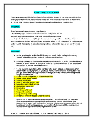
  • 1. 1 ACUTE LYMPHOCYTIC LEUKEMIA BY DR. MAGDI AWAD SASI 2014 ACUTE LYMPHOCYTIC LEUKEMIA Acute lymphoblastic leukemia (ALL) is a malignant (clonal) disease of the bone marrow in which early lymphoid precursors proliferate and replace the normal hematopoietic cells of the marrow. ALL is the most common type of cancer and leukemia in children in the United States. INCIDENCE: Acute leukaemia is an uncommon type of cancer. About 7,600 people are diagnosed with leukaemia each year in the UK. Of these, only about 650 people have acute lymphoblastic leukaemia. Acute lymphoblastic leukaemia((ALL)) is the most common type of cancer to affect children. Approximately 1 in every 2,000 children will develop it. About 85% of cases occur in children aged under 15, with the majority of cases developing in those between the ages of two and five years old. SYMPTOMS:  Acute lymphocytic leukemia (ALL) progress much faster and symptoms may worsen more quickly than chronic lymphocytic leukemia.  Patients with ALL present with either symptoms relating to direct infiltration of the marrow or other organs by leukemic cells, or symptoms relating to the decreased production of normal marrow elements.  Some leukemia symptoms, like night sweats, fever, fatigue and achiness, often resemble flu-like symptoms. If you have the flu, symptoms will likely subside as you get better. Make an appointment to see your doctor if the symptoms persist longer than expected.  Some general symptoms of leukemia include: Fever, chills ,Night sweats Fatigue, weakness Loss of appetite, weight loss Bone/joint pain, Body aches Abdominal discomfort Headaches Shortness of breath Frequent infections Easy bruising or bleeding Petechiae (small red spots under the skin) Fever is one of the most common symptoms of ALL, and patients with ALL often have fever without any other evidence of infection. However, in these patients, one must assume that all fevers are from infections until proved otherwise, because a failure to treat infections promptly and aggressively can be fatal. Infections are still the most common cause of death in patients undergoing treatment for ALL.
  • 2. 2 ACUTE LYMPHOCYTIC LEUKEMIA BY DR. MAGDI AWAD SASI 2014 Patients with ALL often have decreased neutrophil counts, regardless of whether their total white blood cell (WBC) count is low, normal, or elevated. As a result, these individuals are at an increased risk of infection. The prevalence and severity of infections are inversely correlated with the absolute neutrophil count (ANC), which is defined as the number of mature neutrophils plus bands per unit of volume. Infections are common when the absolute neutrophil count is less than 500/μL, and they are especially severe when it is less than 100/μL. Symptoms of anemia are common and include fatigue, dizziness, palpitations, and dyspnea upon even mild exertion. Other patients present with signs of bleeding. Bleeding can be the result of thrombocytopenia due to marrow replacement. Additionally, approximately 10% of patients with ALL have disseminated intravascular coagulation (DIC) at the time of diagnosis. These patients may present with hemorrhagic or thrombotic complications. Some patients present with palpable lymphadenopathy. Others, particularly those with T- cell ALL, present with symptoms related to a large mediastinal mass, such as shortness of breath. Infiltration of the marrow by massive numbers of leukemic cells frequently manifests as bone pain. This pain can be severe and is often atypical in distribution. About 10-20% of ALL patients may present with left upper quadrant fullness and early satiety due to splenomegaly. Although patients may present with symptoms of leukostasis (eg, respiratory distress, altered mental status) because of the presence of large numbers of lymphoblasts in the peripheral circulation, leukostasis is much less common in people with ALL than those with acute myelogenous leukemia (AML), and it occurs only in patients with the highest WBC counts (ie, several hundred thousand per μL). Patients with a high tumor burden, particularly those with severe hyperuricemia, can present in renal failure. Other potential signs and symptoms of ALL may include: Bleeding gums Frequent infections Nosebleeds Easy bruising Swollen lymph nodes around the neck, underarm, stomach or groin Shortness of breath Weight loss HEPATOMEGALLY AND SPLENOMEGALLY:  50% to 75% of patients with ALL & rare in AML.  The visceral involvement can produce symptoms of nausea , abdominal fullness and satiety.  Lymphoadenopathy – ALL ˃AML 80%// 50%  An anterior mediastinal mass ---T cell variant of ALL
  • 3. 3 ACUTE LYMPHOCYTIC LEUKEMIA BY DR. MAGDI AWAD SASI 2014  Practical point—extramedullary leukemia can precede detectable involvement of B.M. EXTRAMEDULLARY TISSUES : Acute leukemia may infiltrate –skin, lung, eye, kidney ,nasopharynx Testicular involvement is common in male with ALL. CHLOROMAS----soft tissues masses of leukemic cells. SYMPTOMS RELATED TO THE EXPANDING MALIGNANT CELL MASS: Bony pain and sternal tenderness. This occur in half of patients. Osteolytic lesions are rare. Renal abnormalities due to: A. Leukemic infiltration B. Ureteral obstruction by urate nephropathy—enlarged LN, uric acid stone, infections ,haemorrhage GIT----bleeding, distention ,satiety ,constipation((due to organomegally or leukemic infiltrate)) . CNS SYMPTOMS: Cells infiltrate in subarachnoid space leads to leukemic meningitis or direct involvement of the brain or spinal cord parenchyma. Neurological symptoms are unusual at the time of diagnosis. CNS is a frequent site of relapse. ((particularly ALL )) Leukemic meningitis— 1st symptoms---------headache and nausea With progression---- papillodema , cranial nerve palsies ,seizures ,altered mentation CSF----------------------- leukemic blast cell, increase protein, decrease glucose.
  • 4. 4 ACUTE LYMPHOCYTIC LEUKEMIA BY DR. MAGDI AWAD SASI 2014 METABOLIC DISTURBANCE : Hypokalemia and hyponatremia due to renal involvement . LDH—lactic dehydrogenase may be increased. Physical Examination:  Patients with ALL commonly have physical signs of anemia, including pallor and a cardiac flow murmur. Fever and other signs of infection, including lung findings of pneumonia, can also occur. Fever should be interpreted as evidence of infection, even in the absence of other signs.  Patients with thrombocytopenia usually demonstrate petechiae, particularly on the lower extremities. A large number of ecchymoses is usually an indicator of a coexistent coagulation disorder such as DIC.  Signs relating to organ infiltration with leukemic cells and, to a lesser degree, lymphadenopathy may be present.  Occasionally, patients have rashes that result from infiltration of the skin with leukemic cells. CLASSIFICATION OF ACUTE LEUKEMIA: 1. FAB classification(French-American-British) :1970 A classification of acute leukemia produced by a three-nation joint collaboration. A schema that divides acute leukemia into lymphoid–ALL or myeloid–AML cell lines. This is based on the microscopic features and cytochemistry of blast cells. Acute lymphoblastic leukemia is subdivided into three types(L1-L3). Of childhood ALL, 70% are predominantly L1, 27% are L2, and 3% or less are L3 or Burkitt cell type. In adults with ALL------ 30% are L1, 65% are L2, and 5% are L3. FAB classification, acute leukemia: Acute lymphocytic leukemia (ALL) L1 Small monotonous lymphocytes L2 Mixed L1- and L3-type lymphocytes L3 Large homogeneous blast cells ALL Disease of children 80% and young adults Boys more than the girl, whites more than non whites
  • 5. 5 ACUTE LYMPHOCYTIC LEUKEMIA BY DR. MAGDI AWAD SASI 2014 L1 85% of children. Mild to moderate basophilia Cells are small , twice diameter of small lymphocytes Homogenous nuclear chromatin. Regular nuclear membrane. Small or no nucleolus. Scanty cytoplasm without vacuoles B cell.—pro B or pre B lineage Common ALL ((CALLA)) Tdt—postive----CD19 & CD10 MPO is always negative L2 15% adults. .Cells are larger, cleafted nuclei. .Variable nuclear chromatin. .An irregular nuclear shape. .Low nuclear cytoplasmic ratio. .More pleomorphic size and shape. .One or more prominent nucleoli. T cell/ B cell-pro B , pre B lineage Tdt positive , basophilia Anterior mediastinal mass High leukocyte count CD4 , CD5 ,CD1 ,CD2 , CD8 L3 Uncommon Large, homogeneous cells with fine stippled chromatin; regular nuclei; prominent nucleoli. ˂5%, Large vasicular nuclei with abundant deeply basophilic cytoplasm The most distinguishing feature is prominent Cytoplasmic vacoulation. Have high mitotic index Leukemic form of burkitts lymphoma 3 – 4 times diameter of lymphocytes Tdt negative Surface immunoglobulin positive,B CELL The vast majority of ALL ((80%)) have B antigen
  • 6. 6 ACUTE LYMPHOCYTIC LEUKEMIA BY DR. MAGDI AWAD SASI 2014 The leukemic blast (( B cell ALL )) express CD19 antigen. T lymphoblasts in patient with T cell ALL seem to be arrested at early intrathymic stages of maturation. 90% of cases have nuclear enzymes terminal deoxynucleotidyl transferase (Tdt). Tdt is absent in mature lymphocytes ,hairy cell leukemia , CLL . PROGNOSIS: Precursor B cell ALL 80% (( best )) T cell and pre B cell ALL (( intermediate )) B cell ( burkitts ) ((worst)) The major immunophenotypic classification of ALL is as follows: B cell lineage- 1. Pro B ALL 2. Pre B ALL 3. B (surface Ig +) ALL (Burkitt's) T cell lineage*- 1. T cell ALL 2. Prothymic 3. Early thymocyte 4. Cortical (common) thymocyte 5. Medullary (mature) thymocyte 6. Mature T (peripheral) cells  * T cell ALL is usually not subclassified  **** 2YSMD do not need to know the different antigens. You should recognize CD3, CD5, and CD7 as pan T markers; CD19 as a pan B marker; CD4 as T-helper, and CD8 as T- suppressor.**** Risk Factors for Acute Lymphoblastic Leukemia For most people, the cause of ALL is unknown. For this reason, there is no known way to prevent it. However, there are a few known risk factors for this type of leukemia. But it is not yet known whether these risk factors are actual causes of the disease: Exposure to high levels of radiation to treat other types of cancer
  • 7. 7 ACUTE LYMPHOCYTIC LEUKEMIA BY DR. MAGDI AWAD SASI 2014 Exposure to certain chemicals such as benzene, a solvent used in oil refineries and other industries and present in cigarette smoke, certain cleaning products, detergents, and paint strippers Infection with human T-cell lymphoma/leukemia virus-1 (HTLV-1) in rarer cases outside the U.S. or Epstein-Barr virus (EBV), a related leukemia more commonly seen in Africa. Having an inherited genetic syndrome such as Down syndrome Being white Being male Laboratory tests for ALL include the following: 1. Complete blood count with differential 2. Peripheral blood smear 3. Coagulation studies 4. Chemistry profile, including lactic dehydrogenase, uric acid, liver function studies, and BUN/creatinine 5. Appropriate cultures (in particular, blood cultures, urine culture ,throat swab, stool culture ) in patients with fever or other signs of infection 6. Chest x-ray 7. Computed tomography 8. Multiple-gated acquisition scanning 9. Electrocardiography 10. Bone marrow aspiration and biopsy (definitive for confirming leukemia) 11. Immunohistochemistry 12. Flow cytometry 13. Cytogenetics 14. Polymerase chain reaction 15. Gene expression profiling National Comprehensive Cancer Network (NCCN) guidelines note that diagnosis of ALL generally requires the following :  Demonstration of ≥20% bone marrow lymphoblasts  Morphologic assessment of Wright/Giemsa–stained bone marrow aspirate smears  Hematoxylin and eosin (H&E)–stained bone marrow core biopsy and clot sections  Comprehensive flow cytometric immunophenotyping For optimal risk stratification and treatment planning in patients with ALL, the NCCN advises that bone marrow or peripheral blood lymphoblasts must be tested for specific recurrent genetic abnormalities, as follows : 1. Cytogenetics – Karyotyping of G-banded metaphase chromosomes 2. Interphase fluorescence in situ hybridization (FISH) 3. Reverse transcriptase polymerase chain reaction (RT-PCR) for fusion genes (eg, BCR-ABL) In addition, flow cytometric DNA index/ploidy testing can be done to assess for hyperdiploidy and hypodiploidy.
  • 8. 8 ACUTE LYMPHOCYTIC LEUKEMIA BY DR. MAGDI AWAD SASI 2014 CBC  A complete blood cell (CBC) count with differential demonstrates anemia and thrombocytopenia to varying degrees in individuals with (ALL). Patients with ALL can have a high, normal, or low white blood cell (WBC) count, but they usually exhibit neutropenia. The prevalence and severity of infections are inversely correlated with the absolute neutrophil count (ANC); infections are common when the absolute neutrophil count is less than 500/μL, and they are especially severe when it is less than 100/μL. Coagulation studies and chemistry profiles:  Abnormalities in the prothrombin time (PT) / activated partial thromboplastin time (aPTT) / fibrinogen / fibrin degradation products may suggest concomitant disseminated intravascular coagulation (DIC), which results in an elevated PT, decreased fibrinogen levels, and the presence of fibrin split products.  A review of the peripheral blood smear confirms the findings of the CBC count. Circulating blasts are usually seen. Schistocytes are sometimes seen if DIC is present.  A chemistry profile is recommended. Most patients with ALL have an elevated lactic dehydrogenase level (LDH), and they frequently have an elevated uric acid level. In addition, liver function tests and blood urea nitrogen (BUN)/creatinine determinations are necessary before the initiation of therapy. Cultures  Appropriate cultures, in particular blood cultures, should be obtained in patients with fever or with other signs of infection without fever. Imaging  Chest x-ray films may reveal signs of pneumonia and/or a prominent mediastinal mass in some cases of T-cell acute lymphoblastic leukemia (ALL).  Computed tomography (CT) scans can further define the degree of lymphadenopathy in some patients, including those with mediastinal masses.  Multiple-gated acquisition (MUGA) scans or electrocardiograms (ECGs) are needed when the diagnosis of acute lymphoblastic leukemia (ALL) is confirmed, because many chemotherapeutic agents used in the treatment of acute leukemia are cardiotoxic.  An ECG is recommended before the initiation of treatment. Bone Marrow Aspiration and Biopsy:  Bone marrow aspiration and biopsy are the definitive diagnostic tests to confirm the diagnosis of leukemia. Immunophenotyping helps to elucidate the subtype.  Aspiration slides should be stained for morphology with either Wright or Giemsa stain. The diagnosis of ALL is made when at least 30% lymphoblasts [FAB] classification) or 20% lymphoblasts ( [WHO] classification) are present in the bone marrow and/or peripheral blood.  In addition, slides should be stained with myeloperoxidase (MPO) (or Sudan black) and terminal deoxynucleotidyl transferase (TdT), unless another method is used, such as flow cytometry.  Bone marrow samples should also be sent for flow cytometry and cytogenetics. Approximately 15% of patients with ALL have a t(9;22) translocation (ie, Philadelphia [Ph] chromosome), but other chromosomal abnormalities may also occur, such as t(4;11), t(2;8), and t(8;14).
  • 9. 9 ACUTE LYMPHOCYTIC LEUKEMIA BY DR. MAGDI AWAD SASI 2014 Giemsa stain Trephine biopsy
  • 10. 10 ACUTE LYMPHOCYTIC LEUKEMIA BY DR. MAGDI AWAD SASI 2014 Histological Features: The older, traditional classification of (ALL) is the French-American-British (FAB) classification. This has now been replaced by the newer World Health Organization (WHO) classification but the FAB system is listed for historical purposes, as follows:  L1 – Small cells with homogeneous chromatin, regular nuclear shape, small or absent nucleolus, and scanty cytoplasm; subtype represents 25-30% of adult cases  L2 – Large and heterogeneous cells, heterogeneous chromatin, irregular nuclear shape, and nucleolus often large; subtype represents 70% of cases (most common)  L3 – Large and homogeneous cells with multiple nucleoli, moderate deep blue cytoplasm, and cytoplasmic vacuolization that often overlies the nucleus (most prominent feature); subtype represents 1-2% of adult cases WHO classification of ALL : B lymphoblastic leukemia/lymphoma: B lymphoblastic leukemia/lymphoma, NOS B lymphoblastic leukemia/lymphoma with recurrent genetic abnormalities  B lymphoblastic leukemia/lymphoma with t(9;22)(q34;q11.2), BCR-ABL1  B lymphoblastic leukemia/lymphoma with t(v;11q23); MLL rearranged  B lymphoblastic leukemia/lymphoma with t(12;21)(p13;q22) TEL-AML1 (ETV6-RUNX1)  B lymphoblastic leukemia/lymphoma with hyperdiploidy  B lymphoblastic leukemia/lymphoma with hypodiploidy  B lymphoblastic leukemia/lymphoma with t(5;14)(q31;q32) IL3-IGH  B lymphoblastic leukemia/lymphoma with t(1;19)(q23;p13.3) TCF3-PBX1 The WHO classifies the L1 and L2 subtypes of ALL as either precursor B lymphoblastic leukemia/lymphoblastic lymphoma or precursor T lymphoblastic leukemia/lymphoblastic lymphoma depending on the cell of origin. The L3 subtype of ALL is included in the group of mature B-cell neoplasms, as the subtype Burkitt lymphoma/leukemia. Immunohistochemistry A negative myeloperoxidase (MPO) stain and a positive terminal deoxynucleotidyl transferase (TdT) is the hallmark of the diagnosis of most cases of (ALL). However, positive confirmation of lymphoid (and not myeloid) lineage should be sought by flow cytometric demonstration of lymphoid antigens, such as CD3 (T-lineage ALL) or CD19 (B- lineage ALL), in order to avoid confusion with some types of myeloid leukemia (eg, M0), which also stain negative with myeloperoxidase . Polymerase Chain Reaction or Cytogenics Although more than 95% of cases of the L1 or L2 subtype of (ALL) are positive for Terminal deoxynucleotidyl transferase (TdT), TdT is not specific for ALL; TdT is absent in L3 (mature B-cell) ALL. However, TdT helps to distinguish ALL from malignancies of more mature lymphocytes (ie, non-Hodgkin lymphoma [NHL]). In cases of acute leukemia that are myeloperoxidase (MPO) negative and TdT positive, the distinction between (AML) and ALL is made on the basis of flow cytometry results. Patients with AML demonstrate myeloid markers such as CD33, whereas patients with ALL demonstrate lymphoid markers. Further confusion arises because some patients with ALL have aberrant expression of myeloid markers, such as CD13. However, if the cells are TdT positive, MPO negative, CD33 negative, and demonstrate lymphoid markers, the leukemia is considered ALL.
  • 11. 11 ACUTE LYMPHOCYTIC LEUKEMIA BY DR. MAGDI AWAD SASI 2014 Cytogenetic abnormalities occur in approximately 70% of cases of ALL in adults. These abnormalities include balanced translocations as occur in cases of AML. However, abnormalities of chromosome number (hypodiploidy, hyperdiploidy) are more common in ALL than in AML. Studies for bcr-abl analysis by (PCR) or cytogenetics may help distinguish patients with Philadelphia chromosome–positive (Ph+ ALL) from those with the lymphoid blastic phase of (CML). Most patients with Ph+ ALL have the p190 type of bcr-abl, whereas patients with lymphoid blastic CML have the p210 type of bcr-abl. Common Cytogenetic Abnormalities in ALL Common Cytogenetic Abnormalities in ALL Abnormality Genes Involved 3-Year Event-Free Survival t(10;14)(q24;q11) HOX11/TCRA 75% 6q Unknown 47% 14q11 TCRA/TCRD 42% 11q23 MLL 18-26% 9p Unknown 22% 12 TEL 20% t(1;19)(q23;p13) PBX1/E2A 20% t(8;14)(q24;q32) t(2;8)(p12;q24) t(8;22)(q24;q11) c-myc/IGH IGK/c-myc c-myc/IGL 17% 80% t(9;22)(q34;q11) bcr-abl 5-10% 66%‡ t(4;11)(q21;q23) AF4-MLL 0-10%
  • 12. 12 ACUTE LYMPHOCYTIC LEUKEMIA BY DR. MAGDI AWAD SASI 2014 Eighty-five percent of cases of ALL are derived from B cells. The primary distinction is among the following (see below):  Early (pro-B) ALL, which is TDT positive, CD10 (CALLA) negative, surface immunoglobulin (Ig) negative  Precursor B ALL, which is TDT positive, CD10 (CALLA) positive, surface Ig negative  Mature B cell (Burkitt) ALL, which is TdT negative, surface Ig positive. Fifteen percent of these cases are derived from T cells. Immunophenotyping of ALL Cells – ALL of B-Cell Lineage (85% of cases of adult ALL) ALL Cells TdT CD19 CD10 CyIg SIg Early B-precursor ALL + + - - - Pre–B-cell ALL + + + + - B-cell ALL - + +/- +/- + ALL = acute lymphoblastic leukemia; Cylg = Cytoplasmic immunoglobulin; SIg =Surface immunoglobulin; TdT = terminal deoxynucleotidyl transferase. These cases are subclassified into different stages corresponding to the phases of normal thymocyte development. The early subtype is surface CD3 negative, cytoplasmic CD3 positive, and either double negative (CD4-, CD8-) or double positive (CD4+, CD8+). The latter subtype is surface CD3 positive, CD1a negative, and positive for either CD4 or CD8, but not both. Immunophenotyping of ALL Cells – ALL of T-Cell Lineage (15% of cases of adult ALL) ALL Cells TdT Surface CD3 CD4/CD8 Early T-precursor ALL + - +/+ or -/- T-cell ALL + + +/- or -/+ Treatment for acute leukemia: Patient need hospital with appropriate blood product support facilities, leukapheresis capabilities, or physicians and nurses familiar with the treatment of patients with leukemia . (ALL) is best treated by physicians who have significant experience in the treatment of patients with acute leukemia(( haematologist)). The four components of ALL treatment are induction, consolidation, maintenance, and central nervous system (CNS) prophylaxis. Patients with ALL require hospital admission for induction chemotherapy, and they require readmission for consolidation chemotherapy or for the treatment of toxic effects of chemotherapy. Surgical intervention may be required for the placement of a central venous catheter, such as a triple lumen, Broviac, or Hickman catheter.
  • 13. 13 ACUTE LYMPHOCYTIC LEUKEMIA BY DR. MAGDI AWAD SASI 2014 Supportive Care - Blood Products Patients with (ALL) have a deficiency in the ability to produce normal blood cells, and they need replacement therapy. This deficiency is temporarily worsened by the addition of chemotherapy. All blood products must be irradiated to prevent transfusion-related graft versus host disease, which is almost invariably fatal. Packed red blood cells are given to patients with a hemoglobin level ˂ 7-8 g/dL or at a higher level if the patient has significant cardiovascular or respiratory compromise. Platelets are transfused if the count is less than 10,000-20,000/μL. Patients with pulmonary or gastrointestinal hemorrhage receives platelet transfusions to maintain a value ˃ 50,000/μL. Patients with central nervous system CNS hemorrhage are transfused to achieve a platelet count of 100,000/μL. Fresh frozen plasma is given to patients with a significantly prolonged prothrombin time (PT). Cryoprecipitate is given if the fibrinogen level is less than 100 g/dL. Supportive Care - Therapy and Prophylaxis for Infection  Antibiotics are given to all febrile patients.  At a minimum, include a third-generation cephalosporin (or equivalent), usually with an amino glycoside.  In addition to this minimum, other antibiotic agents are added to treat specific documented or possible infections.  Patients with persistent fever after 3-5 days of antibacterial antibiotics should have an antifungal antibiotic (liposomal or lipid complex amphotericin, new generation azole or echinocandin) added to their regimen.  Patients with sinopulmonary complaints would receive anti-Aspergillus treatment. Particular care is warranted for patients receiving steroids as part of their treatment, because the signs and symptoms of infection may be subtle or even absent. The use of prophylactic antibiotics in neutropenic patients who are not febrile is controversial. However, most clinicians prescribe them for patients undergoing induction therapy. A commonly used regimen includes the following:  Ciprofloxacin (oral [PO] 500 mg twice daily [bid])  Fluconazole (200 mg PO daily), itraconazole (200 mg PO bid), or posaconazole (200 mg PO three times daily [tid])  Acyclovir (200 mg PO 5 times/d) or valacyclovir (500 mg PO daily) Once patients taking these antibiotics become febrile, they are switched to intravenous antibiotics. Most treatment plans for (ALL) have 3 steps. These are induction, consolidation, and maintenance.
  • 14. 14 ACUTE LYMPHOCYTIC LEUKEMIA BY DR. MAGDI AWAD SASI 2014 1. Induction therapy kills leukemia cells in the blood and bone marrow to induce remission. Treatments include chemotherapy and corticosteroids. Induction usually lasts 4 weeks and is done in a hospital. But ALL people have leukemia cells with a certain gene change. This gene is called the Philadelphia chromosome. These people will be treated with a tyrosine kinase inhibitor. Standard induction therapy typically involves either a four-drug regimen of vincristine, prednisone, anthracycline, and cyclophosphamide or L -asparaginase or a five-drug regimen of vincristine, prednisone, anthracycline, cyclophosphamide, and L -asparaginase given over the course of 4-6 weeks. Using this approach, complete remissions (CRs) are obtained in 65-85% of patients. The rapidity with which a patient's disease enters CR correlates with treatment outcome. In a large French study, patients with greater than 5% blasts in their bone marrow on day 15 had a lower response rate (34% vs 91%), worse disease-free survival, and worse overall survival than patients with low blast counts on day 15 2. Consolidation therapy kills any leukemia cells that may be present even though they don't show up in tests. If these cells regrow, they could cause a relapse. Treatments include more chemotherapy and may include stem cell transplant. This step may also include preventive treatment of the brain or spinal cord with radiation or chemotherapy. Consolidation usually takes several months but doesn't require staying overnight in the hospital.
  • 15. 15 ACUTE LYMPHOCYTIC LEUKEMIA BY DR. MAGDI AWAD SASI 2014 The use of consolidation chemotherapy in (ALL) is supported by several studies. Because most studies have showed a benefit to consolidation therapy, regimens using a standard 4- to 5-drug induction usually include consolidation therapy with Ara-C in combination with an anthracycline or epipodophyllotoxin. 3. Maintenance therapy also prevents any remaining leukemia cells from growing. This may be done using lower doses of chemotherapy than those used during induction or consolidation. Chemotherapy is given with pills and once-a-month intravenous (IV) treatment. Maintenance is often continued for up to 3 years, but during this time, most people are able to go back to being as active as they were before beginning treatment. When there are no signs of leukemia for 5 years, a person is usually considered cured. But if the leukemia doesn't go into remission, or if it comes back within the first few years, treatments may include more chemotherapy, a stem cell transplant, or joining a clinical trial for new treatments. The effectiveness of maintenance chemotherapy in adults with (ALL) has not been studied in a controlled clinical trial. However, several phase II studies without maintenance therapy have shown inferior results compared with historical controls. Although maintenance appears necessary, using a more intensive versus less intensive regimen does not appear to be beneficial. Intensification of maintenance therapy from a 12-month course of a four-drug regimen compared with a 14-month course of a seven-drug regimen did not show a difference in disease-free survival between the two groups. CNS prophylaxis: Patients with (ALL) frequently have meningeal leukemia at the time of relapse. A minority of patients have meningeal disease at the time of initial diagnosis. As a result, central nervous system (CNS) prophylaxis with intrathecal chemotherapy is essential. High-dose systemic chemotherapy reduces CNS relapse. However, early intrathecal chemotherapy is necessary to achieve the lowest risk of CNS relapse. CNS relapse rates were 3% for group 4 hyperfractionated cyclophosphamide, vincristine, doxorubicin, and dexamethasone (hyper-CVAD). All subjects received intrathecal chemotherapy starting in induction. High-risk subjects received 16 intrathecal treatments, and low-risk subjects received four intrathecal treatments. Newer Induction Approache: Standard induction regimens are modeled after pediatric programs and were originally developed when supportive care was significantly inferior to what is available today. Few antibiotics were available, and transfusion capabilities were minimal. Consequently, milder regimens were designed in an attempt to minimize early deaths during induction. With the addition of third-generation cephalosporins and sophisticated blood-banking techniques, the ability to support patients through a pancytopenic phase has increased dramatically. As a result, more intensive induction approaches are used by many physicians. Two notable examples are the Memorial Acute Lymphoblastic Leukemia – 2 (ALL-2) protocol and the hyper-CVAD (cyclophosphamide, vincristine, doxorubicin, and dexamethasone) protocol.
  • 16. 16 ACUTE LYMPHOCYTIC LEUKEMIA BY DR. MAGDI AWAD SASI 2014 A. ALL-2 protocol The ALL-2 protocol uses an intensive, high-dose, mitoxantrone-based, acute myelogenous leukemia (AML)-style induction regimen. Weiss et al reported treatment of 37 subjects with newly diagnosed ALL with this induction regimen followed by a first consolidation with vincristine, prednisone, L - asparaginase, and methotrexate; a second consolidation with Ara-C and etoposide; and then 2 years of maintenance therapy. Of these subjects, 84% achieved CR. The median remission duration was 17 months, and median survival was 20 months.- In a randomized phase III trial comparing the ALL-2 regimen with the L-20 regimen (vincristine, prednisone, cyclophosphamide and doxorubicin), the CR rate was 83% for patients receiving ALL-2 compared with 70% for patients receiving L- 20. Overall survival at 4 years was superior for patients receiving ALL-2 (40%) versus those receiving L-20 (22%). A. Hyper-CVAD regimen The hyper-CVAD regimen is based on the success achieved with short-term, dose- intensive chemotherapy regimens in children. It incorporates hyperfractionated cyclophosphamide and intensive doses of Ara-C and methotrexate in combination with dexamethasone and vincristine. Maintenance therapy with prednisone, vincristine (Oncovin), methotrexate, and mercaptopurine (Purinethol) (ie, POMP protocol) is given to patients with nonmature B-cell ALL. From 1992-2000, 288 patients received hyper-CVAD at MD Anderson Cancer Center, which 17% of patients had the Philadelphia (Ph) chromosome, and 13% had T-cell ALL. Overall, 92% of patients obtained a CR. The 5-year survival and percentage of patients in CR at 5 years were both 38%. Patients with Ph+ ALL had a 92% CR rate but only a 12% 5-year survival. Patients with T-cell ALL had a 75% CR rate and a 48% 5-year survival. Patients with Burkitt ALL had a 93% CR rate and a 67% 5-year survival. Newer modifications of the hyper-CVAD regimen include the addition of imatinib to patients whose leukemia is Philadelphia chromosome positive, and rituxan to patients whose leukemia is CD20 positive. Both of these approaches have resulted in improvements in disease-free survival More advanced regimen in treatment: Treatment of Mature B-Cell ALL Mature B-cell (ALL) is a special type (( 5% of adult ALL)) . The hallmark of mature B-cell ALL is the presence of surface immunoglobulin on the lymphoblasts. Using conventional regimens, only 30- 40% of patients enter complete remission (CR) and few patients survive long term. Newer short-term intensive therapies show improved results. A report of the hyper-CVAD regimen showed that disease in 93% of subjects entered CR, median survival was 16 months, and disease in 67% of subjects alive at 5 years.
  • 17. 17 ACUTE LYMPHOCYTIC LEUKEMIA BY DR. MAGDI AWAD SASI 2014 In a report by Hoelzer et al, with the use of regimens containing intensive cyclophosphamide and intermediate methotrexate or ifosfamide and high-dose methotrexate, CR rates were 63% (cyclophosphamide + intermediate methotrexate) and 74% (ifosfamide + high-dose methotrexate). Disease-free survival rates increased to 50% in the first group and 71% in the second group, and overall survival increased to 50% compared with 0% for historical controls. Although previously these patients were referred for transplantation in first remission, many physicians now defer transplantation for the time of relapse because of these improved results. Burkitt ALL cells are CD20 positive. This allows for the addition of targeted therapy with rituximab. Many studies are have demonstrated improved efficacy, including prolonged survival, when rituximab is added to chemotherapy in these patients. The combination of hyper-CVAD (hyperfractionated cyclophosphamide, vincristine, doxorubicin, and dexamethasone) plus rituxan resulted in an overall 3-year survival of 80% compared with 50% for historical controls treated without rituxan Treatment of Ph Chromosome–Positive ALL PH + In the past, Philadelphia chromosome–positive (Ph+) ALL was treated with the same regimens as other types of ALL, with poor results. However, the tyrosine kinase inhibitor imatinib inhibits the bcr-abl fusion protein of Ph+ ALL and thus allows targeted therapy of this disease. As a single agent, imatinib has limited activity. Imatinib The German Multicenter ALL (GMALL) trial conducted a randomized study of imatinib versus standard induction therapy for patients with Ph-positive ALL older than 55 years and reported the overall complete remission (CR) rate was 96.3% in patients randomly assigned to imatinib and 50% in patients allocated to standard chemotherapy. Severe adverse events were significantly more frequent during standard induction chemotherapy (90% vs 39%). The addition of imatinib to chemotherapy has resulted in significantly improved outcomes. The addition of imatinib to hyper-CVAD (hyperfractionated cyclophosphamide, vincristine, doxorubicin, and dexamethasone) resulted in a 3-year disease-free survival rate of 66% and overall survival of 55% compared with a 14% 3-year disease-free survival rate and 15% overall survival for patients treated with hyper-CVAD without imatinib. Newer tyrosine kinase inhibitors have been developed for patients with chronic myelogenous leukemia (CML) that has become resistant to imatinib. These agents are also being studied in Ph- positive ALL. Nilotinib and dasatinib Nilotinib is a tyrosine kinase inhibitor that has a higher binding affinity and selectivity for the ABL kinase than imatinib. Nilotinib has 20 to 50 times the inhibitory activity against imatinib-sensitive CML cell lines relative to imatinib. Dasatinib is a potent, orally active inhibitor of the BCR-ABL, c-KIT and the SRC family of kinases. Dasatinib is a more potent inhibitor of BCR-ABL and c-KIT than imatinib mesylate, and it is effective in patients with CML that is resistant to or intolerant of imatinib. The Gruppo Italiano Malattie Ematologiche dell'Adulto (GIMEMA) presented the interim results of a prospective study of dasatinib in patients with newly diagnosed Ph-positive ALL. Prednisone was started 7 days before the first dasatinib administration and continued until day 31. Dasatinib was
  • 18. 18 ACUTE LYMPHOCYTIC LEUKEMIA BY DR. MAGDI AWAD SASI 2014 administered for a total of 84 days. At the time of the report, all 23 patients treated showed a complete hematologic response by day 22. Although nilotinib and dasatinib are clearly active in Ph-positive ALL, it is likely that, similar to the results seen with imatinib, these responses will likely not be durable. Therefore each of these agents is currently being studied in combination with standard chemotherapy regimens. That said, these new tyrosine kinase inhibitors are not without their drawbacks and adverse events. Dasatinib has been associated with pleural effusions and pulmonary arterial hypertension while nilotinib has been linked to biochemical changes in liver function and QT-interval prolongation. Development of resistance may also occur with these agents. Ponatinib Ponatinib (Iclusig), a kinase inhibitor, was approved by the US Food and Drug Administration (FDA) in December 2012 for patients with Ph+ ALL that is resistant or intolerant to prior tyrosine kinase inhibitor therapy, including those with the T315I mutation. Because ponatinib has a high risk for thromboembolic events, its use is restricted to patients for whom no other tyrosine kinase inhibitor therapy is indicated. In October 2013, at the FDA’s request, ponatinib was temporarily removed from the market because of safety concerns. The FDA cited an increased risk for life-threatening blood clots and severe narrowing of blood vessels. Ponatinib was returned to the US market within 2 months, but with new safety measures to address the risk for serious cardiovascular and thrombotic events. The revised indications for patients with ALL are now limited to two groups: 1. Adults with T315I-positive Ph+ ALL. 2. Adults with Ph+ ALL for whom no other tyrosine kinase inhibitor therapy is indicated. The revised labeling also states that the optimal dose of ponatinib has not been determined. The recommended starting dose remains at 45 mg PO once daily with or without food, but additional information has been included regarding dose decreases and discontinuations. The boxed warning has been revised to include the risk for heart failure, including fatalities, and the incidence of vascular occlusion (at least 27%). Treatment of the Younger Adult Older children and younger adults with acute lymphoblastic leukemia (ALL) can be referred to either adult or pediatric hematologists. Usually, the patient will receive either an adult or pediatric regimen based on this referral pattern. However, several studies have suggested that younger patients are best treated on pediatric protocols. In a study by the Programme for the Study of Therapeutics for Haematological Malignancies (PETHEMA), adolescents and young adults were treated with a pediatric regimen (ALL-96), demonstrating a response to therapy that was similar to previously reported, although a slight increase in hematologic toxicity was observed in the adult patients.
  • 19. 19 ACUTE LYMPHOCYTIC LEUKEMIA BY DR. MAGDI AWAD SASI 2014 Transplantation: Most authorities agree that allogeneic transplantation should be offered to young patients with high-risk features whose (ALL) is in first remission. Young patients without adverse features should receive induction, consolidation, and maintenance therapy. In these patients, transplantation is reserved for relapse. Older patients whose disease is in complete remission (CR) may be considered for such investigational approaches as allogeneic transplantation with nonmyeloablative chemotherapy (ie, mini-transplants). Previously, patients with mature B-cell ALL would have been referred for transplantation when their disease was in first CR; however, with improving results from more intensive chemotherapy regimens, many clinicians are reserving transplantation for patients who have experienced relapse. Hematopoietic stem cell transplantation (HSCT) seems to be a valuable option for a subgroup of infants with mixed-lineage ALL carrying poor prognostic factors that include age younger than 6 months and either poor response to steroids at day 8 or leukocyte levels of 300 g/L or higher. However, when only high-risk patients were considered (ie, Philadelphia chromosome–positive (Ph+), null ALL; >35 y; white blood cell [WBC] count >30,000/μL; or time to CR > wk , allogeneic BMT proved superior to autologous BMT or chemotherapy with respect to overall survival rates (44% vs 20%) and disease-free survival rates (39% vs 14%). Other phase 2 studies have confirmed a benefit for high-risk patients who undergo allogeneic BMT, with as many as 50% achieving long- term remissions. Stem cell transplantation In the GOELAL02 study, patients with any high-risk feature (age >35 y, non–T-ALL, WBC >30,000, adverse cytogenetics: t[9;22], t[4;11], or t[1;19], or no CR after induction) received either allogeneic or autologous stem cell transplantation. For patients younger than 50 years, the 6-year overall survival rate was improved for patients receiving allogeneic transplantation (75%) compared with those receiving autologous transplantation (40%). The United Kingdom Medical Research demonstrated that matched related allogeneic transplantations for ALL in first complete CR provide the most potent antileukemic therapy and considerable survival benefit for standard-risk patients. However, the transplantation-related mortality for high-risk older patients was unacceptably high and abrogated the reduction in relapse risk. Allogeneic transplantation can also be effective therapy for patients who have experienced relapse after chemotherapy. Yet, although autologous transplantation is relatively safe, it is associated with a high relapse rate, making this modality of little use in patients with ALL. Unrelated donor transplantation For patients without a sibling donor, an alternative is an unrelated donor (URD) transplant. Weisdorf et al found that autologous BMT was associated with a lower transplant-related mortality rate, but URD transplantations had a lower risk of relapse. In patients whose disease was in second CR, URD transplantations resulted in a superior rate of disease-free survival. Although peripheral blood has come to be preferred to bone marrow as the source for stem cells from unrelated donors (about 75% of transplants), a randomized phase III trial found that peripheral-blood stem cells did not yield improved survival as compared with bone-marrow cells and were significantly associated with chronic graft-vs-host disease (GVHD); SOME suggested that peripheral-blood stem cells might be appropriate for patients at higher risk for graft failure and bone-marrow cells for all others
  • 20. 20 ACUTE LYMPHOCYTIC LEUKEMIA BY DR. MAGDI AWAD SASI 2014 Treatment of Relapsed ALL Patients with relapsed (ALL) have an extremely poor prognosis. Most patients are referred for investigational therapies. Young patients who have not previously undergone transplantation are referred for such therapy. Reinduction regimens include the hyper-CVAD (cyclophosphamide, vincristine, doxorubicin, dexamethasone) protocol and high-dose cytosine arabinoside (Ara-C)– based regimens. As noted earlier, the hyper-CVAD regimen is based on hyperfractionated cyclophosphamide and intermediate doses of Ara-C and methotrexate. The complete remission (CR) rate was 44% and median survival was 42 weeks. Arlin et al reported that 8 of 10 patients with relapsed ALL achieved CR with high-dose Ara-C and high-dose mitoxantrone . A similar regimen using a single high dose of idarubicin in combination with Ara-C (the Memorial ALL-3 protocol) resulted in CR rates of 58-78% in patients who experienced relapse. In August 2012, the US Food and Drug Administration (FDA) approved vincristine liposomal (Marqibo) for the treatment of Philadelphia chromosome negative (Ph-) ALL in adults. It is indicated for patients in second or greater relapse or whose disease has progressed following two or more anti-leukemia therapies. This product is a sphingomyelin/cholesterol liposome- encapsulated formulation of vincristine In December 2012, the FDA approved the kinase inhibitor ponatinib for Ph+ ALL that is resistant or intolerant to prior tyrosine kinase inhibitor therapy Novel and Experimental Drug Therapies A number of new drugs are in development for the treatment of ALL. Clofarabine is a novel nucleoside analogue that is approved for the treatment of pediatric patients with refractory or relapsed ALL. This agent inhibits DNA synthesis at both DNA polymerase I and at RNA reductase. Overall response rates average 25%. 506U78 (nelarabine [Arranon]) is a novel purine nucleoside that is a prodrug of guanine arabinoside (ara-G). This agent was approved as an orphan drug by the US Federal Drug Administration (FDA) in October 2005. Complete responses have been reported in 31% of patients and in 54% of patients with T-cell ALL. The dose-limiting toxicity of this drug is neurotoxicity Supportive Care - Growth Factors: The use of granulocyte colony-stimulating factor (G-CSF) during induction chemotherapy for (ALL) is supported by several studies. In a Groupe d'Etude et de Traitement de la Leucemie Aigue Lymphoblastique de l'Adulte (GET- LALA) study, in patients who received G-CSF, GM-CSF, or no growth factor during induction therapy, the median time for neutrophil recovery was 17 days for G-CSF, 18 days for GM-CSF, and 21 days for no growth factors Hyperuricemia and Tumor Lysis Syndrome: Tumor lysis syndrome is a potentially life-threatening complication that may be seen in patients receiving chemotherapy for acute leukemias and high-grade non-Hodgkin lymphomas. This syndrome is characterized by elevated blood levels of uric acid, phosphate, and potassium; decreased levels of calcium; and acute renal failure. Patients with a high tumor burden, particularly those with severe hyperuricemia, can present in renal failure. Allopurinol at 300 mg 1-3 times per day is recommended during induction therapy
  • 21. 21 ACUTE LYMPHOCYTIC LEUKEMIA BY DR. MAGDI AWAD SASI 2014 until blasts are cleared and hyperuricemia resolves. High-risk patients (those with very high lactate dehydrogenase [LDH] or leukemic infiltration of the kidneys) can benefit from rasburicase. In a study by Cortes et al, adults with hyperuricemia or those at high risk for tumor lysis syndrome not only had an improved plasma uric acid response rate with rasburicase alone (0.20 mg/kg/d intravenously [IV], days 1-5) (87%) or in combination with allopurinol (IV rasburicase 0.20 mg/kg/d, days 1-3, followed by oral [PO] allopurinol 300 mg/d, days 3-5) (78%) than with allopurinol alone (300 mg/d PO, days 1-5) (66%), but they also had more rapid control of their plasma uric acid with rasburicase alone (4 h) or with allopurinol (4 h) than with allopurinol alone (27 h) Long-Term Monitoring: Patients with (ALL) are monitored on an outpatient basis for disease status and the effects of chemotherapy. Maintenance therapy for these patients is also administered in an outpatient setting. In addition, all patients should be on trimethoprim-sulfamethoxazole (TMP-SMZ) to prevent Pneumocystis jiroveci pneumonia, and patients may benefit from receiving oral nystatin or clotrimazole troches to reduce the risk of candidiasis. Patients with a high risk of relapse may also need additional antifungal therapy, such as itraconazole. Medication Summary: Antineoplastic agents are used for induction, consolidation, and maintenance therapy and central nervous system (CNS) prophylaxis in patients with acute lymphoblastic leukemia (ALL). Those medications cause severe bone marrow depression, and only physicians specifically trained in their use should administer them. In addition, access to appropriate supportive care is required.Other drug classes used in treatment of ALL include the following:  Corticosteroids may be used during induction, consolidation, and/or maintenance therapy  Tyrosine kinase inhibitors are used in treatment of Philadelphia chromosome – positive (Ph+) ALL  Colony-stimulating factors are used to treat or prevent neutropenia and to mobilize autologous peripheral blood progenitor cells for bone marrow transplantation (BMT) and in management of chronic neutropenia  Prophylactic antibiotics and antifungal drugs are given to prevent infection in patients receiving chemotherapy Dexamethasone is another corticosteroid that acts as an important chemotherapeutic agent in the treatment of ALL. Like prednisone, this agent is used in induction and reinduction therapy and is also given as intermittent pulses during continuation therapy. Antineoplastics: Used for induction, consolidation, maintenance, and central nervous system (CNS) prophylaxis. Cancer chemotherapy is based on an understanding of tumor cell growth and how drugs affect this growth. After cells divide, they enter a period of growth (ie, phase G1), followed by DNA synthesis (ie, phase S), a premitotic phase (ie, G2), then, a mitotic cell division (ie, phase M). Cell-division rates vary for different tumors. Most common cancers grow slowly compared with normal tissues, and the rate may be decreased in large tumors. This difference allows normal cells to recover from chemotherapy more quickly than malignant ones and is the rationale behind current cyclic dosage schedules. Antineoplastic agents interfere with cell reproduction. Some agents act at specific phases of the cell cycle, whereas others (ie, alkylating agents, anthracyclines, cisplatin) are not phase specific. Cellular apoptosis (ie, programmed cell death) is another potential mechanism of many antineoplastic agents.
  • 22. 22 ACUTE LYMPHOCYTIC LEUKEMIA BY DR. MAGDI AWAD SASI 2014 Vincristine (Vincasar PFS) Vincristine is a vinca alkaloid agent that acts by arresting cells in metaphase. Vincristine liposomal (Marqibo) A sphingomyelin/cholesterol liposome-encapsulated formulation of vincristine. Indicated for treatment of Ph-negative ALL for patients in second or greater relapse or whose disease has progressed following 2 or more antileukemia therapies. Asparaginase Erwinia chrysanthemi (Erwinaze) Catalyzes deamidation of asparagine to aspartic acid and ammonia, thereby reducing circulating levels of asparagine. Lack of asparagine synthetase activity results in cytotoxicity specific for leukemic cells that depend on an exogenous source of the amino acid asparagine. Indicated as part of a multiagent chemotherapeutic regimen as a substitute for asparaginase (Elspar). Pegaspargase (Oncaspar, PEG L Asparaginase) Modified version of L-asparaginase. Selective killing of leukemic cells it thought to be due to depletion of plasma asparagine, the amino acid required for protein synthesis. It is indicated as a component of a multi-agent chemotherapeutic regimen for the first line treatment of ALL. It is also indicated for use in patients with hypersensitivity to native forms of L-asparaginase. Methotrexate (Trexall) Methotrexate is an antimetabolite of the folic acid analogue type. This agent inhibits dihydrofolate reductase, resulting in inhibition of DNA synthesis, repair, and cellular replication. Mercaptopurine (Purinethol) Is antimetabolite of the purine analogue type. Its primary effect is inhibition of DNA synthesis. Cyclophosphamide Is an alkylating agent of the nitrogen mustard type , acts by inhibiting cell growth & proliferation Cytarabine Cytosine arabinoside is an antimetabolite that induces activity as a result of activation to cytarabine triphosphate , includes inhibition of DNA polymerase & incorporation into DNA & RNA. Daunorubicin (Cerubidine) Daunorubicin is an anthracycline that inhibits topoisomerase II. This agent also inhibits DNA and RNA synthesis by intercalating between DNA base pairs. Idarubicin (Idamycin) Is a topoisomerase II inhibitor -- inhibits cell proliferation by inhibiting DNA and RNA polymerase.
  • 23. 23 ACUTE LYMPHOCYTIC LEUKEMIA BY DR. MAGDI AWAD SASI 2014 Mitoxantrone (Novantrone) Mitoxantrone is also a topoisomerase II inhibitor. This agent inhibits cell proliferation by intercalating DNA and inhibiting topoisomerase II. Dasatinib (Sprycel) Dasatinib is a multiple tyrosine kinase inhibitor that inhibits the growth of cell lines overexpressing BCR-ABL. This agent is indicated for Philadelphia chromosome–positive (Ph+ ALL) in individuals with resistance to or who were intolerant of previous therapy. Nelarabine (Arranon) Nelarabine is a prodrug of the deoxyguanosine analogue 9-beta-D-arabinofuranosylguanine (ara- G) that is converted to the active 5'-triphosphate, ara-GTP, a T-cell–selective nucleoside analogue. Leukemic blast cells accumulate ara-GTP, which allows for incorporation into DNA, leading to inhibition of DNA synthesis and cell death. This agent is approved by the US Food and Drug Administration (FDA) as an orphan drug to treat persons with T-cell ALL whose disease has not responded to or which has relapsed with at least 2 chemotherapy regimens. Clofarabine (Clolar) Clofarabine is a purine nucleoside antimetabolite that inhibits DNA synthesis and is indicated for relapsed or refractory acute lymphoblastic leukemia in pediatric patients. Pools of cellular deoxynucleotide triphosphate are decreased by inhibiting ribonucleotide reductase and terminating DNA chain elongation and repair. This agent also disrupts mitochondrial membrane integrity. It is indicated for the treatment of patients aged 1-21 years who have relapsed or refractory acute ALL. For adults older than 21 years, base dosing on surface area as in pediatrics. Clofarabine is not indicated for adults older than 21 years. Tyrosine Kinase Inhibitors Philadelphia chromosome-positive (Ph+) ALL is treated with tyrosine kinase inhibitors. These agents provide targeted therapy by inhibiting the BCR-ABL fusion protein. Imatinib (Gleevec) Imatinib is indicated for relapsed or refractory Ph+ ALL. It is also indicated for newly diagnosed PH+ CML in chronic phase and for Ph+ CML in blast crisis, accelerated phase, or chronic phase after failure of interferon-alpha therapy. Nilotinib (Tasigna) Nilotinib is indicated for newly diagnosed Ph+ CML in chronic phase and for the treatment of Ph+ CML (chronic phase, accelerated phase) in patients resistant or intolerant to prior therapy including imatinib. Dasatinib (Sprycel) Dasatinib is indicated for Ph+ ALL with resistance or intolerance to prior therapy. It is also indicated for newly diagnosed Ph+ CML in chronic phase, CML (chronic, accelerated, or plast phase Ph+) with resistance or intolerance to prior therapy including imatinib. Ponatinib (Iclusig) Ponatinib is a kinase inhibitor indicated for patients with CML or Ph+ ALL that is resistant or intolerant to prior tyrosine kinase inhibitor therapy, including those with the T315I mutation.
  • 24. 24 ACUTE LYMPHOCYTIC LEUKEMIA BY DR. MAGDI AWAD SASI 2014 Because ponatinib has a high risk for thromboembolic events, its use is restricted for patients whom no other TKI therapy is indicated. Colony-Stimulating Factors(CSF): (CSF) act as hematopoietic growth factors that stimulate the development of granulocytes. These agents are used to treat or prevent neutropenia when patients receive myelosuppressive cancer chemotherapy and to reduce the period of neutropenia that is associated with bone marrow transplantation (BMT). Colony-stimulating factors are also used to mobilize autologous peripheral blood progenitor cells for BMT and in management of chronic neutropenia. Filgrastim (Neupogen) Activates and stimulates the production, maturation, migration, and cytotoxicity of neutrophils. Pegfilgrastim (Neulasta) Pegfilgrastim is a long-acting filgrastim created by the covalent conjugate of recombinant G-CSF (ie, filgrastim) and monomethoxypolyethylene glycol. As with filgrastim, this agent acts on hematopoietic cells by binding to specific cell surface receptors, thereby activating and stimulating production, maturation, migration, and cytotoxicity of neutrophils. Antimicrobials Prophylactic antimicrobics are given to prevent infection in patients receiving chemotherapy. Trimethoprim-sulfamethoxazole (Septra, Bactrim)-- (TMP-SMZ) Inhibits bacterial growth by inhibiting the synthesis of dihydrofolic acid. All immunocompromised patients should be treated with TMP-SMZ to prevent Pneumocystis carinii pneumonia (PCP). Antifungals These agents may change the permeability of the fungal cell, resulting in a fungicidal effect. Nystatin Nystatin is used to prevent fungal infections in mucositis. This agent is a fungicidal and fungistatic antibiotic from Streptomyces noursei that is effective against various yeasts and yeastlike fungi. Nystatin acts by changing the permeability of the fungal cell membrane after binding to cell membrane sterols, causing cellular contents to leak. Treatment with this agent should continue until 48 hours after the symptoms disappear. Nystatin is not substantially absorbed from the gastrointestinal tract. Clotrimazole Clotrimazole may be used instead of nystatin to prevent fungal infections. It is a broad-spectrum antifungal agent that inhibits yeast growth by altering cell membrane permeability, causing death of fungal cells Itraconazole (Sporanox) Itraconazole has fungistatic activity and is used to prevent fungal infections in high-risk patients. This drug is a synthetic triazole antifungal agent that slows fungal cell growth by inhibiting CYP- dependent synthesis of ergosterol, a vital component of fungal cell membranes. The bioavailability of this drug is greater in the oral solution compared with the capsule formulation. .