Faculty Profile prashantha K EEE dept Sri Sairam college of Engineering
Anatomy of respiratory system.
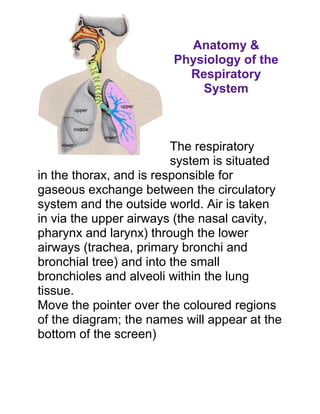
1. Anatomy &
Physiology of the
Respiratory
System
The respiratory
system is situated
in the thorax, and is responsible for
gaseous exchange between the circulatory
system and the outside world. Air is taken
in via the upper airways (the nasal cavity,
pharynx and larynx) through the lower
airways (trachea, primary bronchi and
bronchial tree) and into the small
bronchioles and alveoli within the lung
tissue.
Move the pointer over the coloured regions
of the diagram; the names will appear at the
bottom of the screen)
2. The lungs are divided into lobes; The left
lung is composed of the upper lobe, the
lower lobe and the lingula (a small
remnant next to the apex of the heart), the
right lung is composed of the upper, the
middle and the lower lobes.
Mechanics of Breathing
To take a breath in, the external intercostal
muscles contract, moving the ribcage up
and out. The diaphragm moves down at the
same time, creating negative pressure
within the thorax. The lungs are held to the
thoracic wall by the pleural membranes,
and so expand outwards as well. This
creates negative pressure within the lungs,
and so air rushes in through the upper and
lower airways.
Expiration is mainly due to the natural
elasticity of the lungs, which tend to
collapse if they are not held against the
thoracic wall. This is the mechanism behind
3. lung collapse if there is air in the pleural
space (pneumothorax).
Physiology of Gas Exchange
Each branch of the
bronchial tree eventually
sub-divides to form very
narrow terminal
bronchioles, which
terminate in the alveoli.
There are many millions
of alveloi in each lung,
and these are the areas responsible for
gaseous exchange, presenting a massive
surface area for exchange to occur over.
Each alveolus is very closely associated
with a network of capillaries containing
deoxygenated blood from the pulmonary
artery. The capillary and alveolar walls are
very thin, allowing rapid exchange of gases
4. by passive diffusion along concentration
gradients.
CO2 moves into the alveolus as the
concentration is much lower in the alveolus
than in the blood, and O2 moves out of the
alveolus as the continuous flow of blood
through the capillaries prevents saturation
of the blood with O2 and allows maximal
transfer across the membrane.
Terms Definitions
Contains nasal septum, turbinates,
nasal cavity
and cilia.
Divides nasal cavities into right and
nasal septum
left sides.
Bones that protrude into the nasal
cavity- they increase surface area for
turbinates
filtering dust and dirt particles by the
mucous membrane.
cilia Nose hairs, trap larger dirt particles.
sinuses Cavities in the skull, ducts connect
them to the nasal cavity, lined with
5. mucous membrane to warm and
moisten the air. Give resonance to
voice.
types of Frontal, maxillary, ethmoid, and
sinuses sphenoid.
Throat. Common passageway for air
pharynx
and food. 5" long.
When food is swallowed, this closes
over the opening to the larnyx,
epiglottis
preventing food from entering the
lungs.
Voice box. Triangular chamber below
larynx
pharynx. "Adam's Apple".
glottis Vocal cords within the larynx.
Windpipe. 4.5" long. Walls are
alternate bands of membrane and c-
shaped rings of hyaline cartilage to
trachea keep it open. Lined with ciliated
mucous membrane. Coughing and
expectoration gets rid of dust-laden
mucous.
6. Similar to trachea with ciliated
mucous membrane and hyaline
bronchi
cartilage. Lower end of trachea
divides into right and left this.
bronchial Cartilaginous plates (instead of c-
tubes shaped rings of trachea).
Thinner walls of smooth muscle, lined
with ciliated epithelium. Subdivision
bronchioles
of bronci. At the end, alveolar duct
and cluster of alveoli.
Composed of single layer of epithelial
tissue. Inner surfaces covered with
surfactant to keep from collapsing.
alveoli Each surrounded by capillaries.
Oxygen and carbon dioxide
exchange takes place between these
and capillaries.
Fill thoracic cavity. Tissue is porous
lungs
and spongy- it floats.
apex Upper part of lung.
base Lower part of lung.
7. Larger and shorter (displaced by
right lung
liver) and has three lobes.
Smaller (displaced by heart) and has
left lung
two lobes.
Thin, moist, slippery membrane that
covers lungs. Double-walled sac.
pleura
Space is pleural cavity- filled with
pleural fluid to prevent friciton.
Respiration (external, internal, and
cellular). Production of sound (vocal
cords). Pulmonary venilation.
functions of
Inspiration (intercostal muscles lift
the respiratory
ribs outward, sternum rises and the
system
diaphragm contracts and moves
downward- this increases the volume
of the lungs and the air rushes in).
pulmonary
Breathing.
venilation
1 inspiration and 1 expiration= 1
respiratory respiration. Normal adult= 14-20
movement respirations per minute. Increases
with exercise, body temperature, and
8. certain diseases. Age (newborn= 40-
60 per minute). Sleep= respirations
go down. Emotion can bring
respirations up or down.
Deep breath followed by forceful
coughing expulsion of air to clean lower
respiratory tract.
Spasm of diaphragm and spasmotic
hiccups closure of the glottis- irritation to
diaphragm or phrenic nerve.
Air forced through nose to clear
sneezing
respiratory tract.
Deep prolonged breath that fills the
yawning lungs, increases oxygen within the
blood.
Respiratory center located in medulla
neural factors
oblongata (in the brain). Increase in
of breathing
CO2 and decrease in O2 in the blood
control
will trigger respiratory center.
phrenic nerve Stimulates the diaphragm.
chemical Depends on the levels of CO2 in the
9. factors of blood. Chemoreceptors in aorta and
breathing carotid arteries sensitive to the
control amount of blood oxygen.