Super Vision Talk
•
1 like•293 views

One of my passion is science imagery. Data has become art. I wrote a book based on that idea and have given a presentation based on that book many times.
![Our Sensory Box ,[object Object], Dennis Kunkel](data:image/gif;base64,R0lGODlhAQABAIAAAAAAAP///yH5BAEAAAAALAAAAAABAAEAAAIBRAA7)
Recommended
More Related Content
What's hot
What's hot (19)
Similar to Super Vision Talk
Similar to Super Vision Talk (20)
Super Vision Talk
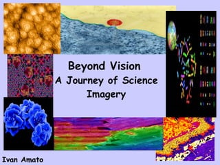
- 1. Beyond Vision A Journey of Science Imagery Ivan Amato
- 3. … our directly visible sliver of the EM spectrum…
- 4. Scientists Makes the Invisible Visible
- 5. Early examples of going beyond vision
- 9. The Science and Nature Gallery 10/30/98 3/8/2001
- 10. Portrait of the fundamentals BNL
- 11. Mandala of Structure Paul Midgely, University of Cambridge
- 12. Atomic Landscapes Joe Stroscio, et. al./ NIST
- 13. Germanium nanowires on a silicon substrate Teresa Clement, Arizona State University
- 14. Screw dislocation in trunk of lead sulfide nanowire yields helical result Mathew J. Bierman University of Wisconsin-Madison (MRS)
- 15. Titania film, 100 nm thick, peels from silicon substrate Martin Wagner, Institute of Thin Films and Interfaces, Juelich, Germany
- 16. Viral geometry Dennis Kunkel
- 17. A tantalum oxide particle with polystyrene contaminant “ walking” on the edge Georff Brennecka, Sandia (MRS)
- 18. Epoxy bristles, 250 nm in diameter, self-assemble,trapping a 2.5 micron polystyrene sphere as they do Boaz Pokroy, Harvard (MRS)
- 19. Inverse opal built with a template of polystyrene spheres of 330 nm diameter Elton Graugnard, Georgia Institute of Technology (MRS)
- 20. Simulation of electrons traveling through a solid in a volume the size of a bacterium Adam Heller Harvard University
- 21. When a surface scientist and visual artist collaborate, you get this. Michael Oliveri, Zhengwei Pan, University of Georgia
- 23. Markus Geisen/2001 Novartis Visions of Science Winner SEM of marine algae with calcite plates.
- 24. Copper Indium Selenide structures, from Angus Rockett’s laboratory.
- 25. Laser light fantastique G.Vander Rhodes, et. al, Boston Univ.
- 26. Cell division fireworks Conley Rieder and Alexey Khadjakov Division of Molecular Medicine Wadsworth Center
- 27. Bone-forming cell growing on a textured metal/metal oxide surface Altmayer, Barth, Shen, and Mathur Leibniz Institute of New Materials Saarbruecken, Germany (MRS)
- 28. Rendition, with visible light emission, in porous silicon of William Blake’s Ancient Days. Ee Jin Teo, National University of Singapore (MRS)
- 30. Components for self-healing polymer systems, from Nancy Sottos’ laboratory; photo by Ben Blaiszik
- 31. Copper indium selenide film with copper selenide plates and indium Selenide needles Olga Volobuzeva, Tallinn University of Technology, Estonia (MRS)
- 32. Snowflake a lá Low Temperature SEM W. Wergin, E. Erbe/USDA
- 33. Peter J. Lee, Applied Superconductivity Center, Florida State University.
- 35. Viewing a Sonic Boom DoD/Ensign John Gay
- 36. Deep water ocean waves encountering current eddies with refraction of waves coded by color Adam Heller, Harvard University
- 37. Rapatronic camera captures “rope tricks” in 1952 nuke test Operation Tumbler-Snapper
- 38. Sprites—upwardly striking, lightning-like atmospheric excitations—can take up mountain-like volumes.
- 39. Earth from above Chile-Bolivia Border Advanced Spaceborne Thermal Emission and Reflection Radiometer (ASTER) Image
- 40. NASA Landsat 7
- 41. Hurricane Isabel, September 17, 2003, as seen from NASA satellites using the Moderate Resolution Imaging Spectroradiometer (MODIS). University of Wisconsin, Space Science and Engineering Center (Liam Gumley)
- 42. Data Fusion: Rick Kohrs, Space Science & Engineering Center, UW
- 43. Cloud features on Uranus as seen on July 11-12, 2004, with a near-IR camera attached to Keck NIRC2 Telescope. Lawrence Sromovsky: UW-Madison Space Science and Engineering Center
- 44. Transition Region and Coronal Explorer (TRACE)
- 45. Hubble Vision
- 48. A black hole screaming Chandra X-ray Observatory J. McClintock and M. Garcia
- 49. WMAP—Map of the universe’s cosmic background radiation
Editor's Notes
- Mosquito Antenna, colorized, SEM
- Galileo, Robert Hooke, Christopher Wren and Thomas Willis, and Marcello Malphigi. Smithsonian images…from Science and the Artist Exhibition
- Star Nursery, Hubble Space Telescope
- Brass, metallography, slip lines between grains.
- Inside Out Opal (Inverse Opal)/Allied Signal/Made of carbon cages/photonics applications.NATURE cover. Eric Heller, Harvard University
- Bubble Chamber. Brookhaven National Laboratory
- Paul Midgely, University of Cambridge, Convergent Beam electron diffraction. Discs and lines reveal lattice parameters and details inside reveal atomic positions in unit cell.
- Joe Strocio, NIST. Cobalt singles and dimers on copper. Electron density visible in green and orange.
- Dennis Kunkel, T4 bacteriophages
- Geisen, Plankton
- Vertical Cavity Surface Emitting Laser, viewed with Near Scanning Optical Microscopy. Bosotn University
- Conley Rieder and Alexey Khadjakov, Mitosis. Stained DNA and tagged-antibody on spindle bodies.
- Stained neurons, optical microscope, Federation of American Societies for Experimental Biology
- Cryogenic confocal electron microscopy. Wergin at USDA.
- 3D sonography
- John Gay aboard USS Constellation. F/A-18 Hornet
- ASTER—Advanced Spaceborne Thermal Emission and Reflection Radiometer. Chile-Bolivia Border…Pampa Luxor Volcano.
- Transition Region and Coronal Explorer, Mosaic showing active regions
- Chandra/NASA/J.McClintock and M. Garcia