SlideShare a Scribd company logo
1 of 45
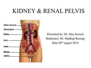
KIDNEY & RENAL PELVIS 
Presented by: Dr. Isha Jaiswal 
Moderator: Dr. Madhup Rastogi 
Date:20th august 2014 
1
KIDNEY : location 
• pair of organs located in the abdominal cavity 
on either side of the spine in a retroperitoneal 
position. 
• Approx. at vertebral level T12 to L3, 
• right kidney being slightly lower than the left. 
• Left kidney is little nearer to median plane than 
right 
• Long axis of kidney is directed downward and 
laterally &runs parallel to the lateral margin of 
the psoas muscle 
• The kidneys are mobile organs that move 
vertically within the retroperitoneum on 
average 0.9 cm to 1.3 cm and as much as 4 cm 
during normal respiration 
2
KIDNEYS :external features 
• Shape :Bean shaped 
• poles: upper & lower 
• Border: medial & lateral 
• Surface: anterior & posterior 
• Size: approx. 11–14 cm in length, 6 cm wide and 3cm thick 
• Weight: around 150 gm. in males & 135 gm. in females
External Features 
• Hilum of the kidney, 
• Concave medial border of 
the kidney 
• Structures enter / leave 
through the hilum (from 
anterior to posterior), 
» Renal vein 
» Renal artery 
» Pelvis 
» Ureter 
» Renal nerves 
» Lymphatics.
Relations of kidney 
 Upper pole: adrenal gland 
 Lower pole: about 2.5 cm above iliac creast 
 Posterior relations: 
 Diaphragm 
 Muscles: psoas major, 
quadratus lumborum, 
transv.abdominis 
 Ribs: 11th &12th ribs on left , 
only 12th on right side 
5
Anterior relations Right kidney: 
 Rt. adrenal gland 
 Liver 
 2nd part duodenum 
 Hepatic flexure colon 
 jejunum 
Left kidney 
 Lt. adrenal gland 
 Spleen 
 Stomach 
 Pancreas 
 splenic flexure colon 
 jejunum 
6
Coverings of kidney 
 fibrous tissue, the 
renal capsule, 
 perinephric fat, 
 renal fascia (of 
Gerota) 
 paranephric fat. 
7
Internal features 
parenchyma, of the kidney is 
divided into two major 
structures: 
Renal cortex: lobules & column 
medulla: pyramid,papilla,calyx
Functional unit of kidney: nephron 
9
10
URINARY DRAINAGE 
Pyramids 
Papillae 
Minor calyces 
Major calyces 
Renal pelvis
Blood supply of 
kidney 
Arterial supply: 
renal artery 
Venous drainage: 
renal vein
• Lymph Drainage : 
• Nerve Supply: 
• Through sympathetic plexus (T10 – L1) fibres 
• Afferent nerves T10 to T12 thoracic nerves. 
13 
The right kidney drains predominantly 
into the paracaval and interaortocaval 
lymph nodes 
left kidney drains exclusively to the 
para-aortic lymph nodes
CANCERS OF KIDNEY 
14
Renal Tumors: incidence 
In U.S in 2011 (ref:parez) 
• 60,920 cases diagnosed(4 % of all new cancers) 
• 13,120 deaths (2% of cancer related death) 
• Approx 88% of solid renal masses are malignant 
• RCC comprise 80-85%of primary kidney tumors 
• Transitional cell carcinoma acoount for 7 % of kidney tumors 
• Rest are lymphoma, sarcomas, oncocytoma 
15
Renal Cell Carcinoma 
• First described by Konig in 1826. 
• In 1883 Grawitz, noted the fatty content of cancer cells similar to that of 
adrenal cells. (Also called as Grawitz’s tumor) 
• All these tumors arise from Renal proximal tubular epithelium 
• The incidence of RCC is increasing & the size decreasing because of 
increased use of abdominal CT scans 
• Male predominance (1.6:1.0 M:F) 
• Highest incidence between age 50-70 
-Median age of diagnosis is 66 years 
-Median age of death 70 years 
16
Risk Factors 
• Tobacco smoking 
contributes to 24-30% of RCC cases 
Tobacco results in a 2-fold increased risk 
• Environmental: 
Cadmium, thorium-di-oxide, 
petroleum 
aresenic 
phenacetin analgesics. 
17
• Occupational: 
leather tanners, shoe workers, asbestos workers, petroleum, 
blast furnace, iron & steel industry 
• Hormonal: diethylistillbestrol, 
• Dietary: fried meats,(vegetables, fruits & alcohol are protective) 
18
• Obesity, 
• HTN, 
• DM 
• ACKD: 
• 50% Pt. on long term dialysis(>3 yrs) 
develop Acquired polycystic kidney disease, out of which 5.8% develops RCC 
19
RCC variant 
It is made up of no. of different types of cancers with different histology, different 
clinical courses and caused by different gene. 
A sarcomatoid variant represents1% to 6% of renal cell carcinoma and these tumors are 
associated with a significantly poorer prognosis. 
BHD=Birt-Hogg-Dubé; FH=fumarate hydratase; VHL=von Hippel-Lindau. 
20 
Clear cell 
75% 
Type 
Incidence (%) 
Associated 
mutations 
VHL 
Papillary type 1 
5% 
c-Met 
Papillary type 2 
10% 
FH 
Chromophobe 
5% 
BHD 
Oncocytoma 
5% 
BHD
Hereditary Renal Cancer Syndromes 
Syndrome Chromosome 
Location (Gene) 
Renal 
Manifestations 
Other Manifestations 
Von Hippel- 
Lindau (VHL) 
3p25 
VHL 
Clear cell renal 
carcinoma: solid 
and/or cystic, 
multiple and 
bilateral 
28%-45% 
Retinal and central nervous system 
hemangioblastomas; pheochromocytomas; 
pancreatic cysts and neuroendocrine 
tumors; endolymphatic sac tumors; 
epididymal and broad ligament 
cystadenomas 
21
Syndrome Chromosome 
Location (Gene) 
Renal Manifestations Other Manifestations 
Hereditary papillary renal 
carcinoma type1(HPRC) 
7q31 
MET 
Papillary renal carcinoma type 
1: solid, multiple and bilateral 
None 
Hereditary 
leiomyomatosis and renal 
cell carcinoma (HLRCC) 
1q42-43 
FH 
Papillary renal carcinoma type 
2, collecting duct carcinoma: 
solitary, aggressive 
Uterine leiomyomas and 
leiomyosarcomas; 
cutaneous leiomyomas 
22
Syndrome Chromosome 
Location (Gene) 
Renal Manifestations Other Manifestations 
Birt-Hogg-Dubé 
syndrome (BHD) 
17p11.2 
BHD 
Hybrid oncocytic renal tumors, 
chromophobe and clear cell renal 
carcinomas, oncocytomas: 
multiple, bilateral 
Benign tumors of hair follicle 
(fibrofolliculomas); lung 
cysts, spontaneous 
pneumothoraces 
Constitutional 
chromosome 3 
translocation 
3p; Not known; 
VHL somatic 
mutations 84% - 
98% 
Clear cell renal carcinoma: 
multiple, bilateral 
None 
23
Spread of renal cancer 
Local Infiltration :through the renal capsule to involve the 
perinephric fat and Gerota's fascia. 
Venous: The tumor may grow directly along the venous channels 
to the renal vein or vena cava. 
Lymph node metastases :involve the renal hilar, para-aortic, 
and paracaval lymph nodes 
Distant metastasis: lung, bone, bone, liver ,adrenal 
24
Natural History 
7% diagnosed incidentally 
45% present with localized disease at time of diagnosis 
25% with locally advanced disease at diagnosis 
30% with metastatic disease at diagnosis 
Lymph node metastases- 9% to 27% (renal hilar, para-aortic 
and paracaval) 
Renal vein – 21% & IVC 4% 
Distant metastases- lung (75%), soft tissue (36%), bone 
(20%), liver (18%), skin (8%) and CNS (8%) 
Ref: DeVita
• CLINICAL FEATURES 
50 % of RCC are now detected incidentally: Radiologist's tumor’ 
Triad of presentation: seen in only 10% pt., poor prognosis 
Pain (80%), 
Hematuria (45%) 
palpable mass (15%) 
26
Other signs and symptoms 
Weight loss (33%) 
 Fever (20%) 
 Hypertension (20%) 
 Hypercalcemia (5%) 
 Night sweats 
 Malaise 
 Varicocele usually left sided, due to obstruction of the 
testicular vein (2% of males) 
 Stauffer’s syndrome: 
Non metastatic hepatic dysfunction reported in 3-20% of cases 
Hepatic function normalizes in 60 to 70% of cases after nephrectomy. 
27
28 
Paraneoplastic syndromes are found in 20% of 
patients with RCC 
Elevated E.S.R. 
Hypertension 
Anemia 
Cachexia 
Pyrexia 
Abnormal liver function 
Hyper calcemia 
Polycythemia 
Neuromyopathy
Clinical presentation 
• History taking: 
• age, Sex,Occupation 
• Chief coimplains: 
 Pain :onset, duration, progress, nature, radiation, 
relation with micturition 
 Renal pain: painless or dull ache, at renal angle 
radiating along subcoastal area towards umbilicus 
along with fever loss of weight malaise 
 Ureteric pain: colic, start at renal angle radiate 
downward along course of ureter,referred to groin 
inner part of thigh, penis etc 
 Bladder pain is midline suprapubic dull, 
 Urethral pain: during or at end of micturition 
29
Haemeturia: 
• Amount, 
• Relation to micturition 
• Association with pain 
Beginning: urethral 
Toward end: vesical 
Throughout: prerenal,renal,vesical 
30
Renal lump 
• History:site onset duration progression 
• Examination 
• Inspection, 
• Palpation 
• Percussion 
• auscultation 
31
Inspection 
in recumbent position 
fulleness in lumbar region 
moves slightly with respiration 
palpation 
murphy’s punch test 
patient sits up 
press over the renal angle to see for tenderness 
32
Palpation of kidney: bimanual method 
• Features of a renal lump 
• Lies in loin 
• Can be moved in loin 
• Reniform shape 
• Ballotable 
• slightly move with respiration 
• fingers can be insinuated between coastal margin and swelling 
33 
• To palpate the left kidney, 
• reach across the client 
• place your left hand under the client’s left flank with your palm upward. 
• Elevate the left flank with your fingers, displacing the kidney upward. 
• Ask the client to take a deep breath 
• use the palmer surface of your right hand to palpate the kidney 
• Repeat the technique for the right kidney
Percussion 
features of a renal lump 
resonant anteriorly due to bowel loops 
dull posteriorly: enlarged kidney displaces colon 
Auscultation: bruit may be heard 
34
Diagnostic Work-Up 
• Laboratory studies 
– CBC, LFT's, alkaline phosphatase, BUN, creatinine, urinalysis 
• Radiographic studies- Increased use of imaging has increased the 
detection of renal lesions most of which are simple cysts. 
– X-Ray KUB region 
– Ultrasonography- Excellent in distinguishing cystic from solid masses 
– Intravenous Urography - Starting point for hematuria evaluations and 
function of contralateral kidney 
– Computed tomography- Provides an excellent assessment of the parenchyma 
and nodal status. 
– Magnetic Resonance Imaging - excellent demonstration of solid renal masses 
and is image test of choice to demonstrate extent of vena caval involvement 
with tumor. Useful in patients with renal insufficiency 
– MRI has no advantage compared with contrast enhanced CT for the diagnosis 
of RCC but it is better for staging of locally advanced cases 
35
Metastatic Work-Up 
• Chest X-ray or Chest CT 
• CT/MRI scan of abdomen or pelvis 
• Bone scan with plan films (for elevated alkaline 
phosphatase or bone pain). 
36
Radiological anatomy 
37
38
Figure : Computed tomography 
demonstrates a right renal carcinoma (m) 
with a large contralateral adrenal 
metastasis (a). 
Figure: CT scan shows large left renal mass 
with calcification (m) invading the left renal 
vein (arrow). 
39
Figure: T1-weighted magnetic 
resonance image demonstrates 
tumor (m) and vascular invasion 
(arrow). Flowing blood (v) in the 
left renal vein is black on this 
scan. 
Figure A: Axial T1-weighted 
image demonstrates a large left 
renal carcinoma with extension 
into the left renal vein (m) with 
protrusion into the IVC (v). B: 
Sagittal T1-weighted image 
shows the relation of the tumor 
thrombus (m) to the IVC (v) in 
the lateral projection. 
40
Renal Cell Carcinoma 
• 3D CT scan showing a left lower pole RCC extending into the 
renal hilum
Renal Cell Carcinoma 
• Multifocal renal cell carcinoma in a patient with Von Hippel Lindau disease. 
Patient had already undergone a right nephrectomy. Contrast-enhanced CT 
scan
Renal cell 
carcinoma of 
left kidney 
involving renal 
vein & inferior 
vena cava 
43
44
Thank you ! 
45

More Related Content

What's hot

Liver resection indications & methods
Liver resection   indications & methodsLiver resection   indications & methods
Liver resection indications & methodsDr Harsh Shah
 
Retroperitoneal mass
Retroperitoneal massRetroperitoneal mass
Retroperitoneal massKundan Singh
 
Multi cystic dysplastic kidney (renal dysplasia)
Multi cystic dysplastic kidney (renal dysplasia)Multi cystic dysplastic kidney (renal dysplasia)
Multi cystic dysplastic kidney (renal dysplasia)SUNIL KUMAR PEDDANA
 
Management of renal cell carcinoma and wilms' tumor
Management of renal cell carcinoma and wilms' tumor Management of renal cell carcinoma and wilms' tumor
Management of renal cell carcinoma and wilms' tumor Anil Gupta
 
Core Needle Biopsy of Breast : Updates
Core Needle Biopsy of Breast : Updates Core Needle Biopsy of Breast : Updates
Core Needle Biopsy of Breast : Updates Diya Das
 
Renal Cell Carcinoma Diagnosis And Management
Renal Cell Carcinoma Diagnosis And ManagementRenal Cell Carcinoma Diagnosis And Management
Renal Cell Carcinoma Diagnosis And ManagementRHMBONCO
 
TESTICULAR CANCERS
TESTICULAR CANCERSTESTICULAR CANCERS
TESTICULAR CANCERSIsha Jaiswal
 
Urolithiasis (urinary stones disease) presentation
Urolithiasis (urinary stones disease) presentationUrolithiasis (urinary stones disease) presentation
Urolithiasis (urinary stones disease) presentationAhmad Kharrouby
 

What's hot (20)

Liver resection indications & methods
Liver resection   indications & methodsLiver resection   indications & methods
Liver resection indications & methods
 
Carcinoma gall bladder
Carcinoma gall bladderCarcinoma gall bladder
Carcinoma gall bladder
 
Retroperitoneal mass
Retroperitoneal massRetroperitoneal mass
Retroperitoneal mass
 
Neoplasm of pancreas
Neoplasm of pancreasNeoplasm of pancreas
Neoplasm of pancreas
 
Renal tumor
Renal tumorRenal tumor
Renal tumor
 
Prostate Biopsy.pptx
Prostate Biopsy.pptxProstate Biopsy.pptx
Prostate Biopsy.pptx
 
Colorectal Cancer
Colorectal CancerColorectal Cancer
Colorectal Cancer
 
Multi cystic dysplastic kidney (renal dysplasia)
Multi cystic dysplastic kidney (renal dysplasia)Multi cystic dysplastic kidney (renal dysplasia)
Multi cystic dysplastic kidney (renal dysplasia)
 
Penile cancer
Penile cancerPenile cancer
Penile cancer
 
Management of renal cell carcinoma and wilms' tumor
Management of renal cell carcinoma and wilms' tumor Management of renal cell carcinoma and wilms' tumor
Management of renal cell carcinoma and wilms' tumor
 
Core Needle Biopsy of Breast : Updates
Core Needle Biopsy of Breast : Updates Core Needle Biopsy of Breast : Updates
Core Needle Biopsy of Breast : Updates
 
Simple renal cysts
Simple renal cystsSimple renal cysts
Simple renal cysts
 
Cancer of the Bladder
Cancer of the BladderCancer of the Bladder
Cancer of the Bladder
 
Renal Cell Carcinoma Diagnosis And Management
Renal Cell Carcinoma Diagnosis And ManagementRenal Cell Carcinoma Diagnosis And Management
Renal Cell Carcinoma Diagnosis And Management
 
Splenectomy
Splenectomy Splenectomy
Splenectomy
 
Cholangiocarcinoma
CholangiocarcinomaCholangiocarcinoma
Cholangiocarcinoma
 
TESTICULAR CANCERS
TESTICULAR CANCERSTESTICULAR CANCERS
TESTICULAR CANCERS
 
Hydronephrosis
HydronephrosisHydronephrosis
Hydronephrosis
 
Renal cell carcinoma
Renal cell carcinomaRenal cell carcinoma
Renal cell carcinoma
 
Urolithiasis (urinary stones disease) presentation
Urolithiasis (urinary stones disease) presentationUrolithiasis (urinary stones disease) presentation
Urolithiasis (urinary stones disease) presentation
 

Viewers also liked

MANAGEMENT OF EARLY STAGE NON SMALL CELL LUNG CARCINOMA
MANAGEMENT OF EARLY STAGE NON SMALL CELL LUNG CARCINOMAMANAGEMENT OF EARLY STAGE NON SMALL CELL LUNG CARCINOMA
MANAGEMENT OF EARLY STAGE NON SMALL CELL LUNG CARCINOMAIsha Jaiswal
 
MANAGEMENT OF CA COLON
MANAGEMENT OF CA COLONMANAGEMENT OF CA COLON
MANAGEMENT OF CA COLONIsha Jaiswal
 
MANAGEMENT OF GLIOMAS
MANAGEMENT OF GLIOMASMANAGEMENT OF GLIOMAS
MANAGEMENT OF GLIOMASIsha Jaiswal
 
BRACHYTHERAPY IN ORAL CAVITY
BRACHYTHERAPY IN ORAL CAVITYBRACHYTHERAPY IN ORAL CAVITY
BRACHYTHERAPY IN ORAL CAVITYIsha Jaiswal
 
Cell survival curve
Cell survival curve Cell survival curve
Cell survival curve Isha Jaiswal
 
Conventional Brachytherapy in carcinoma cervix
Conventional Brachytherapy in carcinoma cervixConventional Brachytherapy in carcinoma cervix
Conventional Brachytherapy in carcinoma cervixIsha Jaiswal
 
radiation therapy in ca breast
radiation therapy in ca breast   radiation therapy in ca breast
radiation therapy in ca breast Isha Jaiswal
 
Management of carcinoma hypopharynx
 Management  of carcinoma hypopharynx  Management  of carcinoma hypopharynx
Management of carcinoma hypopharynx Isha Jaiswal
 
Radiation absorbtion
Radiation absorbtionRadiation absorbtion
Radiation absorbtionIsha Jaiswal
 
RADIOBIOLOGY: oxygen effect & reoxygenation
RADIOBIOLOGY: oxygen effect & reoxygenationRADIOBIOLOGY: oxygen effect & reoxygenation
RADIOBIOLOGY: oxygen effect & reoxygenationIsha Jaiswal
 
Breast: Carcinoma in situ management
Breast: Carcinoma in situ management Breast: Carcinoma in situ management
Breast: Carcinoma in situ management Isha Jaiswal
 
Liver & billary apparatus
Liver & billary apparatusLiver & billary apparatus
Liver & billary apparatusIsha Jaiswal
 
Ovarian & endometrial cancer
Ovarian & endometrial cancerOvarian & endometrial cancer
Ovarian & endometrial cancerIsha Jaiswal
 
management of carcinoma hypopharynx
management of carcinoma hypopharynxmanagement of carcinoma hypopharynx
management of carcinoma hypopharynxIsha Jaiswal
 
RADIATION CARCINOGENESIS
RADIATION CARCINOGENESISRADIATION CARCINOGENESIS
RADIATION CARCINOGENESISIsha Jaiswal
 

Viewers also liked (20)

Renal anatomy
Renal anatomyRenal anatomy
Renal anatomy
 
MANAGEMENT OF EARLY STAGE NON SMALL CELL LUNG CARCINOMA
MANAGEMENT OF EARLY STAGE NON SMALL CELL LUNG CARCINOMAMANAGEMENT OF EARLY STAGE NON SMALL CELL LUNG CARCINOMA
MANAGEMENT OF EARLY STAGE NON SMALL CELL LUNG CARCINOMA
 
MANAGEMENT OF CA COLON
MANAGEMENT OF CA COLONMANAGEMENT OF CA COLON
MANAGEMENT OF CA COLON
 
MANAGEMENT OF GLIOMAS
MANAGEMENT OF GLIOMASMANAGEMENT OF GLIOMAS
MANAGEMENT OF GLIOMAS
 
BRACHYTHERAPY IN ORAL CAVITY
BRACHYTHERAPY IN ORAL CAVITYBRACHYTHERAPY IN ORAL CAVITY
BRACHYTHERAPY IN ORAL CAVITY
 
Cell survival curve
Cell survival curve Cell survival curve
Cell survival curve
 
Conventional Brachytherapy in carcinoma cervix
Conventional Brachytherapy in carcinoma cervixConventional Brachytherapy in carcinoma cervix
Conventional Brachytherapy in carcinoma cervix
 
radiation therapy in ca breast
radiation therapy in ca breast   radiation therapy in ca breast
radiation therapy in ca breast
 
Management of carcinoma hypopharynx
 Management  of carcinoma hypopharynx  Management  of carcinoma hypopharynx
Management of carcinoma hypopharynx
 
Renal cell carcinoma
Renal cell carcinomaRenal cell carcinoma
Renal cell carcinoma
 
Radiation absorbtion
Radiation absorbtionRadiation absorbtion
Radiation absorbtion
 
Atomic structure
Atomic structureAtomic structure
Atomic structure
 
RADIOBIOLOGY: oxygen effect & reoxygenation
RADIOBIOLOGY: oxygen effect & reoxygenationRADIOBIOLOGY: oxygen effect & reoxygenation
RADIOBIOLOGY: oxygen effect & reoxygenation
 
Breast: Carcinoma in situ management
Breast: Carcinoma in situ management Breast: Carcinoma in situ management
Breast: Carcinoma in situ management
 
Liver & billary apparatus
Liver & billary apparatusLiver & billary apparatus
Liver & billary apparatus
 
Ovarian & endometrial cancer
Ovarian & endometrial cancerOvarian & endometrial cancer
Ovarian & endometrial cancer
 
management of carcinoma hypopharynx
management of carcinoma hypopharynxmanagement of carcinoma hypopharynx
management of carcinoma hypopharynx
 
RADIATION CARCINOGENESIS
RADIATION CARCINOGENESISRADIATION CARCINOGENESIS
RADIATION CARCINOGENESIS
 
BREAST CANCER
BREAST CANCERBREAST CANCER
BREAST CANCER
 
Nasopharynx
NasopharynxNasopharynx
Nasopharynx
 

Similar to RENAL ANATOMY & RENAL CELL CANCERS

Urinary system – common pathological correlation
Urinary system – common pathological correlationUrinary system – common pathological correlation
Urinary system – common pathological correlationKochi Chia
 
Renal cell carcinoma
Renal cell carcinomaRenal cell carcinoma
Renal cell carcinomagbanstha
 
Ca kidney [edmond]
Ca kidney [edmond]Ca kidney [edmond]
Ca kidney [edmond]Edmond Wong
 
Non hodgkin lymphoma
Non hodgkin lymphomaNon hodgkin lymphoma
Non hodgkin lymphomaAli Azher
 
Kidney tumors/ renal tumors - malignant benign
Kidney tumors/ renal tumors - malignant benignKidney tumors/ renal tumors - malignant benign
Kidney tumors/ renal tumors - malignant benignRishikRana3
 
osteoblastic osteosarcoma.pptx
osteoblastic osteosarcoma.pptxosteoblastic osteosarcoma.pptx
osteoblastic osteosarcoma.pptxFaiza361083
 
Benign enlargement of prostate
Benign enlargement of prostateBenign enlargement of prostate
Benign enlargement of prostatesunil kumar daha
 
GASTRIC CANCER.pptx
GASTRIC CANCER.pptxGASTRIC CANCER.pptx
GASTRIC CANCER.pptxAishaAkram13
 
Ca Papillary(Thyroid Gland)
Ca Papillary(Thyroid Gland)Ca Papillary(Thyroid Gland)
Ca Papillary(Thyroid Gland)DRASIMSHAHZAD1
 
Ultrasound of the urinary tract - Renal cysts
Ultrasound of the urinary tract - Renal cystsUltrasound of the urinary tract - Renal cysts
Ultrasound of the urinary tract - Renal cystsSamir Haffar
 
testicular carcinoma - Dr ravi.pptx
testicular carcinoma - Dr ravi.pptxtesticular carcinoma - Dr ravi.pptx
testicular carcinoma - Dr ravi.pptxAmandeepSingh952
 
Imaging features of hepatobiliary and pancretic ds
Imaging features of hepatobiliary and pancretic dsImaging features of hepatobiliary and pancretic ds
Imaging features of hepatobiliary and pancretic dsradiologyoffice
 
cystickidneydiseases-161210202710 (1).pdf
cystickidneydiseases-161210202710 (1).pdfcystickidneydiseases-161210202710 (1).pdf
cystickidneydiseases-161210202710 (1).pdfHARYANVITRAVLLER
 

Similar to RENAL ANATOMY & RENAL CELL CANCERS (20)

Urinary system – common pathological correlation
Urinary system – common pathological correlationUrinary system – common pathological correlation
Urinary system – common pathological correlation
 
Torsion of spleen
Torsion of spleenTorsion of spleen
Torsion of spleen
 
Renal cell carcinoma
Renal cell carcinomaRenal cell carcinoma
Renal cell carcinoma
 
Ca kidney [edmond]
Ca kidney [edmond]Ca kidney [edmond]
Ca kidney [edmond]
 
Carcinoma gb
Carcinoma gbCarcinoma gb
Carcinoma gb
 
Renal mass
Renal mass Renal mass
Renal mass
 
Non hodgkin lymphoma
Non hodgkin lymphomaNon hodgkin lymphoma
Non hodgkin lymphoma
 
Esophageal cancer
Esophageal cancerEsophageal cancer
Esophageal cancer
 
Kidney tumors/ renal tumors - malignant benign
Kidney tumors/ renal tumors - malignant benignKidney tumors/ renal tumors - malignant benign
Kidney tumors/ renal tumors - malignant benign
 
Pancreatic Cancer
Pancreatic CancerPancreatic Cancer
Pancreatic Cancer
 
osteoblastic osteosarcoma.pptx
osteoblastic osteosarcoma.pptxosteoblastic osteosarcoma.pptx
osteoblastic osteosarcoma.pptx
 
Benign enlargement of prostate
Benign enlargement of prostateBenign enlargement of prostate
Benign enlargement of prostate
 
Urology
UrologyUrology
Urology
 
GASTRIC CANCER.pptx
GASTRIC CANCER.pptxGASTRIC CANCER.pptx
GASTRIC CANCER.pptx
 
Ca Papillary(Thyroid Gland)
Ca Papillary(Thyroid Gland)Ca Papillary(Thyroid Gland)
Ca Papillary(Thyroid Gland)
 
Ultrasound of the urinary tract - Renal cysts
Ultrasound of the urinary tract - Renal cystsUltrasound of the urinary tract - Renal cysts
Ultrasound of the urinary tract - Renal cysts
 
testicular carcinoma - Dr ravi.pptx
testicular carcinoma - Dr ravi.pptxtesticular carcinoma - Dr ravi.pptx
testicular carcinoma - Dr ravi.pptx
 
Imaging features of hepatobiliary and pancretic ds
Imaging features of hepatobiliary and pancretic dsImaging features of hepatobiliary and pancretic ds
Imaging features of hepatobiliary and pancretic ds
 
Renal cell carcinoma
Renal cell carcinomaRenal cell carcinoma
Renal cell carcinoma
 
cystickidneydiseases-161210202710 (1).pdf
cystickidneydiseases-161210202710 (1).pdfcystickidneydiseases-161210202710 (1).pdf
cystickidneydiseases-161210202710 (1).pdf
 

More from Isha Jaiswal

Physical Models For Time Dose & Fractionation
Physical Models For Time Dose & FractionationPhysical Models For Time Dose & Fractionation
Physical Models For Time Dose & FractionationIsha Jaiswal
 
TIME DOSE & FRACTIONATION
TIME DOSE & FRACTIONATIONTIME DOSE & FRACTIONATION
TIME DOSE & FRACTIONATIONIsha Jaiswal
 
Preoperative versus postoperative chemoradiotherapy for rectal cancer
Preoperative versus postoperative chemoradiotherapy for rectal cancerPreoperative versus postoperative chemoradiotherapy for rectal cancer
Preoperative versus postoperative chemoradiotherapy for rectal cancerIsha Jaiswal
 
Radiotherapy in ca esophagus
Radiotherapy in ca esophagusRadiotherapy in ca esophagus
Radiotherapy in ca esophagusIsha Jaiswal
 
RADIO LOGICAL ANATOMY OF HEAD AND NECK CANCERS
RADIO LOGICAL ANATOMY OF HEAD AND NECK CANCERSRADIO LOGICAL ANATOMY OF HEAD AND NECK CANCERS
RADIO LOGICAL ANATOMY OF HEAD AND NECK CANCERSIsha Jaiswal
 
IMAGING & ITS ROLE IN FEMALE GENITAL CANCER
IMAGING & ITS ROLE IN FEMALE GENITAL CANCERIMAGING & ITS ROLE IN FEMALE GENITAL CANCER
IMAGING & ITS ROLE IN FEMALE GENITAL CANCERIsha Jaiswal
 
Radiological anatomy of lymph node
Radiological anatomy of lymph nodeRadiological anatomy of lymph node
Radiological anatomy of lymph nodeIsha Jaiswal
 
EVOLUTION OF CHEMOTHERAPY IN BREAST CANCER
EVOLUTION OF CHEMOTHERAPY IN BREAST CANCEREVOLUTION OF CHEMOTHERAPY IN BREAST CANCER
EVOLUTION OF CHEMOTHERAPY IN BREAST CANCERIsha Jaiswal
 
carcinoma urinary bladder management
carcinoma urinary bladder management carcinoma urinary bladder management
carcinoma urinary bladder management Isha Jaiswal
 
MANAGEMENT OF HEPATOCELLULAR CARCINOMA
MANAGEMENT OF HEPATOCELLULAR CARCINOMAMANAGEMENT OF HEPATOCELLULAR CARCINOMA
MANAGEMENT OF HEPATOCELLULAR CARCINOMAIsha Jaiswal
 
EBRT IN CARCINOMA CERVIX
EBRT IN CARCINOMA CERVIXEBRT IN CARCINOMA CERVIX
EBRT IN CARCINOMA CERVIXIsha Jaiswal
 
image guided brachytherapy carcinoma cervix
image guided brachytherapy carcinoma cerviximage guided brachytherapy carcinoma cervix
image guided brachytherapy carcinoma cervixIsha Jaiswal
 
Managememt of Carcinoma Nasopharynx
Managememt  of Carcinoma NasopharynxManagememt  of Carcinoma Nasopharynx
Managememt of Carcinoma NasopharynxIsha Jaiswal
 

More from Isha Jaiswal (14)

Physical Models For Time Dose & Fractionation
Physical Models For Time Dose & FractionationPhysical Models For Time Dose & Fractionation
Physical Models For Time Dose & Fractionation
 
TIME DOSE & FRACTIONATION
TIME DOSE & FRACTIONATIONTIME DOSE & FRACTIONATION
TIME DOSE & FRACTIONATION
 
Preoperative versus postoperative chemoradiotherapy for rectal cancer
Preoperative versus postoperative chemoradiotherapy for rectal cancerPreoperative versus postoperative chemoradiotherapy for rectal cancer
Preoperative versus postoperative chemoradiotherapy for rectal cancer
 
Radiotherapy in ca esophagus
Radiotherapy in ca esophagusRadiotherapy in ca esophagus
Radiotherapy in ca esophagus
 
RADIO LOGICAL ANATOMY OF HEAD AND NECK CANCERS
RADIO LOGICAL ANATOMY OF HEAD AND NECK CANCERSRADIO LOGICAL ANATOMY OF HEAD AND NECK CANCERS
RADIO LOGICAL ANATOMY OF HEAD AND NECK CANCERS
 
IMAGING & ITS ROLE IN FEMALE GENITAL CANCER
IMAGING & ITS ROLE IN FEMALE GENITAL CANCERIMAGING & ITS ROLE IN FEMALE GENITAL CANCER
IMAGING & ITS ROLE IN FEMALE GENITAL CANCER
 
Radiological anatomy of lymph node
Radiological anatomy of lymph nodeRadiological anatomy of lymph node
Radiological anatomy of lymph node
 
EVOLUTION OF CHEMOTHERAPY IN BREAST CANCER
EVOLUTION OF CHEMOTHERAPY IN BREAST CANCEREVOLUTION OF CHEMOTHERAPY IN BREAST CANCER
EVOLUTION OF CHEMOTHERAPY IN BREAST CANCER
 
carcinoma urinary bladder management
carcinoma urinary bladder management carcinoma urinary bladder management
carcinoma urinary bladder management
 
MANAGEMENT OF HEPATOCELLULAR CARCINOMA
MANAGEMENT OF HEPATOCELLULAR CARCINOMAMANAGEMENT OF HEPATOCELLULAR CARCINOMA
MANAGEMENT OF HEPATOCELLULAR CARCINOMA
 
EBRT IN CARCINOMA CERVIX
EBRT IN CARCINOMA CERVIXEBRT IN CARCINOMA CERVIX
EBRT IN CARCINOMA CERVIX
 
image guided brachytherapy carcinoma cervix
image guided brachytherapy carcinoma cerviximage guided brachytherapy carcinoma cervix
image guided brachytherapy carcinoma cervix
 
Managememt of Carcinoma Nasopharynx
Managememt  of Carcinoma NasopharynxManagememt  of Carcinoma Nasopharynx
Managememt of Carcinoma Nasopharynx
 
Surface anatomy
Surface anatomySurface anatomy
Surface anatomy
 

Recently uploaded

Choosing the Right CBSE School A Comprehensive Guide for Parents
Choosing the Right CBSE School A Comprehensive Guide for ParentsChoosing the Right CBSE School A Comprehensive Guide for Parents
Choosing the Right CBSE School A Comprehensive Guide for Parentsnavabharathschool99
 
Difference Between Search & Browse Methods in Odoo 17
Difference Between Search & Browse Methods in Odoo 17Difference Between Search & Browse Methods in Odoo 17
Difference Between Search & Browse Methods in Odoo 17Celine George
 
THEORIES OF ORGANIZATION-PUBLIC ADMINISTRATION
THEORIES OF ORGANIZATION-PUBLIC ADMINISTRATIONTHEORIES OF ORGANIZATION-PUBLIC ADMINISTRATION
THEORIES OF ORGANIZATION-PUBLIC ADMINISTRATIONHumphrey A Beña
 
ANG SEKTOR NG agrikultura.pptx QUARTER 4
ANG SEKTOR NG agrikultura.pptx QUARTER 4ANG SEKTOR NG agrikultura.pptx QUARTER 4
ANG SEKTOR NG agrikultura.pptx QUARTER 4MiaBumagat1
 
Activity 2-unit 2-update 2024. English translation
Activity 2-unit 2-update 2024. English translationActivity 2-unit 2-update 2024. English translation
Activity 2-unit 2-update 2024. English translationRosabel UA
 
Full Stack Web Development Course for Beginners
Full Stack Web Development Course  for BeginnersFull Stack Web Development Course  for Beginners
Full Stack Web Development Course for BeginnersSabitha Banu
 
4.16.24 21st Century Movements for Black Lives.pptx
4.16.24 21st Century Movements for Black Lives.pptx4.16.24 21st Century Movements for Black Lives.pptx
4.16.24 21st Century Movements for Black Lives.pptxmary850239
 
MULTIDISCIPLINRY NATURE OF THE ENVIRONMENTAL STUDIES.pptx
MULTIDISCIPLINRY NATURE OF THE ENVIRONMENTAL STUDIES.pptxMULTIDISCIPLINRY NATURE OF THE ENVIRONMENTAL STUDIES.pptx
MULTIDISCIPLINRY NATURE OF THE ENVIRONMENTAL STUDIES.pptxAnupkumar Sharma
 
ENGLISH 7_Q4_LESSON 2_ Employing a Variety of Strategies for Effective Interp...
ENGLISH 7_Q4_LESSON 2_ Employing a Variety of Strategies for Effective Interp...ENGLISH 7_Q4_LESSON 2_ Employing a Variety of Strategies for Effective Interp...
ENGLISH 7_Q4_LESSON 2_ Employing a Variety of Strategies for Effective Interp...JhezDiaz1
 
Global Lehigh Strategic Initiatives (without descriptions)
Global Lehigh Strategic Initiatives (without descriptions)Global Lehigh Strategic Initiatives (without descriptions)
Global Lehigh Strategic Initiatives (without descriptions)cama23
 
Concurrency Control in Database Management system
Concurrency Control in Database Management systemConcurrency Control in Database Management system
Concurrency Control in Database Management systemChristalin Nelson
 
Field Attribute Index Feature in Odoo 17
Field Attribute Index Feature in Odoo 17Field Attribute Index Feature in Odoo 17
Field Attribute Index Feature in Odoo 17Celine George
 
What is Model Inheritance in Odoo 17 ERP
What is Model Inheritance in Odoo 17 ERPWhat is Model Inheritance in Odoo 17 ERP
What is Model Inheritance in Odoo 17 ERPCeline George
 
Virtual-Orientation-on-the-Administration-of-NATG12-NATG6-and-ELLNA.pdf
Virtual-Orientation-on-the-Administration-of-NATG12-NATG6-and-ELLNA.pdfVirtual-Orientation-on-the-Administration-of-NATG12-NATG6-and-ELLNA.pdf
Virtual-Orientation-on-the-Administration-of-NATG12-NATG6-and-ELLNA.pdfErwinPantujan2
 
Daily Lesson Plan in Mathematics Quarter 4
Daily Lesson Plan in Mathematics Quarter 4Daily Lesson Plan in Mathematics Quarter 4
Daily Lesson Plan in Mathematics Quarter 4JOYLYNSAMANIEGO
 
USPS® Forced Meter Migration - How to Know if Your Postage Meter Will Soon be...
USPS® Forced Meter Migration - How to Know if Your Postage Meter Will Soon be...USPS® Forced Meter Migration - How to Know if Your Postage Meter Will Soon be...
USPS® Forced Meter Migration - How to Know if Your Postage Meter Will Soon be...Postal Advocate Inc.
 
Q4-PPT-Music9_Lesson-1-Romantic-Opera.pptx
Q4-PPT-Music9_Lesson-1-Romantic-Opera.pptxQ4-PPT-Music9_Lesson-1-Romantic-Opera.pptx
Q4-PPT-Music9_Lesson-1-Romantic-Opera.pptxlancelewisportillo
 
Food processing presentation for bsc agriculture hons
Food processing presentation for bsc agriculture honsFood processing presentation for bsc agriculture hons
Food processing presentation for bsc agriculture honsManeerUddin
 

Recently uploaded (20)

Choosing the Right CBSE School A Comprehensive Guide for Parents
Choosing the Right CBSE School A Comprehensive Guide for ParentsChoosing the Right CBSE School A Comprehensive Guide for Parents
Choosing the Right CBSE School A Comprehensive Guide for Parents
 
Difference Between Search & Browse Methods in Odoo 17
Difference Between Search & Browse Methods in Odoo 17Difference Between Search & Browse Methods in Odoo 17
Difference Between Search & Browse Methods in Odoo 17
 
THEORIES OF ORGANIZATION-PUBLIC ADMINISTRATION
THEORIES OF ORGANIZATION-PUBLIC ADMINISTRATIONTHEORIES OF ORGANIZATION-PUBLIC ADMINISTRATION
THEORIES OF ORGANIZATION-PUBLIC ADMINISTRATION
 
ANG SEKTOR NG agrikultura.pptx QUARTER 4
ANG SEKTOR NG agrikultura.pptx QUARTER 4ANG SEKTOR NG agrikultura.pptx QUARTER 4
ANG SEKTOR NG agrikultura.pptx QUARTER 4
 
Activity 2-unit 2-update 2024. English translation
Activity 2-unit 2-update 2024. English translationActivity 2-unit 2-update 2024. English translation
Activity 2-unit 2-update 2024. English translation
 
Full Stack Web Development Course for Beginners
Full Stack Web Development Course  for BeginnersFull Stack Web Development Course  for Beginners
Full Stack Web Development Course for Beginners
 
4.16.24 21st Century Movements for Black Lives.pptx
4.16.24 21st Century Movements for Black Lives.pptx4.16.24 21st Century Movements for Black Lives.pptx
4.16.24 21st Century Movements for Black Lives.pptx
 
MULTIDISCIPLINRY NATURE OF THE ENVIRONMENTAL STUDIES.pptx
MULTIDISCIPLINRY NATURE OF THE ENVIRONMENTAL STUDIES.pptxMULTIDISCIPLINRY NATURE OF THE ENVIRONMENTAL STUDIES.pptx
MULTIDISCIPLINRY NATURE OF THE ENVIRONMENTAL STUDIES.pptx
 
ENGLISH 7_Q4_LESSON 2_ Employing a Variety of Strategies for Effective Interp...
ENGLISH 7_Q4_LESSON 2_ Employing a Variety of Strategies for Effective Interp...ENGLISH 7_Q4_LESSON 2_ Employing a Variety of Strategies for Effective Interp...
ENGLISH 7_Q4_LESSON 2_ Employing a Variety of Strategies for Effective Interp...
 
Global Lehigh Strategic Initiatives (without descriptions)
Global Lehigh Strategic Initiatives (without descriptions)Global Lehigh Strategic Initiatives (without descriptions)
Global Lehigh Strategic Initiatives (without descriptions)
 
Concurrency Control in Database Management system
Concurrency Control in Database Management systemConcurrency Control in Database Management system
Concurrency Control in Database Management system
 
Field Attribute Index Feature in Odoo 17
Field Attribute Index Feature in Odoo 17Field Attribute Index Feature in Odoo 17
Field Attribute Index Feature in Odoo 17
 
LEFT_ON_C'N_ PRELIMS_EL_DORADO_2024.pptx
LEFT_ON_C'N_ PRELIMS_EL_DORADO_2024.pptxLEFT_ON_C'N_ PRELIMS_EL_DORADO_2024.pptx
LEFT_ON_C'N_ PRELIMS_EL_DORADO_2024.pptx
 
What is Model Inheritance in Odoo 17 ERP
What is Model Inheritance in Odoo 17 ERPWhat is Model Inheritance in Odoo 17 ERP
What is Model Inheritance in Odoo 17 ERP
 
Virtual-Orientation-on-the-Administration-of-NATG12-NATG6-and-ELLNA.pdf
Virtual-Orientation-on-the-Administration-of-NATG12-NATG6-and-ELLNA.pdfVirtual-Orientation-on-the-Administration-of-NATG12-NATG6-and-ELLNA.pdf
Virtual-Orientation-on-the-Administration-of-NATG12-NATG6-and-ELLNA.pdf
 
Daily Lesson Plan in Mathematics Quarter 4
Daily Lesson Plan in Mathematics Quarter 4Daily Lesson Plan in Mathematics Quarter 4
Daily Lesson Plan in Mathematics Quarter 4
 
USPS® Forced Meter Migration - How to Know if Your Postage Meter Will Soon be...
USPS® Forced Meter Migration - How to Know if Your Postage Meter Will Soon be...USPS® Forced Meter Migration - How to Know if Your Postage Meter Will Soon be...
USPS® Forced Meter Migration - How to Know if Your Postage Meter Will Soon be...
 
FINALS_OF_LEFT_ON_C'N_EL_DORADO_2024.pptx
FINALS_OF_LEFT_ON_C'N_EL_DORADO_2024.pptxFINALS_OF_LEFT_ON_C'N_EL_DORADO_2024.pptx
FINALS_OF_LEFT_ON_C'N_EL_DORADO_2024.pptx
 
Q4-PPT-Music9_Lesson-1-Romantic-Opera.pptx
Q4-PPT-Music9_Lesson-1-Romantic-Opera.pptxQ4-PPT-Music9_Lesson-1-Romantic-Opera.pptx
Q4-PPT-Music9_Lesson-1-Romantic-Opera.pptx
 
Food processing presentation for bsc agriculture hons
Food processing presentation for bsc agriculture honsFood processing presentation for bsc agriculture hons
Food processing presentation for bsc agriculture hons
 

RENAL ANATOMY & RENAL CELL CANCERS

  • 1. KIDNEY & RENAL PELVIS Presented by: Dr. Isha Jaiswal Moderator: Dr. Madhup Rastogi Date:20th august 2014 1
  • 2. KIDNEY : location • pair of organs located in the abdominal cavity on either side of the spine in a retroperitoneal position. • Approx. at vertebral level T12 to L3, • right kidney being slightly lower than the left. • Left kidney is little nearer to median plane than right • Long axis of kidney is directed downward and laterally &runs parallel to the lateral margin of the psoas muscle • The kidneys are mobile organs that move vertically within the retroperitoneum on average 0.9 cm to 1.3 cm and as much as 4 cm during normal respiration 2
  • 3. KIDNEYS :external features • Shape :Bean shaped • poles: upper & lower • Border: medial & lateral • Surface: anterior & posterior • Size: approx. 11–14 cm in length, 6 cm wide and 3cm thick • Weight: around 150 gm. in males & 135 gm. in females
  • 4. External Features • Hilum of the kidney, • Concave medial border of the kidney • Structures enter / leave through the hilum (from anterior to posterior), » Renal vein » Renal artery » Pelvis » Ureter » Renal nerves » Lymphatics.
  • 5. Relations of kidney  Upper pole: adrenal gland  Lower pole: about 2.5 cm above iliac creast  Posterior relations:  Diaphragm  Muscles: psoas major, quadratus lumborum, transv.abdominis  Ribs: 11th &12th ribs on left , only 12th on right side 5
  • 6. Anterior relations Right kidney:  Rt. adrenal gland  Liver  2nd part duodenum  Hepatic flexure colon  jejunum Left kidney  Lt. adrenal gland  Spleen  Stomach  Pancreas  splenic flexure colon  jejunum 6
  • 7. Coverings of kidney  fibrous tissue, the renal capsule,  perinephric fat,  renal fascia (of Gerota)  paranephric fat. 7
  • 8. Internal features parenchyma, of the kidney is divided into two major structures: Renal cortex: lobules & column medulla: pyramid,papilla,calyx
  • 9. Functional unit of kidney: nephron 9
  • 10. 10
  • 11. URINARY DRAINAGE Pyramids Papillae Minor calyces Major calyces Renal pelvis
  • 12. Blood supply of kidney Arterial supply: renal artery Venous drainage: renal vein
  • 13. • Lymph Drainage : • Nerve Supply: • Through sympathetic plexus (T10 – L1) fibres • Afferent nerves T10 to T12 thoracic nerves. 13 The right kidney drains predominantly into the paracaval and interaortocaval lymph nodes left kidney drains exclusively to the para-aortic lymph nodes
  • 15. Renal Tumors: incidence In U.S in 2011 (ref:parez) • 60,920 cases diagnosed(4 % of all new cancers) • 13,120 deaths (2% of cancer related death) • Approx 88% of solid renal masses are malignant • RCC comprise 80-85%of primary kidney tumors • Transitional cell carcinoma acoount for 7 % of kidney tumors • Rest are lymphoma, sarcomas, oncocytoma 15
  • 16. Renal Cell Carcinoma • First described by Konig in 1826. • In 1883 Grawitz, noted the fatty content of cancer cells similar to that of adrenal cells. (Also called as Grawitz’s tumor) • All these tumors arise from Renal proximal tubular epithelium • The incidence of RCC is increasing & the size decreasing because of increased use of abdominal CT scans • Male predominance (1.6:1.0 M:F) • Highest incidence between age 50-70 -Median age of diagnosis is 66 years -Median age of death 70 years 16
  • 17. Risk Factors • Tobacco smoking contributes to 24-30% of RCC cases Tobacco results in a 2-fold increased risk • Environmental: Cadmium, thorium-di-oxide, petroleum aresenic phenacetin analgesics. 17
  • 18. • Occupational: leather tanners, shoe workers, asbestos workers, petroleum, blast furnace, iron & steel industry • Hormonal: diethylistillbestrol, • Dietary: fried meats,(vegetables, fruits & alcohol are protective) 18
  • 19. • Obesity, • HTN, • DM • ACKD: • 50% Pt. on long term dialysis(>3 yrs) develop Acquired polycystic kidney disease, out of which 5.8% develops RCC 19
  • 20. RCC variant It is made up of no. of different types of cancers with different histology, different clinical courses and caused by different gene. A sarcomatoid variant represents1% to 6% of renal cell carcinoma and these tumors are associated with a significantly poorer prognosis. BHD=Birt-Hogg-Dubé; FH=fumarate hydratase; VHL=von Hippel-Lindau. 20 Clear cell 75% Type Incidence (%) Associated mutations VHL Papillary type 1 5% c-Met Papillary type 2 10% FH Chromophobe 5% BHD Oncocytoma 5% BHD
  • 21. Hereditary Renal Cancer Syndromes Syndrome Chromosome Location (Gene) Renal Manifestations Other Manifestations Von Hippel- Lindau (VHL) 3p25 VHL Clear cell renal carcinoma: solid and/or cystic, multiple and bilateral 28%-45% Retinal and central nervous system hemangioblastomas; pheochromocytomas; pancreatic cysts and neuroendocrine tumors; endolymphatic sac tumors; epididymal and broad ligament cystadenomas 21
  • 22. Syndrome Chromosome Location (Gene) Renal Manifestations Other Manifestations Hereditary papillary renal carcinoma type1(HPRC) 7q31 MET Papillary renal carcinoma type 1: solid, multiple and bilateral None Hereditary leiomyomatosis and renal cell carcinoma (HLRCC) 1q42-43 FH Papillary renal carcinoma type 2, collecting duct carcinoma: solitary, aggressive Uterine leiomyomas and leiomyosarcomas; cutaneous leiomyomas 22
  • 23. Syndrome Chromosome Location (Gene) Renal Manifestations Other Manifestations Birt-Hogg-Dubé syndrome (BHD) 17p11.2 BHD Hybrid oncocytic renal tumors, chromophobe and clear cell renal carcinomas, oncocytomas: multiple, bilateral Benign tumors of hair follicle (fibrofolliculomas); lung cysts, spontaneous pneumothoraces Constitutional chromosome 3 translocation 3p; Not known; VHL somatic mutations 84% - 98% Clear cell renal carcinoma: multiple, bilateral None 23
  • 24. Spread of renal cancer Local Infiltration :through the renal capsule to involve the perinephric fat and Gerota's fascia. Venous: The tumor may grow directly along the venous channels to the renal vein or vena cava. Lymph node metastases :involve the renal hilar, para-aortic, and paracaval lymph nodes Distant metastasis: lung, bone, bone, liver ,adrenal 24
  • 25. Natural History 7% diagnosed incidentally 45% present with localized disease at time of diagnosis 25% with locally advanced disease at diagnosis 30% with metastatic disease at diagnosis Lymph node metastases- 9% to 27% (renal hilar, para-aortic and paracaval) Renal vein – 21% & IVC 4% Distant metastases- lung (75%), soft tissue (36%), bone (20%), liver (18%), skin (8%) and CNS (8%) Ref: DeVita
  • 26. • CLINICAL FEATURES 50 % of RCC are now detected incidentally: Radiologist's tumor’ Triad of presentation: seen in only 10% pt., poor prognosis Pain (80%), Hematuria (45%) palpable mass (15%) 26
  • 27. Other signs and symptoms Weight loss (33%)  Fever (20%)  Hypertension (20%)  Hypercalcemia (5%)  Night sweats  Malaise  Varicocele usually left sided, due to obstruction of the testicular vein (2% of males)  Stauffer’s syndrome: Non metastatic hepatic dysfunction reported in 3-20% of cases Hepatic function normalizes in 60 to 70% of cases after nephrectomy. 27
  • 28. 28 Paraneoplastic syndromes are found in 20% of patients with RCC Elevated E.S.R. Hypertension Anemia Cachexia Pyrexia Abnormal liver function Hyper calcemia Polycythemia Neuromyopathy
  • 29. Clinical presentation • History taking: • age, Sex,Occupation • Chief coimplains:  Pain :onset, duration, progress, nature, radiation, relation with micturition  Renal pain: painless or dull ache, at renal angle radiating along subcoastal area towards umbilicus along with fever loss of weight malaise  Ureteric pain: colic, start at renal angle radiate downward along course of ureter,referred to groin inner part of thigh, penis etc  Bladder pain is midline suprapubic dull,  Urethral pain: during or at end of micturition 29
  • 30. Haemeturia: • Amount, • Relation to micturition • Association with pain Beginning: urethral Toward end: vesical Throughout: prerenal,renal,vesical 30
  • 31. Renal lump • History:site onset duration progression • Examination • Inspection, • Palpation • Percussion • auscultation 31
  • 32. Inspection in recumbent position fulleness in lumbar region moves slightly with respiration palpation murphy’s punch test patient sits up press over the renal angle to see for tenderness 32
  • 33. Palpation of kidney: bimanual method • Features of a renal lump • Lies in loin • Can be moved in loin • Reniform shape • Ballotable • slightly move with respiration • fingers can be insinuated between coastal margin and swelling 33 • To palpate the left kidney, • reach across the client • place your left hand under the client’s left flank with your palm upward. • Elevate the left flank with your fingers, displacing the kidney upward. • Ask the client to take a deep breath • use the palmer surface of your right hand to palpate the kidney • Repeat the technique for the right kidney
  • 34. Percussion features of a renal lump resonant anteriorly due to bowel loops dull posteriorly: enlarged kidney displaces colon Auscultation: bruit may be heard 34
  • 35. Diagnostic Work-Up • Laboratory studies – CBC, LFT's, alkaline phosphatase, BUN, creatinine, urinalysis • Radiographic studies- Increased use of imaging has increased the detection of renal lesions most of which are simple cysts. – X-Ray KUB region – Ultrasonography- Excellent in distinguishing cystic from solid masses – Intravenous Urography - Starting point for hematuria evaluations and function of contralateral kidney – Computed tomography- Provides an excellent assessment of the parenchyma and nodal status. – Magnetic Resonance Imaging - excellent demonstration of solid renal masses and is image test of choice to demonstrate extent of vena caval involvement with tumor. Useful in patients with renal insufficiency – MRI has no advantage compared with contrast enhanced CT for the diagnosis of RCC but it is better for staging of locally advanced cases 35
  • 36. Metastatic Work-Up • Chest X-ray or Chest CT • CT/MRI scan of abdomen or pelvis • Bone scan with plan films (for elevated alkaline phosphatase or bone pain). 36
  • 38. 38
  • 39. Figure : Computed tomography demonstrates a right renal carcinoma (m) with a large contralateral adrenal metastasis (a). Figure: CT scan shows large left renal mass with calcification (m) invading the left renal vein (arrow). 39
  • 40. Figure: T1-weighted magnetic resonance image demonstrates tumor (m) and vascular invasion (arrow). Flowing blood (v) in the left renal vein is black on this scan. Figure A: Axial T1-weighted image demonstrates a large left renal carcinoma with extension into the left renal vein (m) with protrusion into the IVC (v). B: Sagittal T1-weighted image shows the relation of the tumor thrombus (m) to the IVC (v) in the lateral projection. 40
  • 41. Renal Cell Carcinoma • 3D CT scan showing a left lower pole RCC extending into the renal hilum
  • 42. Renal Cell Carcinoma • Multifocal renal cell carcinoma in a patient with Von Hippel Lindau disease. Patient had already undergone a right nephrectomy. Contrast-enhanced CT scan
  • 43. Renal cell carcinoma of left kidney involving renal vein & inferior vena cava 43
  • 44. 44