MRI of Knee joint-- hossam massoud
•Download as PPTX, PDF•
185 likes•14,113 views

MRI imaging of knee joint -- from radiological anatomy to pathology. inspired from my dear professor Mamdouh Mahfouz, professor of radio diagnosis - Cairo university.
![Knee joint complaint
Pain Trauma
Swelling Osteoarthritis
Suspected pathology [previous Examination]
Inflammation
Tumors
MRI OF THE KNEE JOINT
INDICATIONS](data:image/gif;base64,R0lGODlhAQABAIAAAAAAAP///yH5BAEAAAAALAAAAAABAAEAAAIBRAA7)
Recommended
More Related Content
What's hot
What's hot (20)
Similar to MRI of Knee joint-- hossam massoud
Similar to MRI of Knee joint-- hossam massoud (20)
Recently uploaded
Recently uploaded (20)
MRI of Knee joint-- hossam massoud
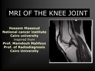
- 1. MRI OF THE KNEE JOINT Hossam Massoud National cancer institute Cairo university inspired from Prof. Mamdouh Mahfouz Prof. of Radiodiagnosis Cairo University
- 2. Knee joint complaint Pain Trauma Swelling Osteoarthritis Suspected pathology [previous Examination] Inflammation Tumors MRI OF THE KNEE JOINT INDICATIONS
- 5. Protocol of examination • Axial T1 localizer • Sagittal T1, PD, T2 • Coronal gradient echo, STIR •If contrast is injected → Axial, Sagittal and coronal T1 WIs Scanning parameters 4mm slice thickness 1mm inter slice gap Field of view [FOV] = 16cm
- 7. How to know the pulse sequence?! T1/ PD T2 Gradient STIR
- 8. PD T1 T2 PD
- 9. Structure or lesionsT2T1 Cortical bone Menisci (medial ,lateral) Ligaments (ACL,PCL,…) Calcification LowLow Common MR appearances
- 10. Common MR appearances Fat (subcutaneous, lipoma,…) Bone marrow LowHigh Fluid (effusion, cyst, ganglion) HighLow Blood (heamoarthrosis) HighHigh T1 T2
- 11. Kinematic MRI
- 13. Where?!Items to be evaluated Sagittal PD Sagittal PD Coronal Axial Sagittal PD Sagittal T1& T2 Sagittal T2 Menisci (medial & lateral) Ligaments - Cruciate (ACL, PCL) - Collateral - Retinacular Tendons ( Quadriceps,Pattelar) Bones Synovial effusion
- 14. Meniscus Medial meniscus Banana- shaped Posterior horn wider, longer, taller than anterior horn Posterior horn tightly attached to the capsule Grade II degeneration more common
- 15. Menisci Lateral meniscus C- shape Posterior and anterior horns are symmetric Anterior horn may be hypo plastic, extremely thin Discoid meniscus and meniscal cysts more common
- 17. AC B A B C
- 18. Lateral meniscus
- 19. Medial meniscus
- 21. Medial & lateral menisci
- 29. Meniscus:
- 30. Meniscus:
- 31. Meniscal Lesions Tear Degeneration Cyst Discoid
- 33. Types of Meniscal degeneration Grade I Grade II Meniscal fraying
- 34. Tear
- 35. Tear
- 39. Meniscal degeneration with free edge fraying
- 40. Meniscal degeneration with free edge fraying
- 41. Types of Meniscal Tears Simple Complex Special types
- 42. Simple Meniscal Tears Horizontal Vertical Radial
- 43. Horizontal tear
- 44. Horizontal tear
- 45. Horizontal tear
- 46. Horizontal tear
- 47. Horizontal tear
- 48. Vertical tear Occurs typically in the outer 1/3 of the posterior horn or body of the meniscus [ rare in the anterior horn ]
- 50. Vertical tear
- 51. Vertical tear
- 52. Radial tear Vertical tear of the free edge of the meniscus [ Root tear ] Ghost meniscus
- 53. Ghost meniscus If there is no history of Meniscal surgery and the posterior horn is absent near the intercondylar notch
- 54. Special Meniscal Tears Flap Bucket handle MC separation
- 55. Flap tear [Oblique ] Should have tow components , horizontal and vertical Common in the medial meniscus Fish mouth
- 58. Decreased height , Free piece Small sized posterior horn Medially displaced fragment Double post. Cruciate ligament sign
- 60. Bucket Handel tear Small sized posterior horn [ sagittal ] Medially displaced fragment[ coronal] Double PCL sign [ sagittal ]
- 64. Flipped meniscus : Double Delta Sign Bucket Handel tear , Lateral meniscus
- 65. Flipped meniscus : Double Delta Sign
- 66. Discoid meniscus Dysplastic meniscus with loss of normal semi lunar shape. 50% or more coverage of the tibial plateau. Meniscal body segment seen in 3 or more sagittal images
- 68. Discoid meniscus
- 69. Discoid meniscus
- 70. Meniscal cyst A Cyst extending from a meniscal tear Common sites : Anterior horn LM , Posterior horn MM
- 74. Ligamentous Lesions ACL PCL Collateral Retinacular
- 78. Primary signs [ In the ligament ] Total discontinuity Abnormal signal Abnormal configuration Abrupt angulation Wavy appearance Abnormal axis Anterior cruciate ligament injury
- 81. Secondary signs [ Outside the ligament ] • Bone contusions [Pivot- shift bruises ] • Anterior translocation of the tibia • Uncovered meniscus sign • Avulsion fracture of the tibial insertion • Segond fracture 70-100% with ACL tear • PCL buckling • PCL line sign Anterior cruciate ligament injury Hyperextension ACL tear with "kissing bone bruises."
- 83. Anterior tibial translocation with” uncovered meniscus sign”
- 84. Segond fracture An elliptical vertically 3x10mm bone fragment parallel to the lateral tibial cortex, about 4mm distal to the plateau. Best seen on AP or tunnel radiographic views 75 -100% association with ACL tear
- 85. Segond fracture in patient with ACL tear. T1- weighted coronal MRI shows a small, low- signal elongated fracture fragment that is parallel to the lateral tibia. The association of Segond fractures with ACL tears approaches 100%.
- 87. Posterior cruciate ligament The major stabilizer of the knee Uniform low signal , no striations Twice strong as the ACL The menisco-femoral ligaments are intimately related to PCL. They connect the posterior horn of the lateral meniscus to the medial femoral condyle Ligament of Humphrey anterior to PCL Ligament of Wrisberg posterior to PCL
- 88. PCL injuries represent about 12% of knee injuries Combined PCL injuries represent 97% With ACL 65% With MCL 50% With MM 30% Posterior cruciate ligament TYPES OF PCL INJURES Complete tear 40% Partial tear 55% Avulsion tear 7%
- 89. NORMAL PCL TORN PCL MR FINDINGS Increased signal due to hemorrhage and edema Diffuse enlargement of PCL
- 91. NORMAL PCL AVULSION TEAR • Involves the tibial insertion • Retracted bone fragment • Bone marrow edema at avulsion site • The actual PCL may be normal
- 93. Collateral ligaments MCL is about 8-11 cm LCL is about 5-7 cm Isolated injuries are rare, usually with ACL and MM
- 94. Collateral ligaments Grade I : microscopic tear Grade II :partial tear Grade III : complete tear GRADING SYSTEM Grade I,II and isolated grade III are treated conservatively, while grade III tears associated with ACL tears are treated by repairing ACL only
- 95. Proton density coronal image shows the normal medial collateral ligament as a thin, taut, well-defined, low-signal structure extending from the medial femoral epicondyle to the medial tibial metaphysis
- 96. Grade I medial collateral ligament tear with surrounding edema (straight arrows) on a T2WI Note the normal thickness and signal of the medial collateral ligament and continued close apposition to the femoral and tibial cortices.
- 97. Grade II medial collateral ligament tear seen on a coronal proton density image shows slight thickening of the medial collateral ligament and separation from the underlying cortices. Bone marrow edema of the lateral tibial plateau is seen due to valgus stress 7 months after conservative treatment
- 98. Grade II medial collateral ligament tear seen on a coronal T1 and STIR images showing slight thickening of the medial collateral ligament and separation from the underlying cortices.
- 99. Grade III medial collateral ligament tear on a coronal fast spin-echo T2-weighted image demonstrates a disrupted ligament that is thickened and retracted with surrounding edema (black arrow).
- 100. Acute grade III tear with a folded ligament (arrow) and surrounding edema on a coronal proton density image.
- 101. Acute tear of the proximal portion of the lateral collateral ligament is seen on this coronal proton density image (white arrow). Note the associated grade II medial collateral ligament tear.
- 103. ? Grade III MCL tear with abnormal signal and edema
- 104. Lateral pressure syndrome Thickening of the lateral retinaculum Lateral knee pain Obese, athletic patients May be associated with chondromalacia
- 105. Patella alta Sequlae of patellofemoral dysplasia Lengthening of the infrapatellar tendon May be associated with chondromalacia Length of patellar tendon/ length of patella > 1.3
- 106. Patella Baja Poliomyelitis Achondroplasia JRA
- 107. Pigmented villo-nodular synovitis Idiopathic Monoarticular disease 1% incidence Hypertrophic synovial masses with hemosiderin laden macrophages bone erosions Intermediate signal in T1 and low signal in T2 with enhancement after contrast injection Typical location posterior to Hoffa’s fat pad Painless swelling , pain with progressive disease Treatment by synovectomy
- 110. PIGMENTED VILLONODULAR SYNOVITIS VERSUS LIPOMA ARBORESCENS
- 112. POPLITEAL CYST Fluid in the bursa which is usually communicating with the joint space Other names Baker’s cyst Gastrocnemius/semimembranosus bursa
- 113. Medial plica syndrome Inflamed synovial plica causing pain , crepitus and pseudolocking Often in adolescents and athletics No measurement for plica thickness Four types of plica Suprapatellar 90% Medial 15 -30% Infrapatellar Lateral [ rare]
- 114. PLICA SYNDROME
- 115. Knee pain with Normal MRI ?! Kinematic Meniscal contusion Plicae
- 116. Plicae Anterior knee pain, clicking, locking Supra patellar, medial patellar, infrapatellar Medial patellar Plicae (Normal variant) 60% of adult knee Medial patellar Plicae
- 117. Supra patellar plica
- 119. Osteochonddritis dissecans Osteochondral fragment in a typical location Young male Lateral aspect of the medial femoral condoyle Variable sized fragment attached or detached Criteria of unstable fragment Large size more than 1cm Fluid between the fragment and donor bone Cystic changes at the donor site Enhancement of the separation line
- 125. Bone infarcts Serpigenous lesions in the bone marrow Variable in size [ Chinese figures ] Double line sign is diagnostic [peripheral hyperintense with hypointense inner border on T2 CAUSES POSTTRAUMATIC STEROIDS COLLAGEN DISEASES ALCOHOLISM PANCREATITIS SPONTANEOUS
- 126. BONE INFARCTS
- 127. BONE INFARCTS
- 130. Quiz Cases
- 131. 1 23 4 56 7 8 Case 1
- 132. 1 2 Case 2
- 133. Case 3
- 134. Case 4
- 135. Case 5
- 136. 1 2 3 4 Case 6
- 137. Case 7
- 138. Case 8
- 139. Case 9
- 140. Case 10 1 2 3 4
- 141. Case 11
- 143. Case 12
- 144. Case 13
- 145. Case 14
- 146. Case 15
- 147. Case 16
- 148. Case 17
- 149. Case 17
- 150. Case 18
- 151. Case 19
- 152. Case 20
- 153. ACL TEAR Case 21
- 154. Case 19