Physiology of Aging3
•
0 likes•389 views

The document discusses age-related changes in visual function based on a lecture. It covers 3 main topics: 1) Optical changes like increased light absorption and scatter in the lens and other structures, reducing retinal illumination. 2) Neural changes in the retina like loss of rods, cones and optic nerve fibers. These neural changes may underlie declines in visual functions. 3) Specific age-related changes in visual functions including reduced acuity, contrast sensitivity, dark adaptation, color vision and oculomotor abilities. Both optical and neural factors contribute to these declines.

Recommended
More Related Content
Viewers also liked
Viewers also liked (20)
Similar to Physiology of Aging3
Similar to Physiology of Aging3 (20)
More from Hossein Mirzaie
More from Hossein Mirzaie (20)
Recently uploaded
Recently uploaded (20)
Physiology of Aging3
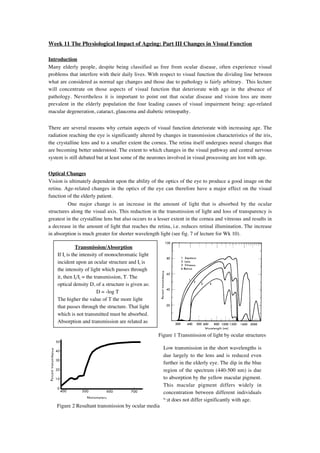
- 1. Week 11 The Physiological Impact of Ageing: Part III Changes in Visual Function Introduction Many elderly people, despite being classified as free from ocular disease, often experience visual problems that interfere with their daily lives. With respect to visual function the dividing line between what are considered as normal age changes and those due to pathology is fairly arbitrary. This lecture will concentrate on those aspects of visual function that deteriorate with age in the absence of pathology. Nevertheless it is important to point out that ocular disease and vision loss are more prevalent in the elderly population the four leading causes of visual impairment being: age-related macular degeneration, cataract, glaucoma and diabetic retinopathy. There are several reasons why certain aspects of visual function deteriorate with increasing age. The radiation reaching the eye is significantly altered by changes in transmission characteristics of the iris, the crystalline lens and to a smaller extent the cornea. The retina itself undergoes neural changes that are becoming better understood. The extent to which changes in the visual pathway and central nervous system is still debated but at least some of the neurones involved in visual processing are lost with age. Optical Changes Vision is ultimately dependent upon the ability of the optics of the eye to produce a good image on the retina. Age-related changes in the optics of the eye can therefore have a major effect on the visual function of the elderly patient. One major change is an increase in the amount of light that is absorbed by the ocular structures along the visual axis. This reduction in the transmission of light and loss of transparency is greatest in the crystalline lens but also occurs to a lesser extent in the cornea and vitreous and results in a decrease in the amount of light that reaches the retina, i.e. reduces retinal illumination. The increase in absorption is much greater for shorter wavelength light (see fig. 7 of lecture for Wk 10). Transmission/Absorption If Ii is the intensity of monochromatic light incident upon an ocular structure and It is the intensity of light which passes through it, then It/Ii = the transmission, T. The optical density D, of a structure is given as: D = -log T The higher the value of T the more light that passes through the structure. That light which is not transmitted must be absorbed. Absorption and transmission are related as Figure 1 Transmission of light by ocular structures Low transmission in the short wavelengths is due largely to the lens and is reduced even further in the elderly eye. The dip in the blue region of the spectrum (440-500 nm) is due to absorption by the yellow macular pigment. This macular pigment differs widely in concentration between different individuals but does not differ significantly with age. Figure 2 Resultant transmission by ocular media
- 2. Another major change in the optics of the eye in later life is increased intra-ocular light scatter which although may not lead to a reduction in the amount of retinal illumination, does cause image degradation due to loss of image contrast via the introduction of veiling luminance. Neural Changes Over the past decade there have been increasing numbers of reports that suggest that age-related changes do occur in the neural components of the retina and the rest of the visual pathway. We are still unclear as to precisely what influence these neural changes have on visual function in the elderly but some of the changes that occur with age are listed below: • loss of rods and cones • ↓ in cone density (in rhesus monkeys) • ↓ human cone photopigment density • structural changes in the outer segments of photoreceptors • ↓ in the number of optic nerve fibres Age-related Changes in Visual Function Acuity The measurement of visual acuity is the standard clinical method of evaluating pattern vision. Acuity is typically assessed using high contrast, high luminance letter charts. There have been many studies that have investigated how acuity changes with age the results of which are summarised in figure 3 below: Figure 3. Mean letter acuity as a function of age as reported in several studies. All studies appear to agree that acuity decreases with increasing age, but there are discrepancies between the studies in terms of the rate of decline, the age of onset of the decline and so forth. These differences are likely to be due in the main to methodological inconsistencies between the studies. Weale (1975) has argued that a large proportion of this acuity loss is due to neural deterioration and cell death in the visual pathway (see below for more on optical vs neural theories). Contrast Sensitivity Spatial Unlike measures of vision such as Snellen acuity, spatial contrast sensitivity testing examines pattern vision at low as well as high contrast for a range of stimulus sizes (or more correctly spatial frequencies). When spatial contrast sensitivity is measured for elderly subjects and compared to the performance in the younger population it is found that older subjects exhibit a loss in sensitivity at higher spatial frequencies (see figure 4). The magnitude of this loss increases with increasing spatial frequency and also when the surrounding light levels decrease.
- 3. 1000 71 yrs 21 yrs Contrast Sensitivity 100 10 Figure 4. Contrast sensitivity as a function of spatial frequency. 1 0.1 1 10 100 Spatial Frequency (cycles/deg) Temporal Temporal contrast sensitivity measurement assesses the sensitivity of the visual system to stimuli which change as a function of time (e.g. flickering or moving). Studies have shown that there is a loss in temporal resolution for luminance modulated uniform fields. With drifting sinusoidal grating stimuli there is a loss of contrast sensitivity with ageing even at low temporal frequencies, for both colour and luminance stimuli (see figure 5). Motion sensitivity also decreases with age. Figure 5. Temporal contrast sensitivity (motion direction discrimination) for colour and luminance stimuli in young and old subjects Optical vs Neural Theories of Sensitivity Loss Although the underlying basis of this loss in sensitivity with age has not be fully explained it would seem that it is not due to refractive error, senile miosis of the pupil, increased light absorption or cognitive factors associated with decision making or setting a threshold. This conclusion has been reached following studies on elderly subjects with intra-ocular lens implants and using laser stimuli which by-pass the eye’s optics yet still indicate a loss of sensitivity at high spatial frequencies. It would appear that neural changes that occur in the retina and visual system are the most likely cause of contrast sensitivity loss with age. Various studies on ageing have tried to answer the question whether there is a selective deterioration of one or other of the two main neural processing pathways, the Parvo- (P) or Magno- (M) cellular pathways (see Spear, 1993). Given the roles played by the two pathways in the visual systems of non-human primates, a deterioration in the high spatial and low temporal frequency domain would suggest a selective P system deficit, whilst the loss of motion sensitivity would suggest a decline in the activity of the M system. The non-specific losses in contrast sensitivity for both colour and luminance patterns (Fiorentini et al., 1996), suggest that ageing affects both P & M systems in generalised manner.
- 4. Colour Vision With regards to the effects of ageing on colour vision there seems to be little doubt that there is a reduction in sensitivity of the short wavelength sensitive (S) cones in later life. Whether this loss extends to the middle (M) and long (L) wavelength sensitive cones is still the matter of debate. The loss of S-cone sensitivity may be due in part to optical factors since we know that absorption for short wavelength sensitive light increases in the elderly crystalline lens. But even when this increased absorption is accounted for S-cone sensitivity is still reduced in the older eye, suggesting that neural age changes must also play some part. Some researchers argue that the S-cone system is more susceptible to damage by the ageing process than either the L or M-cone systems. As a consequence of this reduced S-cone sensitivity elderly patients tend to exhibit more tritan like performance in colour vision tasks. This was demonstrated by Knoblauch et al. (1987) who used the Farnsworth-Munsell 100 hue test to assess the variations of colour vision with age as well as luminance. They showed increases in the error score with age with scores reaching a maximum along the near vertical axis (see figures 6 & 7) indicating tritanopia. Fig 7 Fig 6 Other changes that occur in colour vision include changes in the isoluminant point. When two colours are alternated at a fast rate (15Hz) the relative luminances of the colours can be adjusted until perceived flicker is minimum. At the minimum flicker point the two colours are equi- or iso-luminant. This technique is known as heterochromatic flicker photometry (HFP). When older subjects perform this task they tend to require higher levels of green in order to achieve minimum flicker. FIGURE 8. (A) Contrast sensitivity as a function of colour ratio a horizontal grating 1 c/deg sinusoidally reversed in contrast at 15Hz. The colour ratio 0.47 at which contrast sensitivity was minimum was taken as the equiluminant value. (B) Equihrminant colour ratios of the ten young (o) and ten old subjects (•) participating in the psychophysical experiment, as a function of age. Arrows indicate the means for the 2 age groups. Green iso- Red From Fiorentini et al. (1996) luminant
- 5. Dark Adaptation Dark adaptation is the time dependent increase in visual sensitivity that occurs in darkness following exposure to bright illumination levels and reveals fundamental information about the function of rods and cones. Numerous studies have shown that the elderly have elevated thresholds (i.e. decreased sensitivity) throughout the entire time course of dark adaptation in both the rod and cone portions of the function (see figure 9). The mechanisms that underlie these changes in adaptation in the elderly may be both neural and optical in origin. Threshold Time (mins) Figure 9 Dark adaptation for different age groups Oculomotor Function Age-related changes in dynamic oculomotor control have been reported. • during saccadic eye movements older subjects show an increased latency of saccadic onset. To a lesser extent saccade duration and velocity may decrease with age. • older people also exhibit significantly slower smooth pursuit eye movements for targets moving at speeds greater than 10º/s. • a decrease in the ability to resolve the detail of moving stimuli (Dynamic VA) Binocular Vision There is some indication that binocular summation and stereoacuity decrease with age. However, the conclusions drawn from studies of binocular vision must be treated with some caution since few of the studies carried so far have 1) been rigorous about the ocular health and refractive correction of the participants, 2) yielded direct measures of stereoacuity or 3) used stereo tests that adequately isolated the cue of binocular disparity. Visual Fields Visual sensitivity across the whole visual field is adverse affected by the processes of ageing. Studies have indicated isopter constriction in older adults as well as a generalised loss in sensitivity throughout the whole of the visual field (see figure 10). Fig. 10. Foveal threshold to a small light stimulus as a function of age
- 6. Another approach to examining the visual field is to assess the ‘functional’ or ‘useful’ field of view which involves the localisation and identification of complex stimuli in the periphery. The limits of useful field of view are affected by many factors such as the presence secondary tasks and background distractor stimuli. The impact of these variables is much greater for older people. Visual Electrophysiology The visual evoked potential (VEP) is a composite electrical signal generated by the occipital cortex in response to a visual stimulus. Generally the amplitudes of these responses decrease as a function of age and their latency increases. This is found across a wide range of stimuli that are used to elicit the responses (see figure 11). Fig 11 Mean latencies of the VEPS of young (open columns) and old (hatched columns) subjects Researchers argue that age-related increases in VEP latency are neurally based and are related to decreases in nerve conduction velocities caused by demyelination and retinal deterioration. Objectives: Following this lecture the student should be able to: 1. Describe the major changes that occur in visual function as a result of ageing. 2. Discuss the differential roles of optical and neural factors as reasons for decline in visual function. Further Reading. Weale RA. (1992) The Senescence of Human Vision. Oxford Medical Publications. (Chapters 3 & 4) Library S612.84 Owsley C. & Sloane ME. (1990). Vision and Aging. In: Handbook of Neuropsychology Vol 4. Eds. Boller F. & Grafman J. pp 229-249. Elsevier Science Publishers. Knoblauch, K et al. (1987). Age and illuminance effects in the Farnsworth-Munsell 100 hue test. Applied Optometry 26, 1441-1448. Spear,P. D. (1993). Neural bases of visual deficits during aging. Vision Research, 33, 2589-2609. Fiorentini, A. et al (1996). Visual Ageing: unspecific decline of the responses to luminance and colour. Vision Research, 36, 3557- 3566.